SlideShare ist ein Scribd-Unternehmen logo
1 von 20
Downloaden Sie, um offline zu lesen
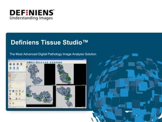
Definiens Tissue Studio™ The Most Advanced Digital Pathology Image Analysis Solution
Definiens Tissue Studio™ overview Enables simple, fast, and accurate ROI detection via the new Definiens Composer Technology™ Provides multi-parametric quantitationon a cell-by-cell basis Handles heterogeneous tissue samples Works with whole slides and TMAs Handles images acquired from any source Operates as a dedicated workstation or as a client server application
Definiens Composer Technology™ What is it? Definiens Composer Technology™ was developed to enable the user to easily teach the system how to identify regions of interest (ROIs).  How does it work? Simply load up to 4 representative images, and use the “paintbrush tool” on a few representative ROIs in your images. Then click the “Learn” button. How does this differ from other approaches?  Training the system with 4 representative images ensures a robust image analysis solution without over fitting. Combines machine learning, pattern recognition and auto adaptive analysis algorithms based on Definiens eCognitionNetwork Language ™. Definiens Tissue Studio™ is faster and more flexible.
ROI Detection and Cell by Cell Analysis across and within selected ROIs Definiens Tissue Studio™ Workflow
Load training images
Define image analysis solution
Load up to four different images:Detect tissue / Background / Segment images
Definiens Composer Technology™:“Paint” on a few representative ROIs
Definiens Composer Technology™:After painting a few ROIs, click the “Learn” button
Definiens Composer Technology™:Computer has now been “trained”
Definiens Composer Technology™:Merge “Object Primitives”
Second Definiens Composer step:Re-classify Islets into “tumor” and “normal”
Select ROIs in tumor islets
ROIs at 20xParameters can be adjusted and previewed here
Detect nuclei
Batch Process:Analyze selected images
Export multi-parametric quantitative features
List of supported instruments Hamamatsu: Nanozoomer Zeiss: Mirax Aperio: Scanscope Applied Imaging: Ariol TissueGnostics Bacus WebSlide DMetrix Generic (tif, jpg, etc.)
Summary Deeper insights  Quantifies nuclear, membrane and cytoplasmic biomarkers on a cell-by-cell basis  Supports all standard IHC tissue stains (blue, brown and red), H&E and IF  Analyzes all slides and TMA images from all major image acquisition devices   Faster results  Identifies regions of interest automatically Operates through a simple, intuitive user interface  Offers unlimited throughput with parallel batch processing  Better decisions  Delivers accurate and reproducible results  Enables multiparametric investigations to identify underlying correlations   Supports translational research through rapid iterative investigation of biomarkers
Questions Contact: Daniel R. Nicolson Director, Global Accounts 484-678-9090 dnicolson@definiens.com

Weitere ähnliche Inhalte

Ähnlich wie Definiens Tissue Studio Advanced Digital Pathology Image Analysis Solution

Automated Face Detection System
Automated Face Detection SystemAutomated Face Detection System
Automated Face Detection SystemAbhiroop Ghatak
 
iVision, an automated imaging system for IHC and ISH on Tissue Microarrays
iVision, an automated imaging system for IHC and ISH on Tissue MicroarraysiVision, an automated imaging system for IHC and ISH on Tissue Microarrays
iVision, an automated imaging system for IHC and ISH on Tissue Microarraysabizarl
 
A DEEP LEARNING APPROACH FOR SEMANTIC SEGMENTATION IN BRAIN TUMOR IMAGES
A DEEP LEARNING APPROACH FOR SEMANTIC SEGMENTATION IN BRAIN TUMOR IMAGESA DEEP LEARNING APPROACH FOR SEMANTIC SEGMENTATION IN BRAIN TUMOR IMAGES
A DEEP LEARNING APPROACH FOR SEMANTIC SEGMENTATION IN BRAIN TUMOR IMAGESPNandaSai
 
An Overview of Software Tools and Platforms for Digital Pathology
An Overview of Software Tools and Platforms for Digital PathologyAn Overview of Software Tools and Platforms for Digital Pathology
An Overview of Software Tools and Platforms for Digital PathologyThe Lifesciences Magazine
 
Machine_Learning_with_MATLAB_Seminar_Latest.pdf
Machine_Learning_with_MATLAB_Seminar_Latest.pdfMachine_Learning_with_MATLAB_Seminar_Latest.pdf
Machine_Learning_with_MATLAB_Seminar_Latest.pdfCarlos Paredes
 
Face recognition technology
Face recognition technologyFace recognition technology
Face recognition technologyranjit banshpal
 
Facial expression recognition projc 2 (3) (1)
Facial expression recognition projc 2 (3) (1)Facial expression recognition projc 2 (3) (1)
Facial expression recognition projc 2 (3) (1)AbhiAchalla
 
"Deep Learning Beyond Cats and Cars: Developing a Real-life DNN-based Embedde...
"Deep Learning Beyond Cats and Cars: Developing a Real-life DNN-based Embedde..."Deep Learning Beyond Cats and Cars: Developing a Real-life DNN-based Embedde...
"Deep Learning Beyond Cats and Cars: Developing a Real-life DNN-based Embedde...Edge AI and Vision Alliance
 
3D localization methods for intracranial electrodes
3D localization methods for intracranial electrodes3D localization methods for intracranial electrodes
3D localization methods for intracranial electrodesBrian Owens
 
The Evolution Of Eclipse 1. 1 )
The Evolution Of Eclipse 1. 1 )The Evolution Of Eclipse 1. 1 )
The Evolution Of Eclipse 1. 1 )Patty Buckley
 
Gaurav Malik Resume 29 Jan 2016
Gaurav Malik Resume 29 Jan 2016Gaurav Malik Resume 29 Jan 2016
Gaurav Malik Resume 29 Jan 2016Gaurav Malik
 
Skin Cancer Detection using Image Processing in Real Time
Skin Cancer Detection using Image Processing in Real TimeSkin Cancer Detection using Image Processing in Real Time
Skin Cancer Detection using Image Processing in Real Timeijtsrd
 
Face detection and recognition
Face detection and recognitionFace detection and recognition
Face detection and recognitionPankaj Thakur
 

Ähnlich wie Definiens Tissue Studio Advanced Digital Pathology Image Analysis Solution (20)

Automated Face Detection System
Automated Face Detection SystemAutomated Face Detection System
Automated Face Detection System
 
EMOTION DETECTION USING AI
EMOTION DETECTION USING AIEMOTION DETECTION USING AI
EMOTION DETECTION USING AI
 
iVision, an automated imaging system for IHC and ISH on Tissue Microarrays
iVision, an automated imaging system for IHC and ISH on Tissue MicroarraysiVision, an automated imaging system for IHC and ISH on Tissue Microarrays
iVision, an automated imaging system for IHC and ISH on Tissue Microarrays
 
A DEEP LEARNING APPROACH FOR SEMANTIC SEGMENTATION IN BRAIN TUMOR IMAGES
A DEEP LEARNING APPROACH FOR SEMANTIC SEGMENTATION IN BRAIN TUMOR IMAGESA DEEP LEARNING APPROACH FOR SEMANTIC SEGMENTATION IN BRAIN TUMOR IMAGES
A DEEP LEARNING APPROACH FOR SEMANTIC SEGMENTATION IN BRAIN TUMOR IMAGES
 
An Overview of Software Tools and Platforms for Digital Pathology
An Overview of Software Tools and Platforms for Digital PathologyAn Overview of Software Tools and Platforms for Digital Pathology
An Overview of Software Tools and Platforms for Digital Pathology
 
Machine_Learning_with_MATLAB_Seminar_Latest.pdf
Machine_Learning_with_MATLAB_Seminar_Latest.pdfMachine_Learning_with_MATLAB_Seminar_Latest.pdf
Machine_Learning_with_MATLAB_Seminar_Latest.pdf
 
Ai use cases
Ai use casesAi use cases
Ai use cases
 
Face recognition technology
Face recognition technologyFace recognition technology
Face recognition technology
 
A guide to Face Detection in Python.pdf
A guide to Face Detection in Python.pdfA guide to Face Detection in Python.pdf
A guide to Face Detection in Python.pdf
 
Facial expression recognition projc 2 (3) (1)
Facial expression recognition projc 2 (3) (1)Facial expression recognition projc 2 (3) (1)
Facial expression recognition projc 2 (3) (1)
 
"Deep Learning Beyond Cats and Cars: Developing a Real-life DNN-based Embedde...
"Deep Learning Beyond Cats and Cars: Developing a Real-life DNN-based Embedde..."Deep Learning Beyond Cats and Cars: Developing a Real-life DNN-based Embedde...
"Deep Learning Beyond Cats and Cars: Developing a Real-life DNN-based Embedde...
 
3D localization methods for intracranial electrodes
3D localization methods for intracranial electrodes3D localization methods for intracranial electrodes
3D localization methods for intracranial electrodes
 
Ch1.pptx
Ch1.pptxCh1.pptx
Ch1.pptx
 
Work Portfolio
Work PortfolioWork Portfolio
Work Portfolio
 
The Evolution Of Eclipse 1. 1 )
The Evolution Of Eclipse 1. 1 )The Evolution Of Eclipse 1. 1 )
The Evolution Of Eclipse 1. 1 )
 
OpenPOWER/POWER9 AI webinar
OpenPOWER/POWER9 AI webinar OpenPOWER/POWER9 AI webinar
OpenPOWER/POWER9 AI webinar
 
Gaurav Malik Resume 29 Jan 2016
Gaurav Malik Resume 29 Jan 2016Gaurav Malik Resume 29 Jan 2016
Gaurav Malik Resume 29 Jan 2016
 
Skin Cancer Detection using Image Processing in Real Time
Skin Cancer Detection using Image Processing in Real TimeSkin Cancer Detection using Image Processing in Real Time
Skin Cancer Detection using Image Processing in Real Time
 
Opencv
OpencvOpencv
Opencv
 
Face detection and recognition
Face detection and recognitionFace detection and recognition
Face detection and recognition
 

Definiens Tissue Studio Advanced Digital Pathology Image Analysis Solution

  • 1. Definiens Tissue Studio™ The Most Advanced Digital Pathology Image Analysis Solution
  • 2. Definiens Tissue Studio™ overview Enables simple, fast, and accurate ROI detection via the new Definiens Composer Technology™ Provides multi-parametric quantitationon a cell-by-cell basis Handles heterogeneous tissue samples Works with whole slides and TMAs Handles images acquired from any source Operates as a dedicated workstation or as a client server application
  • 3. Definiens Composer Technology™ What is it? Definiens Composer Technology™ was developed to enable the user to easily teach the system how to identify regions of interest (ROIs). How does it work? Simply load up to 4 representative images, and use the “paintbrush tool” on a few representative ROIs in your images. Then click the “Learn” button. How does this differ from other approaches? Training the system with 4 representative images ensures a robust image analysis solution without over fitting. Combines machine learning, pattern recognition and auto adaptive analysis algorithms based on Definiens eCognitionNetwork Language ™. Definiens Tissue Studio™ is faster and more flexible.
  • 4. ROI Detection and Cell by Cell Analysis across and within selected ROIs Definiens Tissue Studio™ Workflow
  • 7. Load up to four different images:Detect tissue / Background / Segment images
  • 8. Definiens Composer Technology™:“Paint” on a few representative ROIs
  • 9. Definiens Composer Technology™:After painting a few ROIs, click the “Learn” button
  • 10. Definiens Composer Technology™:Computer has now been “trained”
  • 11. Definiens Composer Technology™:Merge “Object Primitives”
  • 12. Second Definiens Composer step:Re-classify Islets into “tumor” and “normal”
  • 13. Select ROIs in tumor islets
  • 14. ROIs at 20xParameters can be adjusted and previewed here
  • 18. List of supported instruments Hamamatsu: Nanozoomer Zeiss: Mirax Aperio: Scanscope Applied Imaging: Ariol TissueGnostics Bacus WebSlide DMetrix Generic (tif, jpg, etc.)
  • 19. Summary Deeper insights Quantifies nuclear, membrane and cytoplasmic biomarkers on a cell-by-cell basis Supports all standard IHC tissue stains (blue, brown and red), H&E and IF Analyzes all slides and TMA images from all major image acquisition devices   Faster results Identifies regions of interest automatically Operates through a simple, intuitive user interface Offers unlimited throughput with parallel batch processing  Better decisions Delivers accurate and reproducible results Enables multiparametric investigations to identify underlying correlations Supports translational research through rapid iterative investigation of biomarkers
  • 20. Questions Contact: Daniel R. Nicolson Director, Global Accounts 484-678-9090 dnicolson@definiens.com