1. TUBERCULOSIS PLEURAL
EFFUSION - MANAGEMENT
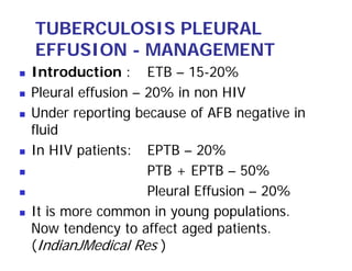
Introduction : ETB – 15-20%
Pleural effusion – 20% in non HIV
Under reporting because of AFB negative in
fluid
In HIV patients: EPTB – 20%
PTB + EPTB – 50%
Pleural Effusion – 20%
It is more common in young populations.
Now tendency to affect aged patients.
(IndianJMedical Res )
2. PATHOGENESIS
Typically 4-7 months following initial
infection with TB.
Rupture of small subpleural focus
Delayed hypersensitivity reaction
Possibility the intense inflammation
obstructs the lymphatic pores in the
parietal pleura which causes
accumulation of protein in pleural
cavity.
3. CLINICAL
MANIFESTATION
Most commonly manifests as an acute
illness
Cough 70% , usually non productive
Chest pain 75%
Usually unilateral
Bil – Occurs in 10%
4. NATURAL HISTORY OF
UNTREATED EFFUSION
90% usually subside spontaneously
Effusion usually resolve and all other
symptoms disappear within 2-4 months
Follow up over time – 40-60% develop
tuberculosis
Size of original effusion and the presence or
absence of small radiologic residual pleural
disease do not correlate with subsequent
tuberculosis
Residual pleural thickening common
sequelae ~ 50%.
5. DIAGNOSIS
Diagnosis depends on the
demonstration of tubercle bacilli –
In sputum
Pleural fluid
Pleural biopsy specimen
Granuloma in pleura
The diagnosis can also be established
with reasonable certaintly by ADA ↑
6. TUBERCULIN TEST
Positive in 60-75% patients, many of negative
patients will become tuberculin positive in 6-8
week
Negative tuberculin – HIV patients &
malnutrition
There may sequestration of PPD reactive
lymphocytes in the pleural space.
A circulating adherent cell suppresses the
sensitized T- lymphocytes
8. RADIOLOGY
Parenchymal infiltrates – 20%
The classic patterns of primary tuberculosis
in the form of non cavitary, lower lobe
parenchymal infiltrates.
CT Scan – 40% parenchyma involvement
Encysted effusion usually occurs during ATT
treatment
Most often encystment occurs in the
costoparietal regions ( Post aspects)
9. PLEURAL FLUID – ANALYSIS
Often protein > 5.0 gm/ dl
Usually more than 50% lymphocytes
If symptoms less than 2 weeks then pleural
fluid may reveal poly.
Eosinophil > 10% usually excludes the
diagnosis of tuberculosis pleuritis.
Mesothelial cells < 3% .
HIV infected patients with tuberculosis
pleuritis may have significant number of
mesothelial cells in the pleural fluids.
10. ADA
Enzyme that catalyzes the conversion of
adenosinie to inosine.
1978 – Pirus et al
ADA-1 – All cells
ADA -2 – Only in monocytes
Majority of ADA in tuberculosis pleural fluid
is ADA -2
Higher the pleural fluid ADA level, the more
likely patient is to have tuberculosis.
11. ADA (contd.)
Level of cut-off value
High Specificity – 83%
Sensitivity – 77-100%
L: N > 0.75 – Specificity ↑
High ADA – empyema
- Rheumatoid arthritis
But do not have pleural fluid
lymphocytosis
Cut off value < 40
40-60
12. ADA ( contd.)
Although the use of a ratio of the ADA -1
to ADA -2 less than 0: 42 will slightly
increase the sensitivity and specificity of
ADA, the separation of ADA into its
isoenzyme is not necessary.
ADA level maintained at ambient
temperature. It decreases with time. But
addition of glycerol, sodium sulfate–Stable
at room temperature for 3 months.
13. INTERFERON – GAMMA
Produced by CD4
Sensitivity & specificity > 94%
It retains diagnostic efficacy in HIV patients
IFN - γ is more specific than ADA chiefly
due to low levels of IFN in lymphoma and
empyma.
Expensive not used routinely.
14. PCR
PCR is certainly not superior to either the
pleural fluid ADA or interferon gamma
levels
Sensitivity ~ 80%
Specificity is not 100%
False positive may occur due to DNA
contamination
Nonviable organism
Latent infection
Reactivation due to imunosuppression caused
by primary malignant involvement of pleura.
15. Lysozyme
Released from lysozyme containing cells –
granulocytes, monocytes, macrophages.
The value of lysozyme levels in the
diagnosis of tuberculosis effusion, however,
is controversial
16. ADA : Lysozyme , has been found to
be useful in differentiating tuberculosis
from empyema. A thresholds value
above 3.3 has been shown to be highly
specific for tuberculosis pleural effusion.
17. PLEURAL FLUIDS STAINS
AND CULTURE
In immunocompetent patients AFB
smear – rarely positive.
In HIV patients AFB smear is positive –
20%
Pleural fluid culture is positive ~ 40%
Use of BACTEC system with bedside
inoculation provides higher yield and
faster results than do conventional
methods.
18. PLEURAL BIOPSY
The demonstration of granuloma in the
parietal pleura suggests TB
Other common causes of granuloma
fungal disease
RA
Sarcoidosis
Acid fast stain is positive in 25%.
Culture for AFB positive – 60% .
Combination of three test. Positive in
91%.
19. Standard recommendation for biopsy
If ADA > 70 L:N > 0.75 – No biopsy
If ADA – 40-70 : L:N > 0.75
Presumptive diagnosis of tuberculosis
Biopsy if clinical picture is not
supportive of tuberculosis
ADA < 40- Clinical picture suggestive of
tuberculosis – Pleural biopsy
20. USG guided ( Tru cut ) biopsy of pleura
Involvement of pleura is not uniform, so
closed biopsy may miss actual site of
lesions.
Closed biopsy with cope or an abrams
needle can obtain adequate specimen and
achieve 57-80% diagnostic rate in
tuberculosis pleuritis.
21. DISADVANTAGES
Air leakage
Needle breakage
May not be diagnostic
Abrams needle wide bore, needs skin
incision
Nonspecific changes upto as high as
68% have been reported in the closed
biopsy.
22. USG GUIDED TRU CUT
BIOPSY
Advantages
It can localize the minimal or loculated
effusion
it can demonstrate focal pleural
thickening
sensitivity for pleural tuberclosis – 86%
23. TREATMENTS
Goals
To prevent the subsequent
development of active tuberculsosis
To relieve symptoms
to prevent development of a fibrothorax
24. ATT
Average patient becomes afebrile
without 2 week
Complete resorption of fluid 6-12 weeks
The incidence of pleural thickening at 6-
12 months -50%
25. CORTICOSTEROIDS
No role of systemic steroids
If more than mildly symptomatic
A therapeutic thoracentesis is
recommended
Severe symptoms & diagnosis has been
established – Steroids
26. Surgical procedure
Large pleural effusion – thoracentesis,
Decortication rarely necessary
Tuberculosis Empyema
ICTD + ATT
27. HIV & TUBERCULOSIS
PLEURAL EFFUSION
Incidence – CD4 < 200- 0 ↓
CD> 200 - ↑
Overall incidence - ±↑↓
Duration of illness longer
Chest pain less common
28. Systemic manifestation – Fever, anemia,
lymphadenopathy, organomegaly – common
Mantoux test often negative (40%)
AFB smear in pleural fluid -25%
If CD4 count < 100. 50% positive fluid AFB smear
positive
Significant number of mesothelial cells in their
pleural fluid
Biopsy – Granuloma on pleural biopsy are less well
formed
In pleural fluid ,the level of albumin are relatively
low
The level of ADA in patients without AIDS are
comparable.