Acute coronary syndrome
•Als PPTX, PDF herunterladen•
210 gefällt mir•16,335 views

Acute coronary syndrome, NSTEMI, STEMI, UA, unstable angina, Cardiology, rounds, IM, Internal Medicine, Cebu, CDUH, Evardone

Empfohlen
Weitere ähnliche Inhalte
Was ist angesagt?
Was ist angesagt? (20)
Ähnlich wie Acute coronary syndrome
Ähnlich wie Acute coronary syndrome (20)
Kürzlich hochgeladen
Kürzlich hochgeladen (20)
Acute coronary syndrome
- 1. ACUTE CORONARY SYNDROME Jose Socrates ‘Dee’ Matuod Evardone Cardiology RIC Level II CDUH-IM JULY 2015
- 2. CASE Profile: 78F, Filipino Hypertensive, Non-DM, Non-smoker, Non-alcoholic beverage drinker CC: CHEST PAIN
- 3. CASE HPI: 2 hours PTA- sudden onset of squeezing, retrosternal chest pain, radiating towards the neck, associated with headache. (+) Vomiting x1, pain score of 8/10, not relieved by rest, about >10 minutes. Admitted at primary hospital and managed as essential hypertension with NSSTWCs on ECG. Subsequently transferred to a Tertiary level for further workup and management
- 4. CASEPhysical Exam Awake, coherent, in mild distress Warm, good turgor and mobility, CRT <2 seconds, Anicteric sclerae, pink palpebral conjunctivae, Equal chest expansion, dec BS on both lower lung fields, with crackles Distinct heart sounds, , tachycardic, regular, no murmur Flat, Normoactive bowel sounds, soft, no organomegaly Strong peripheral pulses, no edema Neurologic: WNL BP: 130/60mmHg, HR: 118 bpm, RR: 26 cpm, 99% O2 mp: 36.5° Neck: No JVD, no murmur
- 5. CASE IMPRESSION: 1) NSTE-ACS (UA vs NSTEMI) 2) HCVD
- 6. CASE DIAGNOSTICS: ECG – Sinus Rhythm with NSSTWCs CXR – Beginning congestion, inflammatory process bilaterally Troponin I – 3.13 (elevated)
- 7. CASE MANAGEMENT: (ER) Aspirin 80 mg 4 tabs chewed, swallowed Clopidogrel 75mg , 4 tabs Clexane 0.4cc Attached to cardiac monitor Advised ICU/CCU admission
- 9. ACUTE CORONARY SYNDROME I: Diagnosis and Management of Patients with Stable Ischemic Heart Disease II: Diagnosis and Management of Patients with Non-ST Elevation Acute Coronary Syndrome III: Diagnosis and Management of Patients with ST Segment Elevation Myocardial Infarction
- 10. ACUTE CORONARY SYNDROME What is ACS? - refers to a spectrum of clinical presentations ranging from those for ST-segment elevation myocardial infarction (STEMI) to presentations found in non–ST- segment elevation myocardial infarction (NSTEMI) or in unstable angina - in general reserved for ischemia precipitated by acute coronary atherothrombosis
- 12. ACUTE CORONARY SYNDROME Ischemic Heart Disease Chronic coronary artery disease (CAD) with STABLE ANGINA Acute Coronary Syndrome STEMI 1) NSTEMI-ACS 2) Unstable Angina
- 13. ACUTE CORONARY SYNDROME Classic manifestation of ischemia is angina pectoris: Pressure, Tightness, Squeezing, Heaviness, Burning ACS most commonly occurs without obvious precipitating factors
- 18. LOCALIZATION OF CORONARY CIRCULATION IN M.I. ANATOMIC ECG LEADS CORONARY ARTERY Septal V1-v2 Proximal LAD Anterior V3-V4 LAD Apical V5-V6 Distal LAD, LCx, or RCA Lateral I, aVL LCx Inferior II, III, aVF RCA(85%), LCx (15%) RV V1-V2 & V4R (most Se) Proximal RCA Posterior ST depression V1-V3 RCA or LCx
- 22. ACUTE CORONARY SYNDROME The American College of Cardiology (ACC) and American Heart Association (AHA) guidelines list the following as pain descriptions uncharacteristic of myocardial ischemia: • Pleuritic pain (i.e., sharp or knifelike pain brought on by respiratory movements or coughing) • Primary or sole location of the discomfort in the middle or lower abdominal region • Pain that may be localized by the tip of one finger, particularly over the left ventricular apex • Pain reproduced with movement or palpation of the chest wall or arms • Constant pain that persists for many hours • Very brief episodes of pain that last a few seconds or less • Pain that radiates into the lower extremities
- 24. Ischemic Heart Disease The major determinants of myocardial oxygen demand (MVO2) are: 1) heart rate, 2) myocardial contractility, and 3) myocardial wall tension (stress) About 75% of the total coronary resistance to flow occurs across three sets of arteries: (1) large epicardial arteries (Resistance 1 = R1), (2) prearteriolar vessels (R2), and (3) arteriolar and intramyocardial capillary vessels (R3)
- 25. 50% Stenosis: there is a limitation of the ability to increase flow to meet increased myocardial demand. 80% Stenosis: myocardial ischemia at rest or with minimal stress Ischemic Heart Disease Coronary Blood Flow Limitation: • spasm, • arterial thrombi, and, • rarely, coronary emboli • as well as by ostial narrowing due to aortitis • Congenital Anomalies
- 27. The severity and duration of the imbalance between myocardial oxygen supply and demand determine whether the damage is : reversible (≤20 min for total occlusion in the absence of collaterals) or permanent, with subsequent myocardial necrosis (>20 min). Ischemic Heart Disease
- 28. ACUTE CORONARY SYNDROME Evaluation of the patient with known or suspected ischemic heart disease
- 29. Requirements : • Two sets of cardiac enzymes at 4-hr intervals should be normal • ECG at the time of arrival and preexercise 12-lead ECG show no significant abnormality • Absence of rest electrocardiographic abnormalities that would preclude accurate assessment of the exercise ECG • From admission to the time that results are available from the second set of cardiac enzymes: patient asymptomatic, lessening chest pain symptoms, or persistent atypical symptoms • Absence of ischemic chest pain at the time of exercise testing Indications and Contraindications for Exercise Electrocardiographic Testing in the Emergency Department Contraindications • New or evolving electrocardiographic abnormalities on the rest tracing • Abnormal cardiac enzyme levels • Inability to perform exercise • Worsening or persistent ischemic chest pain symptoms from admission to the time of exercise testing • Clinical risk profiling indicating that imminent coronary angiography is likely
- 32. STABLE ISCHEMIC HEART DISEASE Laboratory Tests: 1. Fasting lipid profile 2. Fasting glucose and/or glycated hemoglobin (HbA1c) level if available; additional oral glucose tolerance test (OGTT) if both are inconclusive; 3. Complete blood count (CBC); 4. Creatinine level with estimation of glomerular filtration rate (GFR); 5. Biochemical markers of myocardial injury (Troponin T or I) if clinical evaluation suggests an Acute Coronary Syndrome (ACS); 6. Thyroid hormone levels 7. Liver function tests early after beginning statin therapy.
- 33. ISCHEMIC HEART DISEASE Pre-Test Probability (PTP) Assessment Diamond and Forrester pre-test probability of cad by age, sex and symptoms
- 35. ISCHEMIC HEART DISEASE ESTABLISHING DIAGNOSIS Non-invasive stress testing Stress Imaging
- 36. ISCHEMIC HEART DISEASE ESTABLISHING DIAGNOSIS
- 37. ISCHEMIC HEART DISEASE ESTABLISHING DIAGNOSIS Exercise ECG (Treadmill Exercise Test or TET) its simplicity, lower cost and widespread availability, the TET is the initial test of choice to identify inducible ischemia in the majority of patients with intermediate PTP who are able to exercise The low sensitivity of the TET (45% to 50%) despite a high specificity (85% to 90%) is the reason why it is not recommended in patients with a PTP greater than 65% In the latter case,a stress imaging study is more appropriate.
- 38. ISCHEMIC HEART DISEASE CORONARY ARTERIOGRAPHY Coronary arteriography is indicated in: (1) patients with chronic stable angina pectoris who are severely symptomatic despite medical therapy and are being considered for revascularization, i.e., a percutaneous coronary intervention (PCI) or coronary artery bypass grafting (CABG); (2) patients with troublesome symptoms that present diagnostic difficulties in whom there is a need to confirm or rule out the diagnosis of IHD; (3) patients with known or possible angina pectoris who have survived cardiac arrest; (4) patients with angina or evidence of ischemia on noninvasive testing with clinical or laboratory evidence of ventricular dysfunction; and (5) patients judged to be at high risk of sustaining coronary events based on signs of severe ischemia on noninvasive testing, regardless of the presence or severity of symptoms
- 39. ISCHEMIC HEART DISEASE MANAGEMENT Lifestyle Modification and Treatment of Risk Factors 1) Healthy Diet 2) Physical Activity 3) Hypertension 4) Smoking 5) DM 6) Weight Management 7) Lipid Management
- 40. ISCHEMIC HEART DISEASE MANAGEMENT Pharmacologic Therapy to Improve Prognosis Whether or not revascularization is being considered, receive the following medications to improve prognosis, thereby reducing the risk for MI and death: 1. Aspirin low-dose (80 to 160 mg/day) 2. Clopidogrel in case of aspirin intolerance (75 mg/day) 3. Statins irrespective of LDL-cholesterol levels 4. Beta blockers post-MI 5. ACEIs or ARBs (especially in patients with concomitant HF, hypertension or diabetes)
- 41. Pharmacologic Therapy to Improve Prognosis
- 42. Pharmacologic Therapy to Improve Prognosis
- 43. Pharmacologic Therapy to Improve Prognosis
- 45. Non-ST-Segment Elevation Acute Coronary Syndrome (Non-ST-Segment Elevation Myocardial Infarction and Unstable Angina)
- 46. NSTE-ACS Pathophysiology: 1. imbalance between oxygen supply and oxygen demand resulting from a partially occluding thrombus forming on a disrupted atherothrombotic coronary plaque or on eroded coronary artery endothelium 2. dynamic obstruction 3. severe mechanical obstruction 4. increased myocardial oxygen demand
- 47. NSTE-ACS
- 48. NSTE-ACS
- 49. NSTE-ACS DIAGNOSIS: Clinical : (1) it occurs at rest (or with minimal exertion), lasting >10 minutes; (2) it is of relatively recent onset (i.e., within the prior 2 weeks); and/or (3) it occurs with a crescendo pattern (i.e., distinctly more severe, prolonged, or frequent than previous episodes) NSTEMI - elevated levels of biomarkers of cardiac necrosis
- 50. NSTE-ACS DIAGNOSIS: 3 major noninvasive tools are used in the evaluation of NSTEMI-ACS: 1. the electrocardiogram (ECG), 2. cardiac biomarkers, and 3. stress testing GOALS : (1) recognize or exclude myocardial infarction (MI) using cardiac biomarkers, preferably cTn; (2) detect rest ischemia (using serial or continuous ECGs); and (3) detect significant coronary obstruction at rest with CCTA and myocardial ischemia using stress testing
- 55. MEDICAL TREATMENT • Bed rest • Continuous ECG monitoring • Ambulation only if No recurrence of ischemia (symptoms or ECG changes) Does not develop an elevation of a biomarker of necrosis for 12–24 h ANTI-ISCHEMIC ANTITHROMBOTIC
- 56. NSTE-ACS Drugs Commonly Used in Intensive Medical Management of Patients with UA and NSTEMI
- 60. NSTE-ACS ORAL ANTIPLATELETS Aspirin Initial dose of 325 mg nonenteric formulation followed by 75–100 mg/d of an enteric or a nonenteric formulation Clopidogrel Loading dose of 300–600 mg followed by 75 mg/d Prasugrel Pre-PCI: Loading dose 60 mg followed by 10 mg/d Ticagrelor Loading dose of 180 mg followed by 90 mg twice daily Intravenous Antiplatelet Therapy Abciximab 0.25 mg/kg bolus followed by infusion of 0.125 μg/ kg per min (maximum 10 μg/min) for 12–24 h Eptifibatide 180 μg/kg bolus followed 10 min later by second bolus of 180 μg with infusion of 2.0 μg/kg per min for 72–96 h following first bolus Tirofiban 5 μg/kg per min followed by infusion of 0.15 μg/kg per min for 48– 96 h
- 61. NSTE-ACS HEPARINS Unfractionated heparin (UFH) Bolus 70–100 U/kg (maximum 5000 U) IV followed by infusion of 12–15 U/kg per h (initial maximum 1000 U/h) titrated to ACT 250–300 s Enoxaparin 1 mg/kg SC every 12 h; the first dose may be preceded by a 30-mg IV bolus; renal adjustment to 1 mg/kg once daily if creatine clearance <30 cc/min Fondaparinux 2.5 mg SC qd Bivalirudin Initial IV bolus of 0.75 mg/kg and an infusion of 1.75 mg/kg per h.
- 62. NSTE-ACS Class I Recommendations for Use of an Early Invasive Strategy in Patients with Non-ST-Segment Elevation Acute Coronary Syndrome: Class I (Level of Evidence: A) Indications 1. Recurrent angina at rest/low-level activity despite treatment 2. Elevated TnT or TnI 3. New ST-segment depression 4. CHF symptoms, rales, MR 5. EF <0.40 6. Sustained VT 7. PCI <6 months, prior CABG 8. High-risk findings from noninvasive testing 9. Hemodynamic instability 10. Mild-to-moderate renal dysfunction 11. Diabetes mellitus 12. High TIMI Risk Score (>3)b
- 63. NSTE-ACS PRINZMETAL’S VARIANT ANGINA - syndrome of severe ischemic pain that usually occurs at rest and is associated with transient ST segment elevation - focal spasm of an epicardial coronary artery, leading to severe transient myocardial ischemia and occasionally infarction - may be related to hypercontractility of vascular smooth muscle due to adrenergic vasoconstrictors, leukotrienes, or serotonin - has decreased substantially during the past few decades - PVA are generally younger and have fewer coronary risk factors - The clinical diagnosis of PVA is made by the detection of transient ST-segment elevation with rest pain - Focal spasm is most common in the right coronary artery
- 64. NSTE-ACS PRINZMETAL’S VARIANT ANGINA Diagnosis: Hyperventilation or intracoronary acetylcholine has been used to provoke focal coronary stenosis on angiography or to provoke rest angina with ST-segment elevation to establish the diagnosis TREATMENT Nitrates and Calcium Channel Blockers Aspirin- may actually increase the severity of ischemic episodes PROGNOSIS 1. Survival at 5 years is excellent (∼90–95%) 2. Nonfatal MI occurs in up to 20% of patients by 5 years 3. There is a tendency for symptoms 4. and cardiac events to diminish over time
- 65. NSTE-ACS CORONARY ANGIOGRAPHY IS NOT RECOMMENDED in patients with • extensive co-morbidities (e.g., liver or pulmonary failure; cancer); • in whom the risks of revascularization are not likely to outweigh the benefits; • in patients with acute chest pain and a low likelihood of ACS; • or in patients who will not consent to revascularization regardless of the findings.
- 66. NSTE-ACS Revascularization by PCI PCI IS RECOMMENDED for NSTE-ACS patients with 1- to 2-vessel CAD, with or without significant proximal left anterior descending CAD, but with a large area of viable myocardium and high-risk criteria on noninvasive testing.
- 67. NSTE-ACS Revascularization by CABG Surgery CABG IS RECOMMENDED for patients with 1. significant left main disease, and is the preferred revascularization strategy for patients with 2. multi-vessel coronary disease; 3. vessels with lesions not favorable for PCI; 4. depressed systolic function (LVEF lower than 50%); and diabetes.
- 68. NSTE-ACS Drug/Medication Management Strongly RECOMMENDED for patients with 1. aspirin indefinitely, and ticagrelor 90 mg twice daily or clopidogrel 75 mg daily, for 12 months 2. underwent stenting, with aspirin indefinitely, plus ticagrelor 90 mg twice daily or prasugrel 10 mg daily or clopidogrel 75 mg daily, for 12 months in patients with drug-eluting stents, and 6 months in patients with bare metal stents 3. beta blockers be continued indefinitely for all NSTE- ACS patients unless contraindicated
- 72. STEMI Criteria for Acute Myocardial Infarction • Detection of a rise and/or fall of cardiac biomarker values (preferably cardiac troponin [cTn]) with at least one value above the 99th percentile upper reference limit (URL) and with at least one of the following: • Symptoms of ischemia • New or presumed new significant ST-segment T-wave (ST-T) changes or new left bundle branch block (LBBB) • Development of pathologic Q waves in the electrocardiogram (ECG) • Imaging evidence of new loss of viable myocardium or new regional wall motion abnormality • Identification of an intracoronary thrombus by angiography or autopsy • Cardiac death with symptoms suggestive of myocardial ischemia and presumed new ischemic ECG changes of new LBBB, but death occurred before cardiac biomarkers were obtained or before cardiac biomarker values would be increased.
- 73. STEMI Criteria for Acute Myocardial Infarction • Percutaneous coronary intervention (PCI)–related MI is arbitrarily defined by elevation of cTn values (>5 × 99th percentile URL) in patients with normal baseline values (≤99th percentile URL) or a rise of cTn values >20% if the baseline values are elevated and are stable or falling. In addition, either • Coronary artery bypass grafting (CABG)–related MI is arbitrarily defined by elevation of cardiac biomarker values (>10 × 99th percentile URL) in patients with normal baseline cTn values (≤99th percentile URL). In addition, either (i) symptoms suggestive of myocardial ischemia, or (ii) new ischemic ECG changes, or (iii) angiographic findings consistent with a procedural complication, or (iv) imaging demonstration of new loss of viable myocardium or new regional wall motion abnormality are required. • Stent thrombosis associated with MI when detected by coronary angiography or autopsy in the setting of myocardial ischemia and with a rise and/ or fall of cardiac biomarker values with at least one value above the 99th percentile URL. (i) new pathologic Q waves or new LBBB, or (ii) angiographic documented new graft or new native coronary artery occlusion, or (iii) imaging evidence of new loss of viable myocardium or new regional wall motion abnormality.
- 74. STEMI Classification of Myocardial Infarction Type I: Spontaneous Myocardial Infarction Type 2: Myocardial Infarction Secondary to an Ischemic Imbalance Type 3: Myocardial Infarction Resulting in Death When Biomarker Values Are Unavailable Type 4a: Myocardial Infarction Related to Percutaneous Coronary Intervention (PCI) Type 4b: Myocardial Infarction Related to Stent Thrombosis Type 5: Myocardial Infarction Related to Coronary Artery Bypass Grafting (CABG
- 76. STEMI the goals for the management of patients with suspected STEMI include: 1. control of cardiac discomfort, 2. rapid identification of patients who are candidates for urgent reperfusion therapy, 3. triage of lower-risk patients to the appropriate location in the hospital, and 4. avoidance of inappropriate discharge of patients with STEMI MANAGEMENT IN THE EMERGENCY DEPARTMENT Aspirin is essential in the management of patients with suspected STEMI and is effective across the entire spectrum of acute coronary syndromes OXYGEN O2 should be administered by nasal prongs or face mask (2–4 L/min) for the first 6–12 h after infarction
- 77. STEMI 1) Sublingual nitroglycerin Up to three doses of 0.4 mg should be administered at about 5-min intervals 2) Morphine 3) Intravenous beta blockers/ Oral Beta blockers CONTROL OF DISCOMFORT in the first 24 h for patients who do not have any of the following: (1) signs of heart failure, (2) evidence of a low-output state, (3) increased risk for cardiogenic shock, or (4) other relative contraindications to beta blockade (PR interval greater than 0.24 seconds, second- or third-degree heart block, active asthma, or reactive airway disease). Fifteen minutes after the last intravenous dose, an oral regimen is initiated of 50 mg every 6 h for 48 h, followed by 100 mg every 12 h
- 78. STEMI
- 79. STEMI Initial ER Management • Aspirin 160 to 320 mg tablet (non-enteric coated, chewed); • Clopidogrel 300 to 600 mg whether or not fibrinolysis will be given; • Clopidgrel 600 mg or prasugrel 60 mg or ticagrelor 180 mg when a patient will undergo PCI; • Nitrates, either via sublingual or intravenous(IV) routes. Nitrates are contraindicated in patients with hypotension or those who took a phosphodiesterase 5 (PDE5) inhibitor within 24 hrs (48 hrs for tadalafil); • Morphine 2 to 4 mg IV for relief of chest pain, and; • Supplemental oxygen MAY BE RECOMMENDED during the first 6 hours to patients with arterial oxygen saturation of less than 90%.
- 80. STEMI In-hospital Treatment Reperfusion therapy IS RECOMMENDED to all eligible patients with STEMI with symptom onset within the prior 12 hours. Early revascularization, the goal being 12 hours, is a primary treatment goal in patients with STEMI
- 81. STEMI
- 82. STEMI Fibrinolysis 15-mg bolus followed byTPA • 50 mg intravenously over the first 30 min, • followed by 35 mg over the next 60 min 1.5 million units (MU) intravenously over 1 hStreptokinase given as a single weight-based intravenous bolus of 0.53 mg/kg over 10 s Tenecteplase (TNK) double-bolus regimen consisting of a 10-MU bolus given over 2–3 min, followed byReteplase (rPA) • second 10-MU bolus 30 min later
- 83. STEMI
- 84. STEMI
- 85. STEMI TIMI FLOW GRADE — The degree of perfusion in the infarct-related artery (IRA) is typically described by the TIMI flow grade: ●TIMI 0 refers to the absence of antegrade flow beyond a coronary occlusion. ●TIMI 1 flow is faint antegrade coronary flow beyond the occlusion, although filling of the distal coronary bed is incomplete. ●TIMI 2 flow is delayed or sluggish antegrade flow with complete filling of the distal territory. ●TIMI 3 flow is normal flow which fills the distal coronary bed completely. TIMI
- 86. STEMI Primary Percutaneous Coronary Intervention (PCI) 1. RECOMMENDED in patients with STEMI and ischemic symptoms of less than 12 hours’ duration 2. RECOMMENDED in patients with STEMI and ischemic symptoms of less than 12 hours’ duration who have contraindications to fibrinolytic therapy, irrespective of the time delay from first medical contact. 3. RECOMMENDED in patients with STEMI and cardiogenic shock or acute severe heart failure (HF), irrespective of time delay from MI onset 4. MAY BE RECOMMENDED in patients with STEMI if there is clinical and/or ECG evidence of ongoing ischemia between 12 and 24 hours after symptom onset
- 87. STEMI Coronary Artery Bypass Grafting (CABG) 1. IS RECOMMENDED in failed PCI with persistent pain or hemodynamic instability in patients with coronary anatomy suitable for surgery 2. IS RECOMMENDED in persistent or recurrent ischemia refractory to medical therapy in patients who have coronary anatomy suitable for surgery, and are not candidates for PCI or fibronolytic therapy 3. RECOMMENDED in patients with STEMI at the time of operative repair of mechanical defects.
- 88. STEMI Antiplatelets and Antithrombotics 1. It IS RECOMMENDED that aspirin should be used indefinitely in all patients with STEMI with a dosage of 80 to 100 mg/day. 2. It IS RECOMMENDED that clopidogrel 75 mg per day orally should be added to aspirin in patients with STEMI and maintained for at least 14 days 3. It IS RECOMMENDED that patients who are truly intolerant to aspirin can instead receive clopidogrel 75 mg per day as long-term secondary prevention
- 89. STEMI Duration of Dual Antiplatelet Therapy and Antithrombotic Combination Therapies after STEMI 1. Combination Therapies after STEMI DAPT by combining aspirin and an ADP-receptor blocker (clopidogrel, prasugrel or ticagrelor) IS RECOMMENDED in patients with STEMI who are undergoing primary PCI (for up to 12 months) or (clopidogrel) fibrinolysis (for up to 12 months, although the data available pertain only to one month of DAPT), and in those who have not undergone reperfusion therapy (for at least 1 month and up to 12 months).
- 90. STEMI Lipid Lowering Agents 1. High-dose statins are RECOMMENDED in all patients during the first 24 hours of admission for STEMI, irrespective of the patient’s cholesterol concentration, in the absence of contraindications (allergy, active liver disease). 2. Atorvastatin or Rosuvastatin are recommended during the early phase of therapy up to at least four weeks. 3. It IS RECOMMENDED to give high-dose rosuvastatin (20 to 40 mg) or atorvastatin (40 to 80 mg) therapy before emergency percutaneous coronary intervention to 1. reduce periprocedural inflammatory response, 2. to reduce myocardial dysfunction, and 3. to prevent contrast-induced nephropathy
- 91. THANK YOU
- 94. STABLE ISCHEMIC HEART DISEASE Recommended: Echocardiography Ambulatory ECG Monitoring
- 97. STABLE ISCHEMIC HEART DISEASE
- 98. STABLE ISCHEMIC HEART DISEASE
- 99. STABLE ISCHEMIC HEART DISEASE
- 101. ACUTE CORONARY SYNDROME RISK FACTORS
- 102. STEMI
- 103. STEMI
- 104. ACUTE CORONARY SYNDROME Definition of Myocardial Infarction
- 105. ACUTE CORONARY SYNDROME Classification of Myocardial Infarction
Hinweis der Redaktion
- the term acute coronary syndrome, which encompasses unstable angina, NSTEMI, and STEMI, is in general reserved for ischemia precipitated by acute coronary atherothrombosis
- the term acute coronary syndrome, which encompasses unstable angina, NSTEMI, and STEMI, is in general reserved for ischemia precipitated by acute coronary atherothrombosis. Acute coronary syndrome (ACS) refers to a spectrum of clinical presentations ranging from those for ST-segment elevation myocardial infarction (STEMI) to presentations found in non–ST-segment elevation myocardial infarction (NSTEMI) or in unstable angina. It is almost always associated with rupture of an atherosclerotic plaque and partial or complete thrombosis of the infarct-related artery.
- the term acute coronary syndrome, which encompasses unstable angina, NSTEMI, and STEMI, is in general reserved for ischemia precipitated by acute coronary atherothrombosis
- The classic manifestation of ischemia is angina, which is usually described as a heavy chest pressure or squeezing, a burning feeling, or difficulty breathing (see Chapter 11). The discomfort often radiates to the left shoulder, neck, or arm. It typically builds in intensity over a period of a few minutes. The pain may begin with exercise or psychological stress, but ACS most commonly occurs without obvious precipitating factors. Myocardial ischemia causing chest discomfort, termed angina pectoris, is a primary clinical concern in patients presenting with chest symptoms. Myocardial ischemia is precipitated by an imbalance between myocardial oxygen requirements and myocardial oxygen supply, resulting in insufficient delivery of oxygen to meet the heart’s metabolic demands. Myocardial oxygen consumption may be elevated by increases in heart rate, ventricular wall stress, and myocardial contractility, whereas myocardial oxygen supply is determined by coronary
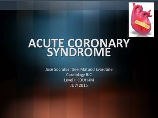
- This picture shows the main symptoms of a heart attack. People who are having a heart attack often have only some of these symptoms. The pain, pressure, and discomfort caused by a heart attack mostly affect the left side of the body (shown in darker red) but can also affect the right. If you think you are having a heart attack, call 9-1-1 for an ambulance. Do not try to get yourself to the hospital.
- Figure 12.9 Coronary Circulation The coronary arteries supply arterial blood to the muscular wall of the heart. The pressure gradient for flow through the arteries is affected by tissue pressure in the wall of the heart during systole, particularly in the left coronary circulation. Thus, while flow through both right and left coronary arteries is related to aortic pressure, these compressive forces reduce left coronary flow during systole. During diastole, flow is greatly enhanced in the left coronary circulation by the actions of vasodilator metabolites that build up during systole. The metabolite adenosine is believed to be particularly important in this enhancement of coronary flow.
- Figure 12.9 Coronary Circulation The coronary arteries supply arterial blood to the muscular wall of the heart. The pressure gradient for flow through the arteries is affected by tissue pressure in the wall of the heart during systole, particularly in the left coronary circulation. Thus, while flow through both right and left coronary arteries is related to aortic pressure, these compressive forces reduce left coronary flow during systole. During diastole, flow is greatly enhanced in the left coronary circulation by the actions of vasodilator metabolites that build up during systole. The metabolite adenosine is believed to be particularly important in this enhancement of coronary flow.
- Figure 12.9 Coronary Circulation The coronary arteries supply arterial blood to the muscular wall of the heart. The pressure gradient for flow through the arteries is affected by tissue pressure in the wall of the heart during systole, particularly in the left coronary circulation. Thus, while flow through both right and left coronary arteries is related to aortic pressure, these compressive forces reduce left coronary flow during systole. During diastole, flow is greatly enhanced in the left coronary circulation by the actions of vasodilator metabolites that build up during systole. The metabolite adenosine is believed to be particularly important in this enhancement of coronary flow.
- Criteria for Acute Myocardial Infarction The term acute myocardial infarction (MI) should be used when there is evidence of myocardial necrosis in a clinical setting consistent with acute myocardial ischemia. Under these conditions, any one of the following criteria meets the diagnosis for MI: • Detection of a rise and/or fall of cardiac biomarker values (preferably cardiac troponin [cTn]) with at least one value above the 99th percentile upper reference limit (URL) and with at least one of the following: • Symptoms of ischemia • New or presumed new significant ST-segment T-wave (ST-T) changes or new left bundle branch block (LBBB) • Development of pathologic Q waves in the electrocardiogram (ECG) • Imaging evidence of new loss of viable myocardium or new regional wall motion abnormality • Identification of an intracoronary thrombus by angiography or autopsy • Cardiac death with symptoms suggestive of myocardial ischemia and presumed new ischemic ECG changes of new LBBB, but death occurred before cardiac biomarkers were obtained or before cardiac biomarker values would be increased. • Percutaneous coronary intervention (PCI)–related MI is arbitrarily defined by elevation of cTn values (>5 × 99th percentile URL) in patients with normal baseline values (≤99th percentile URL) or a rise of cTn values >20% if the baseline values are elevated and are stable or falling. In addition, either (i) symptoms suggestive of myocardial ischemia, or (ii) new ischemic ECG changes, or (iii) angiographic findings consistent with a procedural complication, or (iv) imaging demonstration of new loss of viable myocardium or new regional wall motion abnormality are required. • Stent thrombosis associated with MI when detected by coronary angiography or autopsy in the setting of myocardial ischemia and with a rise and/ or fall of cardiac biomarker values with at least one value above the 99th percentile URL. • Coronary artery bypass grafting (CABG)–related MI is arbitrarily defined by elevation of cardiac biomarker values (>10 × 99th percentile URL) in patients with normal baseline cTn values (≤99th percentile URL). In addition, either (i) new pathologic Q waves or new LBBB, or (ii) angiographic documented new graft or new native coronary artery occlusion, or (iii) imaging evidence of new loss of viable myocardium or new regional wall motion abnormality
- The American College of Cardiology (ACC) and American Heart Association (AHA) guidelines list the following as pain descriptions uncharacteristic of myocardial ischemia7: • Pleuritic pain (i.e., sharp or knifelike pain brought on by respiratory movements or coughing) • Primary or sole location of the discomfort in the middle or lower abdominal region • Pain that may be localized by the tip of one finger, particularly over the left ventricular apex • Pain reproduced with movement or palpation of the chest wall or arms • Constant pain that persists for many hours • Very brief episodes of pain that last a few seconds or less • Pain that radiates into the lower extremities
- The major determinants of myocardial oxygen demand (MVO2) are heart rate, myocardial contractility, and myocardial wall tension (stress). Blood flows through the coronary arteries in a phasic fashion, with the majority occurring during diastole. About 75% of the total coronary resistance to flow occurs across three sets of arteries: (1) large epicardial arteries (Resistance 1 = R1), (2) prearteriolar vessels (R2), and (3) arteriolar and intramyocardial capillary vessels (R3). In the absence of significant flow-limiting atherosclerotic obstructions, R1 is trivial; the major determinant of coronary resistance is found in R2 and R3.
- Coronary blood flow also can be limited by spasm (see Prinzmetal’s angina in Chap. 294), arterial thrombi, and, rarely, coronary emboli as well as by ostial narrowing due to aortitis. Congenital abnormalities such as the origin of the left anterior descending coronary artery from the pulmonary artery may cause myocardial ischemia and infarction in infancy, but this cause is very rare in adults. When a stenosis reduces the diameter of an epicardial artery by 50%, there is a limitation of the ability to increase flow to meet increased myocardial demand. When the diameter is reduced by ∼80%, blood flow at rest may be reduced, and further minor decreases in the stenotic orifice area can reduce coronary flow dramatically to cause myocardial ischemia at rest or with minimal stress.
- Cascade of mechanisms and manifestations of ischemia.
- The severity and duration of the imbalance between myocardial oxygen supply and demand determine whether the damage is reversible (≤20 min for total occlusion in the absence of collaterals) or permanent, with subsequent myocardial necrosis (>20 min).
- Figure 293-2 Evaluation of the patient with known or suspected ischemic heart disease. On the left of the figure is an algorithm for identifying patients who should be referred for stress testing and the decision pathway for determining whether a standard treadmill exercise with electrocardiogram (ECG) monitoring alone is adequate. A specialized imaging study is necessary if the patient cannot exercise adequately (pharmacologic challenge is given) or if there are confounding features on the resting ECG (symptom-limited treadmill exercise may be used to stress the coronary circulation). Panels B–E, continued on the next page, are examples of the data obtained with ECG monitoring and specialized imaging procedures. CMR, cardiac magnetic resonance; EBCT, electron beam computed tomography; ECHO, echocardiography; IHD, ischemic heart disease; MIBI, methoxyisobutyl isonitrite; MR, magnetic resonance; PET, positron emission tomography. A. Lead V4 at rest (top panel) and after 4.5 min of exercise (bottom panel). There is 3 mm (0.3 mV) of horizontal ST-segment depression, indicating a positive test for ischemia. (Modified from BR Chaitman, in E Braunwald et al [eds]: Heart Disease, 8th ed, Philadelphia, Saunders, 2008.) B. A 45-year-old avid jogger who began experiencing classic substernal chest pressure underwent an exercise echo study. With exercise the patient’s heart rate increased from 52 to 153 beats/min. The left ventricular chamber dilated with exercise, and the septal and apical portions became akinetic to dyskinetic (red arrow). These findings are strongly suggestive of a significant flow-limiting stenosis in the proximal left anterior descending artery, which was confirmed at coronary angiography. (Modified from SD Solomon, in E. Braunwald et al [eds]: Primary Cardiology, 2nd ed, Philadelphia, Saunders, 2003.) C. Stress and rest myocardial perfusion single-photon emission computed tomography images obtained with 99m-technetium sestamibi in a patient with chest pain and dyspnea on exertion. The images demonstrate a medium-size and severe stress perfusion defect involving the inferolateral and basal inferior walls, showing nearly complete reversibility, consistent with moderate ischemia in the right coronary artery territory (red arrows). (Images provided by Dr. Marcello Di Carli, Nuclear Medicine Division, Brigham and Women’s Hospital, Boston, MA.) D. A patient with a prior myocardial infarction presented with recurrent chest discomfort. On cardiac magnetic resonance (CMR) cine imaging, a large area of anterior akinesia was noted (marked by the arrows in the top left and right images, systolic frame only). This area of akinesia was matched by a larger extent of late gadolinium-DTPA enhancements consistent with a large transmural myocardial infarction (marked by arrows in the middle left and right images). Resting (bottom left) and adenosine vasodilating stress (bottom right) first-pass perfusion images revealed reversible perfusion abnormality that extended to the inferior septum. This patient was found to have an occluded proximal left anterior descending coronary artery with extensive collateral formation. This case illustrates the utility of different modalities in a CMR examination in characterizing ischemic and infarcted myocardium. DTPA, diethylenetriamine penta-acetic acid. (Images provided by Dr. Raymond Kwong, Cardiovascular Division, Brigham and Women’s Hospital, Boston, MA.) E. Stress and rest myocardial perfusion PET images obtained with rubidium-82 in a patient with chest pain on exertion. The images demonstrate a large and severe stress perfusion defect involving the mid and apical anterior, anterolateral, and anteroseptal walls and the left ventricular apex, showing complete reversibility, consistent with extensive and severe ischemia in the mid-left anterior descending coronary artery territory (red arrows). (Images provided by Dr. Marcello Di Carli, Nuclear Medicine Division, Brigham and Women’s Hospital, Boston, MA.)
- TABLE 50-7 Indications and Contraindications for Exercise Electrocardiographic Testing in the Emergency Department Requirements before exercise electrocardiographic testing that should be considered in the ED setting: • Two sets of cardiac enzymes at 4-hr intervals should be normal • ECG at the time of arrival and preexercise 12-lead ECG show no significant abnormality • Absence of rest electrocardiographic abnormalities that would preclude accurate assessment of the exercise ECG • From admission to the time that results are available from the second set of cardiac enzymes: patient asymptomatic, lessening chest pain symptoms, or persistent atypical symptoms • Absence of ischemic chest pain at the time of exercise testing Contraindications to exercise electrocardiographic testing in the ED setting: • New or evolving electrocardiographic abnormalities on the rest tracing • Abnormal cardiac enzyme levels • Inability to perform exercise • Worsening or persistent ischemic chest pain symptoms from admission to the time of exercise testing • Clinical risk profiling indicating that imminent coronary angiography is likely
- Laboratory Tests It IS RECOMMENDED that the following initial laboratory tests be performed to establish CV risk factors, identify possible causes of ischemia and determine prognosis: 1. Fasting lipid profile (total cholesterol, high density lipoprotein [HDL] cholesterol, low density lipoprotein [LDL] cholesterol and triglyceride levels); 2. Fasting glucose and/or glycated hemoglobin (HbA1c) level if available; additional oral glucose tolerance test (OGTT) if both are inconclusive; 3. Complete blood count (CBC); 4. Creatinine level with estimation of glomerular filtration rate (GFR); 5. Biochemical markers of myocardial injury (Troponin T or I) if clinical evaluation suggests an Acute Coronary Syndrome (ACS); 6. Thyroid hormone levels if with clinical suspicion of thyroid disorder, and; 7. Liver function tests early after beginning statin therapy.
- High: > 90% pre-test probability Intermediate: between 10% and 90% pre-test probability Low: between 5% and 10% pre-test probability Very low: < 5% pre-test probability Adapted from Diamond GA, Forrester JS. (1982). Probability of CAD.8
- A stress imaging study IS RECOMMENDED as the initial diagnostic and prognostic test, if facilities, resources and local expertise permit, in patients within the higher range of PTP; patients with LVEF less than 50% without typical angina; patients with resting ECG abnormalities and especially symptomatic patients with prior revascularization (percutaneous coronary intervention [PCI] or coronary artery bypass grafting [CABG]). Exercise testing rather than pharmacological testing is recommended whenever possible.
- An exercise ECG (TET) IS RECOMMENDED as the initial diagnostic and prognostic test, if resources and local expertise for a stress imaging study are not available, in patients with intermediate PTP who have normal resting ECGs and are able to exercise. 30 Because of its simplicity, lower cost and widespread availability, the TET is the initial test of choice to identify inducible ischemia in the majority of patients with intermediate PTP who are able to exercise. The low sensitivity of the TET (45% to 50%) despite a high specificity (85% to 90%) is the reason why it is not recommended in patients with a PTP greater than 65%. In the latter case, a stress imaging study is more appropriate.
- Examples of other indications for coronary arteriography include the following: 1. Patients with chest discomfort suggestive of angina pectoris but a negative or nondiagnostic stress test who require a definitive diagnosis for guiding medical management, alleviating psychological stress, career or family planning, or insurance purposes. 2. Patients who have been admitted repeatedly to the hospital for a suspected acute coronary syndrome (Chaps. 294 and 295), but in whom this diagnosis has not been established and in whom the presence or absence of CAD should be determined. 3. Patients with careers that involve the safety of others (e.g., pilots, firefighters, police) who have questionable symptoms or suspicious or positive noninvasive tests and in whom there are reasonable doubts about the state of the coronary arteries. 4. Patients with aortic stenosis or hypertrophic cardiomyopathy and angina in whom the chest pain could be due to IHD. 5. Male patients >45 years and females >55 years who are to undergo a cardiac operation such as valve replacement or repair and who may or may not have clinical evidence of myocardial ischemia. 6. Patients after myocardial infarction, especially those who are at high risk after myocardial infarction because of the recurrence of angina or the presence of heart failure, frequent ventricular premature contractions, or signs of ischemia on the stress test. 7. Patients with angina pectoris, regardless of severity, in whom noninvasive testing indicates a high risk of coronary events (poor exercise performance or severe ischemia). 8. Patients in whom coronary spasm or another nonatherosclerotic cause of myocardial ischemia (e.g., coronary artery anomaly, Kawasaki disease) is suspected.
- 1. Increase polyunsaturated fatty acid consumption, mainly from oily fish: 2 to 3 servings of oily fish per week may help prevent CV disease (CVD); 2. Saturated fatty acids must comprise less than 10% of the total energy intake; protein sources that are low in saturated fats should replace those high in these harmful fats; 3. Limit salt intake to less than 5 grams per day; added salt should be limited; 4. Consume 30 to 45 grams of fiber per day (wholegrain products, fruits and vegetables); simple carbohydrates that have a high glycemic load should be avoided in favor of carbohydrate sources that are high in fiber; 5. Consume 200 grams of vegetables per day (2 to 3 servings or 1 to 4 cups per day); 6. Eat 200 grams of fruits per day (2 to 3 servings or 1 to 2.5 cups per day), and; 7. Limit consumption of alcoholic beverages to two glasses per day (20 grams) for men, and one glass per day (10 grams) for women
- An exercise ECG (TET) IS RECOMMENDED as the initial diagnostic and prognostic test, if resources and local expertise for a stress imaging study are not available, in patients with intermediate PTP who have normal resting ECGs and are able to exercise. 30 Because of its simplicity, lower cost and widespread availability, the TET is the initial test of choice to identify inducible ischemia in the majority of patients with intermediate PTP who are able to exercise. The low sensitivity of the TET (45% to 50%) despite a high specificity (85% to 90%) is the reason why it is not recommended in patients with a PTP greater than 65%. In the latter case, a stress imaging study is more appropriate.
- Patients considered high risk for mortality are those with high-risk coronary anatomy and those with moderate or severe LV dysfunction. High-risk coronary anatomy includes: at least 50% stenosis of the left main coronary artery and/or significant (at least 70% stenosis) 3-vessel CAD; significant proximal LAD stenosis; and a Synergy between PCI with Taxus and Cardiac Surgery (SYNTAX) score of 33 or higher. Moderate or severe LV dysfunction is defined as LVEF of less than 45% and 35%, respectively. These subset of patients have a better prognosis with CABG than with medical treatment.
- PATHOPHYSIOLOGY NSTE-ACS is most commonly caused by an imbalance between oxygen supply and oxygen demand resulting from a partially occluding thrombus forming on a disrupted atherothrombotic coronary plaque or on eroded coronary artery endothelium. Severe ischemia or myocardial necrosis may occur consequent to the reduction of coronary blood flow caused by the thrombus and by downstream embolization of platelet aggregates and/or atherosclerotic debris. Other causes of NSTE-ACS include: (1) dynamic obstruction (e.g., coronary spasm, as in Prinzmetal’s variant angina [see “Prinzmetal’s Variant Angina” later]); (2) severe mechanical obstruction due to progressive coronary atherosclerosis; and (3) increased myocardial oxygen demand produced by conditions such as fever, tachycardia, and thyrotoxicosis in the presence of fixed epicardial coronary obstruction. More than one of these processes may be involved.
- Figure 294-1 Trends of incidence of ST-segment elevation myocardial infarction (STEMI) and non-ST-segment elevation myocardial infarction (NSTEMI) and of frequency of use of troponin assay to diagnose acute myocardial infarction. NRMI, National Registry of Myocardial Infarction.
- 294-3 Algorithm for evaluation and management of patients with suspected acute coronary syndrome (ACS). Follow-up studies refer to ST deviations and elevation of troponin levels. cTn, cardiac troponin; ECG, electrocardiogram; LV, left ventricular. (Modified from JL Anderson et al: J Am Coll Cardiol 61:e179, 2013.)
- The diagnosis of NSTE-ACS is based largely on the clinical presentation. Typically, chest discomfort is severe and has at least one of three features: (1) it occurs at rest (or with minimal exertion), lasting >10 minutes; (2) it is of relatively recent onset (i.e., within the prior 2 weeks); and/or (3) it occurs with a crescendo pattern (i.e., distinctly more severe, prolonged, or frequent than previous episodes). The diagnosis of NSTEMI is established if a patient with these clinical features develops evidence of myocardial necrosis, as reflected in abnormally elevated levels of biomarkers of cardiac necrosis (see below).
- DIAGNOSTIC EVALUATION In addition to the clinical examination, three major noninvasive tools are used in the evaluation of NSTEMI-ACS: the electrocardiogram (ECG), cardiac biomarkers, and stress testing. CCTA is an additional emerging option (Fig. 294-2). The goals are to: (1) recognize or exclude myocardial infarction (MI) using cardiac biomarkers, preferably cTn; (2) detect rest ischemia (using serial or continuous ECGs); and (3) detect significant coronary obstruction at rest with CCTA and myocardial ischemia using stress testing (Chap 270e
- MEDICAL TREATMENT Patients should be placed at bed rest with continuous ECG monitoring for ST-segment deviation and cardiac arrhythmias. Ambulation is permitted if the patient shows no recurrence of ischemia (symptoms or ECG changes) and does not develop an elevation of a biomarker of necrosis for 12–24 h. Medical therapy involves simultaneous anti-ischemic and antithrombotic
- Drugs Commonly Used in Intensive Medical Management of Patients with Unstable Angina and Non-ST-Segment Elevation Myocardial Infarction
- In 1959 Prinzmetal et al. described a syndrome of severe ischemic pain that usually occurs at rest and is associated with transient ST-segment elevation. Prinzmetal’s variant angina (PVA) is caused by focal spasm of an epicardial coronary artery, leading to severe transient myocardial ischemia and occasionally infarction. The cause of the spasm is not well defined, but it may be related to hypercontractility of vascular smooth muscle due to adrenergic vasoconstrictors, leukotrienes, or serotonin. For reasons that are not clear, the prevalence of PVA has decreased substantially during the past few decades.
- Nitrates and calcium channel blockers are the main therapeutic agents. Aspirin may actually increase the severity of ischemic episodes, possibly as a result of the sensitivity of coronary tone to modest changes in the synthesis of prostacyclin. The response to beta blockers is variable. Coronary revascularization may be helpful in patients who also have discrete, flow-limiting, proximal fixed obstructive lesions. Many patients with PVA pass through an acute, active phase, with frequent episodes of angina and cardiac events during the first 6 months after presentation. Survival at 5 years is excellent (∼90–95%). Patients with no or mild fixed coronary obstruction tend to experience a more benign course than do patients with associated severe obstructive lesions. Nonfatal MI occurs in up to 20% of patients by 5 years. Patients with PVA who develop serious arrhythmias during spontaneous episodes of pain are at a higher risk for sudden cardiac death. In most patients who survive an infarction or the initial 3- to 6-month period of frequent episodes, there is a tendency for symptoms and cardiac events to diminish over time.
- Coronary angiography IS NOT RECOMMENDED in patients with extensive co-morbidities (e.g., liver or pulmonary failure; cancer); in whom the risks of revascularization are not likely to outweigh the benefits; in patients with acute chest pain and a low likelihood of ACS; or in patients who will not consent to revascularization regardless of the findings. The benefits of coronary angiography should be balanced by its risks. The benefits of coronary angiography are highest among high risk patients. It is not recommended for patients whose risks may outweigh the benefits, such as in those with extensive comorbidities, or those with a low probability of ACS. In these patients, invasive evaluation can be delayed without increased risk. Non-invasive assessments of inducible ischemia may be performed before hospital discharge.
- Statement 20: Revascularization by PCI PCI IS RECOMMENDED for NSTE-ACS patients with 1- to 2-vessel CAD, with or without significant proximal left anterior descending CAD, but with a large area of viable myocardium and high-risk criteria on noinvasive testing. If lesions are difficult to assess, fractional flow reserve measurements may be used in order to decide on the treatment strategy.
- Statement 21: Revascularization by CABG Surgery CABG IS RECOMMENDED for patients with significant left main disease, and is the preferred revascularization strategy for patients with multi-vessel coronary disease; vessels with lesions not favorable for PCI; depressed systolic function (LVEF lower than 50%); and diabetes. The general consensus in other guidelines is that CABG is most beneficial in patients with disease of the left main coronary artery; involvement of multiple vessels; and other high-risk patients such as those with ventricular dysfunction or diabetes.38
- STATEMENT 24: Drug/Medication Management It IS STRONGLY RECOMMENDED to maintain patients who were treated medically with aspirin indefinitely, and ticagrelor 90 mg twice daily or clopidogrel 75 mg daily, for 12 months. It IS STRONGLY RECOMMENDED to maintain patients who underwent stenting, with aspirin indefinitely, plus ticagrelor 90 mg twice daily or prasugrel 10 mg daily or clopidogrel 75 mg daily, for 12 months in patients with drug-eluting stents, and 6 months in patients with bare metal stents. It IS STRONGLY RECOMMENDED that beta blockers be continued indefinitely for all NSTE-ACS patients unless contraindicated. In those with moderate or severe systolic dysfunction, the dosing regimen must be titrated slowly.
- Figure 295-3 Major components of time delay between onset of symptoms from ST-segment elevation myocardial infarction and restoration of flow in the infarct-related artery. Plotted sequentially from left to right are the times for patients to recognize symptoms and seek medical attention, transportation to the hospital, in-hospital decision making, implementation of reperfusion strategy, and restoration of flow once the reperfusion strategy has been initiated. The time to initiate fibrinolytic therapy is the “door-to-needle” (D-N) time; this is followed by the period of time required for pharmacologic restoration of flow. More time is required to move the patient to the catheterization laboratory for a percutaneous coronary interventional (PCI) procedure, referred to as the “door-to-balloon” (D-B) time, but restoration of flow in the epicardial infarct–related artery occurs promptly after PCI. At the bottom is a variety of methods for speeding the time to reperfusion along with the goals for the time intervals for the various components of the time delay. (Adapted from CP Cannon et al: J Thromb Thrombol 1:27, 1994.)
- Definition of Myocardial Infarction Criteria for Acute Myocardial Infarction The term acute myocardial infarction (MI) should be used when there is evidence of myocardial necrosis in a clinical setting consistent with acute myocardial ischemia. Under these conditions, any one of the following criteria meets the diagnosis for MI: • Detection of a rise and/or fall of cardiac biomarker values (preferably cardiac troponin [cTn]) with at least one value above the 99th percentile upper reference limit (URL) and with at least one of the following: • Symptoms of ischemia • New or presumed new significant ST-segment T-wave (ST-T) changes or new left bundle branch block (LBBB) • Development of pathologic Q waves in the electrocardiogram (ECG) • Imaging evidence of new loss of viable myocardium or new regional wall motion abnormality • Identification of an intracoronary thrombus by angiography or autopsy • Cardiac death with symptoms suggestive of myocardial ischemia and presumed new ischemic ECG changes of new LBBB, but death occurred before cardiac biomarkers were obtained or before cardiac biomarker values would be increased. • Percutaneous coronary intervention (PCI)–related MI is arbitrarily defined by elevation of cTn values (>5 × 99th percentile URL) in patients with normal baseline values (≤99th percentile URL) or a rise of cTn values >20% if the baseline values are elevated and are stable or falling. In addition, either (i) symptoms suggestive of myocardial ischemia, or (ii) new ischemic ECG changes, or (iii) angiographic findings consistent with a procedural complication, or (iv) imaging demonstration of new loss of viable myocardium or new regional wall motion abnormality are required. • Stent thrombosis associated with MI when detected by coronary angiography or autopsy in the setting of myocardial ischemia and with a rise and/ or fall of cardiac biomarker values with at least one value above the 99th percentile URL. • Coronary artery bypass grafting (CABG)–related MI is arbitrarily defined by elevation of cardiac biomarker values (>10 × 99th percentile URL) in patients with normal baseline cTn values (≤99th percentile URL). In addition, either (i) new pathologic Q waves or new LBBB, or (ii) angiographic documented new graft or new native coronary artery occlusion, or (iii) imaging evidence of new loss of viable myocardium or new regional wall motion abnormality. Criteria for Prior Myocardial Infarction Any one of the following criteria meets the diagnosis for prior MI: • Pathologic Q waves with or without symptoms in the absence of nonischemic causes. • Imaging evidence of a region of loss of viable myocardium that is thinned and fails to contract, in the absence of a nonischemic cause. • Pathologic findings of a prior M
- Definition of Myocardial Infarction Criteria for Acute Myocardial Infarction The term acute myocardial infarction (MI) should be used when there is evidence of myocardial necrosis in a clinical setting consistent with acute myocardial ischemia. Under these conditions, any one of the following criteria meets the diagnosis for MI: • Detection of a rise and/or fall of cardiac biomarker values (preferably cardiac troponin [cTn]) with at least one value above the 99th percentile upper reference limit (URL) and with at least one of the following: • Symptoms of ischemia • New or presumed new significant ST-segment T-wave (ST-T) changes or new left bundle branch block (LBBB) • Development of pathologic Q waves in the electrocardiogram (ECG) • Imaging evidence of new loss of viable myocardium or new regional wall motion abnormality • Identification of an intracoronary thrombus by angiography or autopsy • Cardiac death with symptoms suggestive of myocardial ischemia and presumed new ischemic ECG changes of new LBBB, but death occurred before cardiac biomarkers were obtained or before cardiac biomarker values would be increased. • Percutaneous coronary intervention (PCI)–related MI is arbitrarily defined by elevation of cTn values (>5 × 99th percentile URL) in patients with normal baseline values (≤99th percentile URL) or a rise of cTn values >20% if the baseline values are elevated and are stable or falling. In addition, either (i) symptoms suggestive of myocardial ischemia, or (ii) new ischemic ECG changes, or (iii) angiographic findings consistent with a procedural complication, or (iv) imaging demonstration of new loss of viable myocardium or new regional wall motion abnormality are required. • Stent thrombosis associated with MI when detected by coronary angiography or autopsy in the setting of myocardial ischemia and with a rise and/ or fall of cardiac biomarker values with at least one value above the 99th percentile URL. • Coronary artery bypass grafting (CABG)–related MI is arbitrarily defined by elevation of cardiac biomarker values (>10 × 99th percentile URL) in patients with normal baseline cTn values (≤99th percentile URL). In addition, either (i) new pathologic Q waves or new LBBB, or (ii) angiographic documented new graft or new native coronary artery occlusion, or (iii) imaging evidence of new loss of viable myocardium or new regional wall motion abnormality. Criteria for Prior Myocardial Infarction Any one of the following criteria meets the diagnosis for prior MI: • Pathologic Q waves with or without symptoms in the absence of nonischemic causes. • Imaging evidence of a region of loss of viable myocardium that is thinned and fails to contract, in the absence of a nonischemic cause. • Pathologic findings of a prior M
- Type I: Spontaneous Myocardial Infarction Spontaneous myocardial infarction related to atherosclerotic plaque rupture, ulceration, fissuring, erosion, or dissection with resulting intraluminal thrombus in one or more of the coronary arteries leading to decreased myocardial blood flow or distal platelet emboli with ensuing myocyte necrosis. The patient may have underlying severe coronary artery disease (CAD) but on occasion nonobstructive or no CAD. Type 2: Myocardial Infarction Secondary to an Ischemic Imbalance In instances of myocardial injury with necrosis where a condition other than CAD contributes to an imbalance between myocardial oxygen supply and/ or demand, e.g., coronary endothelial dysfunction, coronary artery spasm, coronary embolism, tachy-brady-arrhythmias, anemia, respiratory failure, hypotension, and hypertension with or without left ventricular hypertrophy. Type 3: Myocardial Infarction Resulting in Death When Biomarker Values Are Unavailable Cardiac death with symptoms suggestive of myocardial ischemia and presumed new ischemic electrocardiogram (ECG) changes or new left bundle branch block (LBBB), but death occurring before blood samples could be obtained or before cardiac biomarker could rise, or in rare cases, cardiac biomarkers were not collected. Type 4a: Myocardial Infarction Related to Percutaneous Coronary Intervention (PCI) Myocardial infarction associated with PCI is arbitrarily defined by elevation of cardiac troponin (cTn) values >5 × 99th percentile upper reference limit (URL) in patients with normal baseline values (≤99th percentile URL) or a rise of cTn values >20% if the baseline values are elevated and are stable or falling. In addition, either (i) symptoms suggestive of myocardial ischemia, or (ii) new ischemic ECG changes or new LBBB, or (iii) angiographic loss of patency of a major coronary artery or a side branch or persistent slow or no flow or embolization, or (iv) imaging demonstration of new loss of viable myocardium or new regional wall motion abnormality is required. Type 4b: Myocardial Infarction Related to Stent Thrombosis Myocardial infarction associated with stent thrombosis is detected by coronary angiography or autopsy in the setting of myocardial ischemia and with a rise and/or fall of cardiac biomarker values with at least one value above the 99th percentile URL. Type 5: Myocardial Infarction Related to Coronary Artery Bypass Grafting (CABG) Myocardial infarction associated with CABG is arbitrarily defined by elevation of cardiac biomarker values >10 × 99th percentile URL in patients with normal baseline cTn values (≤99th percentile URL). In addition, either (i) new pathologic Q waves or new LBBB, or (ii) angiographic documented new graft or new native coronary artery occlusion, or (iii) imaging evidence of new loss of viable myocardium or new regional wall motion abnormality.
- In the Emergency Department, the goals for the management of patients with suspected STEMI include control of cardiac discomfort, rapid identification of patients who are candidates for urgent reperfusion therapy, triage of lower-risk patients to the appropriate location in the hospital, and avoidance of inappropriate discharge of patients with STEMI. Many aspects of the treatment of STEMI are initiated in the Emergency Department and then continued during the in-hospital phase of management (Fig. 295-4). The overarching goal is to minimize the time from first medical contact to initiation of reperfusion therapy. This may involve transfer from a non-PCI hospital to one that is PCI capable, with a goal of initiating PCI within 120 min of first medical contact (Fig. 295-4). Aspirin is essential in the management of patients with suspected STEMI and is effective across the entire spectrum of acute coronary syndromes (Fig. 295-1). Rapid inhibition of cyclooxygenase-1 in platelets followed by a reduction of thromboxane A2 levels is achieved by buccal absorption of a chewed 160–325-mg tablet in the Emergency Department. This measure should be followed by daily oral administration of aspirin in a dose of 75–162 mg. In patients whose arterial O2 saturation is normal, supplemental O2 is of limited if any clinical benefit and therefore is not cost-effective. However, when hypoxemia is present, O2 should be administered by nasal prongs or face mask (2–4 L/min) for the first 6–12 h after infarction; the patient should then be reassessed
- Sublingual nitroglycerin can be given safely to most patients with STEMI. Up to three doses of 0.4 mg should be administered at about 5-min intervals. In addition to diminishing or abolishing chest discomfort, nitroglycerin may be capable of both decreasing myocardial oxygen demand (by lowering preload) and increasing myocardial oxygen supply (by dilating infarct-related coronary vessels or collateral vessels). In patients whose initially favorable response to sublingual nitroglycerin is followed by the return of chest discomfort, particularly if accompanied by other evidence of ongoing ischemia such as further ST-segment or T-wave shifts, the use of intravenous nitroglycerin should be considered. Therapy with nitrates should be avoided in patients who present with low systolic arterial pressure (<90 mmHg) or in whom there is clinical suspicion of RV infarction (inferior infarction on ECG, elevated jugular venous pressure, clear lungs, and hypotension). Nitrates should not be administered to patients who have taken a phosphodiesterase-5 inhibitor for erectile dysfunction within the preceding 24 h, because it may potentiate the hypotensive effects of nitrates. An idiosyncratic reaction to nitrates, consisting of sudden marked hypotension, sometimes occurs but can usually be reversed promptly by the rapid administration of intravenous atropine. Morphine is a very effective analgesic for the pain associated with STEMI. However, it may reduce sympathetically mediated arteriolar and venous constriction, and the resulting venous pooling may reduce cardiac output and arterial pressure. These hemodynamic disturbances usually respond promptly to elevation of the legs, but in some patients, volume expansion with intravenous saline is required. The patient may experience diaphoresis and nausea, but these events usually pass and are replaced by a feeling of well-being associated with the relief of pain. Morphine also has a vagotonic effect and may cause bradycardia or advanced degrees of heart block, particularly in patients with inferior infarction. These side effects usually respond to atropine (0.5 mg intravenously). Morphine is routinely administered by repetitive (every 5 min) intravenous injection of small doses (2–4 mg), rather than by the subcutaneous administration of a larger quantity, because absorption may be unpredictable by the latter route. Intravenous beta blockers are also useful in the control of the pain of STEMI. These drugs control pain effectively in some patients, presumably by diminishing myocardial O2 demand and hence ischemia. More important, there is evidence that intravenous beta blockers reduce the risks of reinfarction and ventricular fibrillation (see “Beta- Adrenoceptor Blockers” below). However, patient selection is important when considering beta blockers for STEMI. Oral beta blocker therapy should be initiated in the first 24 h for patients who do not have any of the following: signs of heart failure, (2) evidence of a low-output state, (3) increased risk for cardiogenic shock, or (4) other relative contraindications to beta blockade (PR interval greater than 0.24 seconds, second- or third-degree heart block, active asthma, or reactive airway disease). A commonly employed regimen is metoprolol, 5 mg every 2–5 min for a total of three doses, provided the patient has a heart rate >60 beats/min, systolic pressure >100 mmHg, a PR interval <0.24 s, and rales that are no higher than 10 cm up from the diaphragm. Fifteen minutes after the last intravenous dose, an oral regimen is initiated of 50 mg every 6 h for 48 h, followed by 100 mg every 12 h. Unlike beta blockers, calcium antagonists are of little value in the acute setting, and there is evidence that short-acting dihydropyridines may be associated with an increased mortality risk.
- Figure 295-4 Reperfusion therapy for patients with ST-segment elevation myocardial infarction (STEMI). The bold arrows and boxes are the preferred strategies. Performance of percutaneous coronary intervention (PCI) is dictated by an anatomically appropriate culprit stenosis. *Patients with cardiogenic shock or severe heart failure initially seen at a non–PCI-capable hospital should be transferred for cardiac catheterization and revascularization as soon as possible, irrespective of time delay from myocardial infarction (MI) onset (Class I, LOE: B). †Angiography and revascularization should not be performed within the first 2 to 3 hours after administration of fibrinolytic therapy. CABG, coronary artery bypass graft; DIDO, door-in–door-out; FMC, first medical contact; LOE, level of evidence; STEMI, ST-elevation myocardial infarction. (Adapted with permission from P O’Gara et al: Circulation 127:e362, 2013.)
- Statement 5: Initial ER Management Unless contraindicated, it IS RECOMMENDED that the following routine treatment measures be administered in STEMI patients upon arrival at the ER: • Aspirin 160 to 320 mg tablet (non-enteric coated, chewed); • Clopidogrel 300 to 600 mg whether or not fibrinolysis will be given; • Clopidgrel 600 mg or prasugrel 60 mg or ticagrelor 180 mg when a patient will undergo PCI; • Nitrates, either via sublingual or intravenous(IV) routes. Nitrates are contraindicated in patients with hypotension or those who took a phosphodiesterase 5 (PDE5) inhibitor within 24 hrs (48 hrs for tadalafil); • Morphine 2 to 4 mg IV for relief of chest pain, and; • Supplemental oxygen MAY BE RECOMMENDED during the first 6 hours to patients with arterial oxygen saturation of less than 90%.
- Statement 6: In-hospital Treatment Reperfusion therapy IS RECOMMENDED to all eligible patients with STEMI with symptom onset within the prior 12 hours. Early revascularization, the goal being 12 hours, is a primary treatment goal in patients with STEMI. 2 Delayed reperfusion is associated with poorer myocardial salvage and outcomes
- Figure 295-4 Reperfusion therapy for patients with ST-segment elevation myocardial infarction (STEMI). The bold arrows and boxes are the preferred strategies. Performance of percutaneous coronary intervention (PCI) is dictated by an anatomically appropriate culprit stenosis. *Patients with cardiogenic shock or severe heart failure initially seen at a non–PCI-capable hospital should be transferred for cardiac catheterization and revascularization as soon as possible, irrespective of time delay from myocardial infarction (MI) onset (Class I, LOE: B). †Angiography and revascularization should not be performed within the first 2 to 3 hours after administration of fibrinolytic therapy. CABG, coronary artery bypass graft; DIDO, door-in–door-out; FMC, first medical contact; LOE, level of evidence; STEMI, ST-elevation myocardial infarction. (Adapted with permission from P O’Gara et al: Circulation 127:e362, 2013.)
- Fibrinolysis If no contraindications are present (see below), fibrinolytic therapy should ideally be initiated within 30 min of presentation (i.e., doorto- needle time ≤30 min). The principal goal of fibrinolysis is prompt restoration of full coronary arterial patency. The fibrinolytic agents tissue plasminogen activator (tPA), streptokinase, tenecteplase (TNK), and reteplase (rPA) have been approved by the U.S. Food and Drug Administration for intravenous use in patients with STEMI. These drugs all act by promoting the conversion of plasminogen to plasmin, which subsequently lyses fibrin thrombi. Although considerable emphasis was first placed on a distinction between more fibrin-specific agents, such as tPA, and non-fibrin-specific agents, such as streptokinase, it is now recognized that these differences are only relative, as some degree of systemic fibrinolysis occurs with the former agents. TNK and rPA are referred to as bolus fibrinolytics since their administration does not require a prolonged intravenous infusion. Allergic reactions to streptokinase occur in ∼2% of patients who receive it. While a minor degree of hypotension occurs in 4–10% of patients given this agent, marked hypotension occurs, although rarely, in association with severe allergic reactions. Hemorrhage is the most frequent and potentially the most serious complication. Because bleeding episodes that require transfusion are more common when patients require invasive procedures, unnecessary venous or arterial interventions should be avoided in patients receiving fibrinolytic agents. Hemorrhagic stroke is the most serious complication and occurs in ∼0.5–0.9% of patients being treated with these agents. This rate increases with advancing age, with patients >70 years experiencing roughly twice the rate of intracranial hemorrhage as those <65 years. Large-scale trials have suggested that the rate of intracranial hemorrhage with tPA or rPA is slightly higher than with streptokinase.
- When assessed angiographically, flow in the culprit coronary artery is described by a simple qualitative scale called the Thrombolysis in Myocardial Infarction (TIMI) grading system: grade 0 indicates complete occlusion of the infarct-related artery; grade 1 indicates some penetration of the contrast material beyond the point of obstruction but without perfusion of the distal coronary bed; grade 2 indicates perfusion of the entire infarct vessel into the distal bed, but with flow that is delayed compared with that of a normal artery; and grade 3 indicates full perfusion of the infarct vessel with normal flow. The latter is the goal of reperfusion therapy, because full perfusion of the infarct-related coronary artery yields far better results in terms of limiting infarct size, maintenance of LV function, and reduction of both short- and long-term mortality rates. Additional methods of angiographic assessment of the efficacy of fibrinolysis include counting the number of frames on the cine film required for dye to flow from the origin of the infarct-related artery to a landmark in the distal vascular bed (TIMI frame count) and determining the rate of entry and exit of contrast dye from the microvasculature in the myocardial infarct zone (TIMI myocardial perfusion grade). These methods have an even tighter correlation with outcomes after STEMI than the more commonly employed TIMI flow grade
- Primary Percutaneous Coronary Intervention (PCI) Primary PCI IS RECOMMENDED in patients with STEMI and ischemic symptoms of less than 12 hours’ duration. Primary PCI IS RECOMMENDED in patients with STEMI and ischemic symptoms of less than 12 hours’ duration who have contraindications to fibrinolytic therapy, irrespective of the time delay from first medical contact. Primary PCI IS RECOMMENDED in patients with STEMI and cardiogenic shock or acute severe heart failure (HF), irrespective of time delay from MI onset. It MAY BE RECOMMENDED in patients with STEMI if there is clinical and/or ECG evidence of ongoing ischemia between 12 and 24 hours after symptom onset. Primary PCI is the recommended method of reperfusion when it can be performed in a timely fashion by experienced operators within 90 minutes. Based on the 2-year report of the PHA ACS registry, the median door-toballoon time was 120 minutes, which was 30 minutes longer than what is being recommended by international guidelines.4 Two of the primary reasons that caused delays were financial constraints and delay in securing consent. Proposals or new approaches to address these two reasons could be adopted by hospitals to ultimately improve door-to-balloon time.
- Statement 12: Coronary Artery Bypass Grafting (CABG) CABG IS RECOMMENDED in failed PCI with persistent pain or hemodynamic instability in patients with coronary anatomy suitable for surgery. It IS RECOMMENDED in persistent or recurrent ischemia refractory to medical therapy in patients who have coronary anatomy suitable for surgery, and are not candidates for PCI or fibronolytic therapy. It IS RECOMMENDED in patients with STEMI at the time of operative repair of mechanical defects. Emergency CABG within 6 hours of symptom onset MAY BE RECOMMENDED in patients with STEMI who do not have cardiogenic shock and are not candidates for PCI or fibrinolytic therapy. The use of mechanical circulatory support MAY BE RECOMMENDED in patients with STEMI who are hemodynamically stable and require urgent CABG.
- Antiplatelets and Antithrombotics It IS RECOMMENDED that aspirin should be used indefinitely in all patients with STEMI with a dosage of 80 to 100 mg/day. It IS RECOMMENDED that clopidogrel 75 mg per day orally should be added to aspirin in patients with STEMI and maintained for at least 14 days. It IS RECOMMENDED that patients who are truly intolerant to aspirin can instead receive clopidogrel 75 mg per day as long-term secondary prevention. Antiplatelets can interfere with a number of platelet functions, including aggregation and platelet-mediated vascular constriction, thus giving benefit to patients with ACS. Aspirin block the enzyme cyclooxygenase that mediates the first step in the biosynthesis of prostaglandins and thromboxanes from arachidonic acid, which are potent stimulators of platelet aggregation. The platelet P2Y12 receptor blockers (clopidogrel, ticagrelor, prasugrel) block the binding of ADP to a specific platelet P2Y12, thereby inhibiting activation of the glycoprotein IIb/IIIa complex and platelet aggregation.
- Duration of Dual Antiplatelet Therapy and Antithrombotic Combination Therapies after STEMI DAPT by combining aspirin and an ADP-receptor blocker (clopidogrel, prasugrel or ticagrelor) IS RECOMMENDED in patients with STEMI who are undergoing primary PCI (for up to 12 months) or (clopidogrel) fibrinolysis (for up to 12 months, although the data available pertain only to one month of DAPT), and in those who have not undergone reperfusion therapy (for at least 1 month and up to 12 months). DAPT treatment for 12 months after stenting and following STEMI has 104 PHA Clinical Practice Guidelines for the Management of Coronary Artery Disease traditionally been recommended by consensus in Western guidelines.2,3 It is important to inform patients and their primary physicians about the need to avoid premature discontinuation of DAPT
- Lipid Lowering Agents High-dose statins are RECOMMENDED in all patients during the first 24 hours of admission for STEMI, irrespective of the patient’s cholesterol concentration, in the absence of contraindications (allergy, active liver disease). Atorvastatin or rosuvastatin are recommended during the early phase of therapy up to at least four weeks. It IS RECOMMENDED to give high-dose rosuvastatin (20 to 40 mg) or atorvastatin (40 to 80 mg) therapy before emergency percutaneous coronary intervention to reduce periprocedural inflammatory response, to reduce myocardial dysfunction, and to prevent contrast-induced nephropathy.
- Figure 295-5 Algorithm for assessment of need for implantation of a cardioverter-defibrillator. The appropriate management is selected based on measurement of left ventricular ejection fraction and assessment of the New York Heart Association (NYHA) functional class. Patients with depressed left ventricular function at least 40 days after ST-segment elevation myocardial infarction (STEMI) are referred for insertion of an implantable cardioverter-defibrillator (ICD) if the left ventricular ejection fraction (LVEF) is <30–40% and they are in NYHA class II–III or if the LVEF is <30–35% and they are in NYHA class I functional status. Patients with preserved left ventricular function (LVEF >40%) do not receive an ICD regardless of NYHA functional class. All patients are treated with medical therapy after STEMI. VF, ventricular fibrillation; VT, ventricular tachycardia. (Adapted from data contained in DP Zipes et al: ACC/AHA/ESC 2006 guidelines for management of patients with ventricular arrhythmias and the prevention of sudden cardiac death; a report of the American College of Cardiology/American Heart Association Task Force and the European Society of Cardiology Committee for Practice Guidelines [Writing Committee to Develop Guidelines for Management of Patients with Ventricular Arrhythmias and the Prevention
- There are several reasons why echocardiography should be performed in all patients with a first presentation of suspected SIHD. First of all, disorders such as aortic stenosis or hypertrophic obstructive cardiomyopathy can be ruled out as alternative causes of angina. Secondly, regional wall motion abnormalities may be detected, which increase the likelihood of CAD. The segmental wall motion abnormalities seen with ischemia or infarction correspond closely with the coronary artery blood supply to the myocardium. Resting two-dimensional and Doppler transthoracic echocardiography also provide information on global ventricular function, an important prognostic parameter in patients with SIHD. Estimation of LV systolic function is important in all patients for risk stratification as stated in current foreign guidelines (ESC and ACC/AHA).2,
- Ischemia also causes characteristic changes in the electrocardiogram (ECG) such as repolarization abnormalities, as evidenced by inversion of T waves and, when more severe, displacement of ST segments (Chap. 268). Transient T-wave inversion probably reflects nontransmural, intramyocardial ischemia; transient ST-segment depression often reflects patchy subendocardial ischemia; and ST-segment elevation is thought to be caused by more severe transmural ischemia. Another important consequence of myocardial ischemia is electrical instability, which may lead to isolated ventricular premature beats or even ventricular tachycardia or ventricular fibrillation (Chap. 277). Most patients who die suddenly from IHD do so as a result of ischemia-induced ventricular tachyarrhythmias (
- Criteria for Acute Myocardial Infarction The term acute myocardial infarction (MI) should be used when there is evidence of myocardial necrosis in a clinical setting consistent with acute myocardial ischemia. Under these conditions, any one of the following criteria meets the diagnosis for MI: • Detection of a rise and/or fall of cardiac biomarker values (preferably cardiac troponin [cTn]) with at least one value above the 99th percentile upper reference limit (URL) and with at least one of the following: • Symptoms of ischemia • New or presumed new significant ST-segment T-wave (ST-T) changes or new left bundle branch block (LBBB) • Development of pathologic Q waves in the electrocardiogram (ECG) • Imaging evidence of new loss of viable myocardium or new regional wall motion abnormality • Identification of an intracoronary thrombus by angiography or autopsy • Cardiac death with symptoms suggestive of myocardial ischemia and presumed new ischemic ECG changes of new LBBB, but death occurred before cardiac biomarkers were obtained or before cardiac biomarker values would be increased. • Percutaneous coronary intervention (PCI)–related MI is arbitrarily defined by elevation of cTn values (>5 × 99th percentile URL) in patients with normal baseline values (≤99th percentile URL) or a rise of cTn values >20% if the baseline values are elevated and are stable or falling. In addition, either (i) symptoms suggestive of myocardial ischemia, or (ii) new ischemic ECG changes, or (iii) angiographic findings consistent with a procedural complication, or (iv) imaging demonstration of new loss of viable myocardium or new regional wall motion abnormality are required. • Stent thrombosis associated with MI when detected by coronary angiography or autopsy in the setting of myocardial ischemia and with a rise and/ or fall of cardiac biomarker values with at least one value above the 99th percentile URL. • Coronary artery bypass grafting (CABG)–related MI is arbitrarily defined by elevation of cardiac biomarker values (>10 × 99th percentile URL) in patients with normal baseline cTn values (≤99th percentile URL). In addition, either (i) new pathologic Q waves or new LBBB, or (ii) angiographic documented new graft or new native coronary artery occlusion, or (iii) imaging evidence of new loss of viable myocardium or new regional wall motion abnormality
- TABLE 295-2 Classification of Myocardial Infarction Type I: Spontaneous Myocardial Infarction Spontaneous myocardial infarction related to atherosclerotic plaque rupture, ulceration, fissuring, erosion, or dissection with resulting intraluminal thrombus in one or more of the coronary arteries leading to decreased myocardial blood flow or distal platelet emboli with ensuing myocyte necrosis. The patient may have underlying severe coronary artery disease (CAD) but on occasion nonobstructive or no CAD. Type 2: Myocardial Infarction Secondary to an Ischemic Imbalance In instances of myocardial injury with necrosis where a condition other than CAD contributes to an imbalance between myocardial oxygen supply and/ or demand, e.g., coronary endothelial dysfunction, coronary artery spasm, coronary embolism, tachy-brady-arrhythmias, anemia, respiratory failure, hypotension, and hypertension with or without left ventricular hypertrophy. Type 3: Myocardial Infarction Resulting in Death When Biomarker Values Are Unavailable Cardiac death with symptoms suggestive of myocardial ischemia and presumed new ischemic electrocardiogram (ECG) changes or new left bundle branch block (LBBB), but death occurring before blood samples could be obtained or before cardiac biomarker could rise, or in rare cases, cardiac biomarkers were not collected. Type 4a: Myocardial Infarction Related to Percutaneous Coronary Intervention (PCI) Myocardial infarction associated with PCI is arbitrarily defined by elevation of cardiac troponin (cTn) values >5 × 99th percentile upper reference limit (URL) in patients with normal baseline values (≤99th percentile URL) or a rise of cTn values >20% if the baseline values are elevated and are stable or falling. In addition, either (i) symptoms suggestive of myocardial ischemia, or (ii) new ischemic ECG changes or new LBBB, or (iii) angiographic loss of patency of a major coronary artery or a side branch or persistent slow or no flow or embolization, or (iv) imaging demonstration of new loss of viable myocardium or new regional wall motion abnormality is required. Type 4b: Myocardial Infarction Related to Stent Thrombosis Myocardial infarction associated with stent thrombosis is detected by coronary angiography or autopsy in the setting of myocardial ischemia and with a rise and/or fall of cardiac biomarker values with at least one value above the 99th percentile URL. Type 5: Myocardial Infarction Related to Coronary Artery Bypass Grafting (CABG) Myocardial infarction associated with CABG is arbitrarily defined by elevation of cardiac biomarker values >10 × 99th percentile URL in patients with normal baseline cTn values (≤99th percentile URL). In addition, either (i) new pathologic Q waves or new LBBB, or (ii) angiographic documented new graft or new native coronary artery occlusion, or (iii) imaging evidence of new loss of viable myocardium or new regional wall motion abnormality.