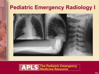
A P L S Pediatric Emergency Radiology 1
•Als PPT, PDF herunterladen•
51 gefällt mir•10,031 views

This document is too short to summarize meaningfully with only 3 sentences. It contains an abbreviated word that provides little context on its own to understand the intent or essential information being conveyed.
![[object Object],[object Object],[object Object],[object Object],[object Object],[object Object],[object Object],[object Object],[object Object],[object Object],[object Object],[object Object],[object Object],[object Object],[object Object],[object Object],[object Object],[object Object],[object Object],[object Object]](data:image/gif;base64,R0lGODlhAQABAIAAAAAAAP///yH5BAEAAAAALAAAAAABAAEAAAIBRAA7)
Empfohlen
Weitere ähnliche Inhalte
Was ist angesagt?
Was ist angesagt? (20)
Ähnlich wie A P L S Pediatric Emergency Radiology 1
Ähnlich wie A P L S Pediatric Emergency Radiology 1 (20)
Mehr von Dang Thanh Tuan
Mehr von Dang Thanh Tuan (20)
Kürzlich hochgeladen
Kürzlich hochgeladen (20)
A P L S Pediatric Emergency Radiology 1
- 3. X-ray diagnosis? 14-month-old girl with vomiting. Identify the target sign in the RUQ again. The crescent sign is formed by the intussusceptum (lead point) protruding into a gas-filled pocket. Identify crescent sign in LUQ again. Intussusception Target sign in RUQ. Target sign in RUQ. Crescent sign in LUQ. Crescent sign in LUQ. Target sign in RUQ. Crescent sign in LUQ. Intussusception
- 4. X-ray diagnosis? 13-month-old boy with vomiting. The crescent sign may not be crescent shaped. The gas-filled pocket may be large, as in this case. Intussusception Ha5 Crescent sign: Note the intussusceptum lead point ascending into the hepatic flexure. Crescent sign: Note the intussusceptum lead point ascending into the hepatic flexure.
- 5. X-ray diagnosis? 11-month-old boy with vomiting. Bowel obstruction with right-sided mass effect: Intussusception Right image: Absence of gas in RUQ and RLQ (suggests a mass effect on right). Poor distribution of gas in general (suggests bowel obstruction). Left image: Absence of hepatic angle (suggests RUQ mass). Absence of gas in RLQ (suggests RLQ mass). Two dilated (smooth) bowel segments (suggests bowel obstruction).
- 6. X-ray diagnosis? 11-month-old girl with vomiting. Identify the target and crescent signs again. RUQ target sign. LUQ crescent sign. Absence of the subhepatic angle. Intussusception Ha 6 RUQ target sign. LUQ crescent sign. Absence of the subhepatic angle. RUQ target sign. LUQ crescent sign. Absence of the subhepatic angle.
- 7. X-ray diagnosis? 7-month-old girl with skull fracture, lethargy, and vomiting. Intussusception Possible target sign in RUQ. Paucity of bowel gas suggestive of right-sided mass and bowel obstruction.
- 8. X-ray diagnosis? 7-month-old girl with vomiting. Intussusception Target sign Absence of hepatic angle Paucity of gas Target sign Absence of hepatic angle. Paucity of gas.
- 9. X-ray diagnosis? 7-month-old boy with vomiting. Suspected I ntussusception RUQ air fluid levels. RUQ bowel loops are smooth (bowel obstruction). Paucity of gas in RLQ.
- 11. X-ray diagnosis? 1-month-old girl spitting up. Air fluid levels: None Gas distribution: Good Normal abdominal radiographs Bowel obstruction criteria: Gas distribution Bowel distention Air fluid levels Bowel distention: Lots of gas, but no distention. Haustra and plicae are preserved. Looks like bag of popcorn, instead of bag of sausages. Bowel walls are NOT smooth (hose-like). Distention criterion is more related to smoothness of bowel walls rather than volume of gas.
- 12. X-ray diagnosis? 9-day-old boy with vomiting. Gas distribution: Fair Bowel distention: No smooth walls Air fluid levels: Many, but they are all small with no J turns ( hairpin loops, candy canes ) ILEUS, No Definite Bowel Obstruction Bowel obstruction criteria: Gas distribution Bowel distention Air fluid levels
- 13. Paucity of gas on the right suggestive of a mass. Residual barium present. While preparing for an ultrasound, the child drinks a bottle and her behavior normalizes. Radiologist identifies an occult diagnosis. Congenital Dislocated Hip X-ray diagnosis? 5-month-old girl discharged yesterday following barium enema reduction of intussusception. Vomited once today. Shenton’s arc. A more focused view of occult diagnostic finding Congenital dislocated hip (CDH). Shenton’s arc is discontinuous.
- 14. Thigh or knee pain could originate from a hip problem. Hip evaluation is required. X-ray diagnosis? 10-year-old obese boy with right thigh and knee pain Slipped Capital Femoral Epiphysis (SCFE) of the Right Hip Right hip physis appears to be wide compared to the left hip. Klein’s line: Superior aspect of the metaphysis to see if it intersects the epiphysis Abnormal: Line misses epiphysis Normal: Line intersects epiphysis
- 15. X-ray diagnosis? Bilateral SCFE Moderate slip Severe slip
- 16. X-ray diagnosis? 6-year-old boy with nausea and abdominal pain. Identify it again Appendicitis Fecalith (appendicolith)
- 17. Find the fecalith (appendicolith) Fecaliths can vary in appearance. This one is small and opaque. This fecalith is faint and oval in shape This fecalith can be seen faintly in the radiograph of the appendix specimen. It is very faint on the abdominal film. There are two or more potential fecaliths here This fecalith is round with a dense opaque dot in it. This fecalith is fairly large This is the last fecalith on this slide
- 18. X-ray diagnosis? 6-year-old boy with abdominal pain Pneumonia
- 19. X-ray diagnosis? 15-month-old boy with fever, coughing, tachypnea. LLL & RML Pneumonia RML infiltrate LLL infiltrate
- 20. X-ray diagnosis? 2 month old with a VSD presents with recurrent seizures. VSD, Thymic, & Parathyroid Aplasia: DiGeorge Syndrome Cardiomegaly (CHF) No thymic shadow Hypocalcemia found on labs X-ray diagnosis? 2 month old with a VSD presents with recurrent seizures. Normal thymus shadows in young infants Cardiomegaly (CHF) No thymic shadow Normal newborn thymus occupies the space anterior to the heart
- 21. X-ray diagnosis? Ventilated infant with sudden deterioration Pneumopericardium Revealing the Thymus “Sail Sign” Air in pericardium reveals shape of infant thymus.
- 22. X-ray diagnosis? 6-month-old boy with cough and congestion. No fever. O 2 Sat 100% on room air. Prominent Thymus Partially Obscuring a RUL Infiltrate: Pneumonia Normal newborn thymus occupies space anterior to heart Prominent asymmetric thymus Infiltrate
- 23. X-ray diagnosis? 18-month-old girl with mild BPD (former premie). Presents with fever, cough, dyspnea. RML Atelectasis RML atelectasis
- 24. X-ray diagnosis? 9-year-old boy with fever, headache, nausea, and coughing. Round Pneumonia: “ Cannonball” Pneumonia Round infiltrate. Spherical consolidation.
- 25. No definite abnormalities X-ray diagnosis? 17-month-old coughing after choking on a chocolate/almond bar Bilateral Air Trapping Bilateral Bronchial Foreign Bodies Nuts + Choking = Bronchoscopy More views: Expiratory view Lateral neck Inspiratory view Expiratory view Insp and Exp views look very similar = air trapping Right side down Left side down Heart should move downward. But in both views, it stays in place, due to bilateral air trapping.
- 26. X-ray diagnosis? 18-month-old girl with fever, noisy breathing, and barking cough. Identify the: Epiglottis Vallecula Vocal cords Trachea Prevertebral soft tissue Retropharyngeal Abscess (also called prevertebral abscess) Clinical symptoms may mimic croup. Epiglottis (E) Vallecula (V) Vocal cords (C) Trachea (T) Prevertebral soft tissue (P) E V C T P Epiglottis - normal Vallecula - normal Trachea - slightly narrow or normal Prevertebral soft tissue (P) - wide and bulging (should be half the width of vertebral body) P
- 27. X-ray diagnosis? 2-year-old boy with fever, stridor, tripoding and NO cough. Identify the: Epiglottis Vallecula Vocal cords Trachea Prevertebral soft tissue Epiglottitis Epiglottis (E) - wide (thumb-like) Vallecula - shallow Trachea - normal Prevertebral soft tissue - normal E E Epiglottis (E) Vallecula (V) Vocal cords (C) Trachea (T) Prevertebral soft tissue (P) V C T P
- 28. X-ray diagnosis? 15-month-old boy with fever, mild stridor, and barking cough. Identify the: Epiglottis Vallecula Vocal cords Trachea Prevertebral soft tissue Croup Epiglottis (E) Vallecula (V) Vocal cords (C) Trachea (T) Prevertebral soft tissue (P) P E V C T Epiglottis - normal Vallecula - normal Trachea (T) - narrow, subglottic edema Prevertebral soft tissue - normal T
- 29. X-ray diagnosis? 6-year-old girl with mild neck pain. No recent trauma. But she was thrown into a swimming pool 30 hours ago with no complaint of neck pain at that time. She is now brought in to the ED on a spine board. Probable C2-C3 Pseudosubluxation Ha29 Swischuk line criterion: Line drawn between posterior arch of C1 and posterior arch of C3. The posterior arch of C2 should be within 1 to 2 mm of this line. Deviation from this line suggests a C2 pedicle fracture; however, this criterion is not perfect. C2 C3 C1 Malalignment of C2 and C3. Is it a true subluxation or is it a pseudosubluxation? C2 C3 C2-C3 pseudosubluxation characteristics: Minimal / mild trauma Minimal / mild pain No signs of a fracture Neck is positioned in flexion (not lordotic), often due to a spine board. Swischuk line criterion. C2 C3
- 30. Probable C2-C3 Pseudosubluxation C2-C3 pseudosubluxation characteristics: Minimal / mild trauma Minimal / mild pain No signs of a fracture Neck is positioned in flexion (not lordotic), often due to a spine board. Swischuk line criterion. X-ray diagnosis? 2-year-old boy who fell off his tricycle is brought in on a spine board. Ha30 Swischuk line: Line drawn between the posterior arch of C1 and the posterior arch of C3. The posterior arch of C2 should be within 1 to 2 mm of this line. C2 C3 C1
- 31. X-ray diagnosis? 7-year-old girl unrestrained in a car crash brought in on a spine board. Fracture of the C2 Pedicle “ Hangman Fracture” Ha31 Swischuk line: satisfactory C2 C3 C1 Fracture of C2 pedicle: Despite a satisfactory Swischuk line. There is very slight subluxation of C2 on C3 due to the fracture.
- 32. X-ray diagnosis? 7-year-old boy injured his head and neck diving into shallow water. No definite abnormalities. His collar is temporarily removed for an odontoid (open mouth) view. Jefferson Fracture (C1 ring) Ha32 It’s hard to see anything with this poor odontoid view. The odontoid is not visible. This odontoid view is still useful to identify the lateral masses (ring of C1) relative to C2 as outlined here. The LMs should be directly over the base of C2. C2 C2 C1 C1 The lateral masses are displaced outward indicating that the ring of C1 has fractured and burst open. LM LM This CT scan shows a Jefferson fracture (C1 ring fracture) sustained when a blow to the top of the head places a load on the long axis of the spine, bursting open the ring of C1. Two normal odontoid views. The lateral masses of C1 are aligned with the base of C2. LM C2 Two normal odontoid views. The lateral masses of C1 are aligned with the base of C2. LM LM LM LM O O C2 C2 C2 C2 Better quality open mouth (odontoid) view demonstrating a Jefferson fracture.
- 33. X-ray diagnosis? 9-year-old boy who fell onto his forearm. Visible forearm deformity. Mid-ulna angulated fracture. Anything else? Monteggia Injury Ulna fracture often results in radial head dislocation. Check radius-capitellum line confirming alignment. Radius should line up with capitellum (C). Misalignment indicates radial head dislocation. C C Abnormal Normal
- 34. X-ray diagnosis? Elbow injury. Elbow evaluation: High yield places to look: Posterior fat pad Anterior fat pad Anterior humerus line Radius-capitellum line Supracondylar region Radial head Olecranon Elbow Joint Effusion Probable occult supracondylar fracture. Anterior fat pad (+) Posterior fat pad (+) Radius-capitellum line (normal) Olecranon Anterior humerus line should bisect capitellum (+) Supracondylar region Radial head
- 35. X-ray diagnosis? Elbow injury Radial Head Fracture Posterior fat pad Anterior fat pad Both unable to assess (true lateral view required) Anterior humerus line: misses capitellum (not a true lateral view) Radius-capitellum line: normal Radial head: Fracture Olecranon: OK Supracondylar region: OK
- 36. X-ray diagnosis? Elbow injury Supracondylar Fracture Supracondylar region: cortex disrupted Posterior fat pad (+) Anterior fat pad (+) Olecranon fossa cortex is fractured
- 37. X-ray diagnosis? Elbow injury Joint Effusion, Olecranon Fracture, Monteggia Injury (radial head dislocation) Posterior fat pad (+) Anterior fat pad (+) Radius-capitellum line is not pointing at capitellum Olecranon fracture
- 38. X-ray diagnosis? 10-year-old boy, wrist injury Displaced Salter-Harris Type 1 Fracture of the Distal Radius Physis Tenderness is elicited over distal radius Salter-Harris type 1 fracture of distal radius physis should be suspected clinically displa non-displa ced ced The epiphysis is displaced
- 39. Hey you !! What kind of Salter-Harris fracture type is this?? Who ME? M = metaphysis E = epiphysis W h o M E ? SH type II M etaphysis and physis SH type III E piphysis and physis SH type IV Metaphysis and Epiphysis SH type V: Physis. Not evident on X-ray. Relies on clinical findings and history of injury mechanism. Tender Calcaneus fracture Fell off 2nd floor onto her feet.
- 40. X-ray diagnosis? 6-week-old boy with “sudden” left thigh swelling and no history of trauma. Severe femur fracture without explanation. Older forearm and tibia fractures. Child Abuse Ha40 Obvious oblique femur fracture with a thinner fracture in the distal half of the femur. Child abuse is suspected. - A skeletal survey is ordered. - Left forearm and right tibia/fibula are shown here. Elbow/Forearm Tib/Fib Proximal radius fracture with periosteal elevation (hard to see). Healing tibia fracture with periosteal elevation.
- 41. X-ray diagnosis? 2 month old who is crying without apparent cause. Osteogenesis imperfecta is suspected. Occult types tend to be autosomal dominant (family history will be positive.) Severe lethal types tend to be recessive. Mid femur fracture. Osteogenesis imperfecta. Family history of “frequent fractures” may be a useful question in fracture patients. Ha41 Obvious mid femur fracture is noted. Child abuse is suspected. - Another view shows the oblique fracture line. - Further questioning about trauma is negative except for bumping him against a door while carrying him in a padded infant carrier. The parents tell you that this couldn’t have been hard enough to cause a fracture. Family history: - Father: 4 fractures, 2 of which occurred with minor trauma. - PGF: 4 fractures from “playing around” - Mother: Scoliosis - 2 aunts: Scoliosis A skeletal survey is done and no other fractures are found. The upper extremities are shown here. Ostepenia is NOT evident. Severe osteogenesis imperfecta. Lethal form in infancy. Severe osteopenia. Multiple rib fractures Crumpled long bones at birth.
Hinweis der Redaktion
- Pediatric Emergency Radiology I Note to instructor: Each slide contains multiple “clicks.” Each click is identified by number in the lecture notes. The last click on each slide is indicated by yellow type.
- Objectives: Intussusception, bowel obstruction, congenital hip dislocation, slipped capital femoral epiphysis, pneumonia, thymus shadow, appendicitis - fecaliths, bronchial foreign body, croup, epiglottitis, retropharyngeal abscess, c-spine pseudosubluxation, Hangman fracture, Jefferson fracture, elbow fractures, Monteggia injury, Salter-Harris fractures, child abuse
- X-ray diagnosis? 14-month-old girl with vomiting. 1) Target sign in RUQ is identified. 2) X-ray is displayed again for viewers to identify the target sign in RUQ. 3) Target sign in RUQ is identified again. 4) Crescent sign in LUQ is identified. 5) The crescent sign is formed by the intussusceptum (lead point) protruding into a gas-filled pocket. X-ray is displayed again for viewers to identify the crescent sign in LUQ. 6) Crescent sign in LUQ is identified again. 7) Target and crescent signs are identified. 8) Intussusception
- X-ray diagnosis? 13-month-old boy with vomiting. 1) Crescent sign in the RUQ. The lead point ascends into the hepatic flexure. 2) X-ray is displayed again. 3) The crescent sign, sometimes called the meniscus sign, may not be crescent or meniscus shaped. It depends on the shape of the gas-filled pocket. In this case, the gas-filled pocket is large, so its shape is not crescent-like. 4) Crescent sign in RUQ is identified again. 5) Intussusception
- X-ray diagnosis? 11-month-old boy with vomiting. 1) In the left image, absence of the hepatic angle (liver edge) suggests the presence of a RUQ mass. Absence of gas in the RLQ suggests a RLQ mass. There are two dilated (smooth wall) bowel segments, suggesting a bowel obstruction. 2) In the right image, there is absence of gas in the RUQ and RLQ, suggestive of a mass on the right. Poor gas distribution in general suggests a bowel obstruction. No air fluid levels are seen. 3) Bowel obstruction with right-sided mass effect: Intussusception
- X-ray diagnosis? 11-month-old girl with vomiting. 1) RUQ target sign and LUQ crescent sign. Absence of the subhepatic angle (liver edge). 2) X-ray is again displayed to identify the target and crescent signs. 3) Signs are identified again. 4) Intussusception
- X-ray diagnosis? 7-month-old girl with a skull fracture, lethargy, and vomiting. The lethargy and vomiting were initially attributed to a brain injury. Possible target sign in the RUQ. Paucity of bowel gas suggestive of a right-sided mass and a bowel obstruction. Intussusception The skull fracture and child abuse are probably unrelated to the intussusception.
- X-ray diagnosis? 7-month-old girl with vomiting 1) Possible target sign in the RUQ. Absence of the hepatic angle. Paucity of gas. 2) Intussusception
- X-ray diagnosis? 7-month-old boy with vomiting. 1) RUQ air fluid levels. The blue and red lines show the air fluid levels in the same loops (hairpin turns, candy canes). RUQ bowel loops are smooth, suggesting a bowel obstruction. Paucity of gas in the RLQ. 2) Suspected intussusception. Bowel obstructions that present with a paucity of gas (as opposed to excessive bowel gas), which occur in infants, are often due to intussusception.
- X-ray diagnosis? 17-day-old boy with vomiting. 1) Bowel obstruction criteria include: Gas distribution, bowel distention, and presence of air fluid levels. 2) Gas distribution is good. 3) Bowel walls are smooth, resembling hoses or sausage. This indicates that the bowel is distended. It is the smoothness that indicates distention rather than a measured bowel diameter. 4) Air fluid level on the upright view. The red and yellow lines indicate the air fluid levels in the same bowel loop (hairpin turns, candy canes). These are more indicative of a bowel obstruction than small air fluid levels. 5) Bowel obstruction. 6) AIM is a useful mnemonic device for the differential diagnosis of bowel obstruction: A: Adhesions, appendicitis I: Intussusception, incarcerated inguinal hernia M: Malrotation (midgut volvulus), Meckel diverticulum
- X-ray diagnosis? 1-month-old girl spitting up. 1) Bowel obstruction criteria: Gas distribution, bowel distention, air fluid levels 2) Gas distribution: Good 3) Air fluid levels: None 4) Bowel distention: Lots of gas, but no distention. Haustra and plicae are preserved. The supine view looks like a bag of popcorn instead of a bag of sausages. The bowel walls are not smooth (not hose-like or sausage-like). 5) Normal abdominal radiographs.
- X-ray diagnosis? 9-day-old boy with vomiting. 1) Bowel obstruction criteria: Gas distribution, bowel distention, air fluid levels. 2) Gas distribution: Fair 3) Bowel distention: No smooth walls. 4) Air fluid levels: Many, but they are all small with no J turns (hairpin turns, candy canes). These small air fluid levels are less suggestive of a bowel obstruction. 5) Ileus, no definite bowel obstruction.
- X-ray diagnosis? 5-month-old girl discharged from the hospital yesterday following barium enema reduction of an intussusception. She vomited once today. 1) Paucity of gas on the right suggestive of a mass. A small amount of residual barium is present. 2) While preparing for abdominal ultrasonography, the child drinks a bottle and her behavior normalizes. 3) The radiologist identifies an occult diagnosis on the abdominal series unrelated to the previous intussusception. 4) A more focused view of the occult diagnostic finding on this view. 5) Congenital dislocated hip (of the patient’s left hip). Shenton arc is the easiest criterion to assess the hip’s location until the femur head ossifies. Other criteria are listed in the text. Shenton arc (also called Shenton line) is drawn as an oval starting with the obturator foramen. It is discontinuous on the left hip on this radiograph. 6) Shenton arc pointed out on the original abdominal series. 7) Congenital dislocated hip can be diagnosed by looking for Shenton arc on all abdominal radiographs.
- X-ray diagnosis? 10-year-old obese boy with right thigh and knee pain. 1) Thigh or knee pain could originate from a hip problem. Evaluation of the hip is required for all complaints of knee or thigh pain. In this case, the patient’s right hip is tender on palpation. 2) The physis of the right femur head is wider compared to the left hip. 3) The Klein line is used to assess the position of the femoral head epiphysis. Drawn from the superior aspect of the femur metaphysis, the Klein line should intersect part of the femoral epiphysis. If the line misses the epiphysis, this suggests that the epiphysis has slipped in the inferior direction. 4) This closeup of the Klein line shows the abnormal line on the right hip (left image), which misses the epiphysis. The left hip (right image) shows a normal Klein line, which intersects part of the epiphysis. 5) Slipped capital femoral epiphysis (SCFE) of the right hip.
- X-ray diagnosis? 1) Moderate slip of the right hip. Severe slip of the left hip. 2) Bilateral SCFE.
- X-ray diagnosis? 6-year-old boy with nausea and abdominal pain. 1) Fecalith (appendicolith) in the RLQ. 2) Circle is removed to identify the fecalith again. 3) Closeup view of the fecalith. 4) Appendicitis
- Find the fecalith (appendicolith). 1) Fecaliths can vary in appearance. This one is small and very radiopaque. 2) Next image (#2). 3) This fecalith is faint and oval in shape. 4) Next image (#3). 5) There are two or more potential fecaliths here. 6) Next image (#4). 7) This fecalith can be seen faintly in the radiograph of the appendix specimen removed during appendectomy. It is very faint on the original abdominal film 8) Next image (#5). 9) This fecalith is round with a dense opaque dot in it. 10) Next image (#6). 11) This fecalith is fairly large. 12) Next image (#7). 13) This is the last fecalith on this slide.
- X-ray diagnosis? 6-year-old boy with abdominal pain. Hint: Abdominal pain is in the epigastrium region. 1) Pneumonia, which is evident on the lung portion of the abdominal series. Pneumonia is a common cause of abdominal pain. Pulmonary infiltrates are frequently evident on abdominal radiographs.
- X-ray diagnosis? 15-month-old boy with fever, coughing, tachypnea. 1) RML infiltrate. LLL infiltrate. 2) LLL and RML pneumonia.
- X-ray diagnosis? 2-month-old with a VSD presents with recurrent seizures beginning today. 1) No thymic shadow. Cardiomegaly (CHF) due to the VSD. Hypocalcemia found on labs. 2) Normal thymus shadows demonstrated in these two normal neonates. 3) In normal neonates, the thymus occupies the mediastinal space anterior to the heart. A solid tissue density should be present here in normal neonates. 4) This AP and lateral chest x-ray shows cardiomegaly, but no thymic shadow on the AP view and on the lateral view. 5) VSD, thymic aplasia (on chest x-ray), parathyroid aplasia (causing hypocalcemia): DiGeorge syndrome.
- X-ray diagnosis? This is a neonate on a ventilator who develops sudden deterioration (precipitous drop in oxygen saturation, bradycardia, hypotension). 1) Air in the pericardium outlines the heart and reveals the shape of the infant’s thymus. 2) Pneumopericardium revealing the thymus “sail sign.” The sail sign is more commonly associated with pneumomediastinum in infants. In this case, the pneumopericardium constrasts with the lower portion of the thymus, exaggerating the “sail sign.”
- X-ray diagnosis? 6-month-old with cough and congestion. No fever. Oxygen saturation is 100% on room air. 1) The PA view shows a prominent asymmetric thymus in the right upper chest. The lateral view shows a normal thymus, which occupies the mediastinal space anterior to the heart. 2) There is a pulmonary infiltrate in the RUL, which is partially obscured by the prominent thymus. 3) Pneumonia. The prominent thymus obscures the RUL infiltrate.
- X-ray diagnosis? 18-month-old girl with mild BPD (former premie). She now presents with fever, cough, and dyspnea. 1) RML atelectasis best seen on the lateral view. 2) RML atelectasis
- X-ray diagnosis? 9-year-old boy with fever, headache, nausea, and coughing. 1) Round infiltrate, spherical consolidation. 2) Round pneumonia. Also called “cannonball” pneumonia.
- X-ray diagnosis? 17-month-old coughing after choking on a chocolate and almond candy bar. 1) No definite abnormalities seen on this PA and lateral view. A foreign body series is ordered. 2) The lateral neck looks ok and the expiratory PA view looks clear. No asymmetry is noted. 3) Comparing the inspiratory and expiratory views. Note that they look very similar. This implies that the expiratory view was actually taken during inspiration, or it implies that the patient is unable to exhale well due to air trapping. Bilateral air trapping implies the presence of a tracheal foreign body or bilateral bronchial foreign bodies. 4) Lateral decubitus views are shown here, both right and left side down view. The heart should swing toward the dependent side in a normal chest. However, it is evident that the heart remains in the “normal” anatomic position despite the dependent view. This suggests air trapping. This view is more reliable since it does not really depend as much on timing of the radiograph or patient cooperation. Since both sides fail to compress, this suggests bilateral air trapping. 5) Bilateral air trapping indicative of bilateral bronchial foreign bodies. A history of choking on nuts is highly predictive of bronchial foreign bodies. Bronchoscopy should be considered even in the absence of radiographic findings. Nuts + choking = bronchoscopy.
- X-ray diagnosis? 18-month-old girl with fever, noisy breathing, and a barking cough. 1) Identify the major structures: Epiglottis, vallecula, vocal cords, trachea, prevertebral soft tissue 2) The letters indicate the major structures. 3) The epiglottis and vallecula are normal. The tracheal shadow is slightly narrow. The prevertebral soft tissue is very wide and bulging anteriorly. It should normally be half the width of a vertebral body. 4) Retropharyngeal abscess (also called prevertebral abscess). Symptoms may mimic croup.
- X-ray diagnosis? 2-year-old boy with fever, stridor and tripoding (leaning anteriorly on his upper extremities), and NO cough. 1) Identify the important structures: Epiglottis, vallecula, vocal cords, trachea, prevertebral soft tissue 2) The letters identify the structures. 3) The epiglottis is wide (thumb-like). The vallecula is very shallow due to the epiglottitis. The vocal cords are very thick. The trachea and the prevertebral soft tissue are normal. 4) Epiglottitis
- X-ray diagnosis? 15-month-old boy with fever, mild stridor, and a barking cough. 1) Identify the important structures: Epiglottis, vallecula, vocal cords, trachea, prevertebral soft tissue 2) The letters identify the structures. 3) The epiglottis and vallecula are normal. The prevertebral soft tissue is normal. The trachea is narrow below the cords (subglottic edema). 4) Croup
- X-ray diagnosis? 6-year-old girl with mild neck pain. 1) No recent trauma today, but she was thrown into a swimming pool 30 hours ago with no complaint of neck pain at that time. She was brought into the ED on a spine board. 2) Malalignment of C2 on C3. Is it a true subluxation, or is it a pseudosubluxation? 3) The characteristics of C2-C3 pseudosubluxation are: a) Minimal/mild trauma, b) Minimal/mild pain, c) No signs of a fracture, d) Neck is in flexion and NOT in the usual lordotic position, which often occurs with a spine board. Young children have large occiputs, which usually puts the neck in flexion on a spine board, e) Swischuk line criterion. 4) Swischuk line criterion. A line is drawn as shown from the arch of C1 to the arch of C3. The arch of C2 should be within 1 to 2 mm of this line. Deviation from this line suggests a pedicle fracture of C2. It should be noted that this criterion is not perfect. 5) Probably C2-C3 pseudosubluxation.
- X-ray diagnosis? 2-year-old boy who fell off his tricycle is brought in on a spine board. 1) C2-C3 pseudosubluxation criteria listed on the slide, and all are met. Note that the neck is flexed (NOT lordotic). 2) The Swischuk line is drawn here, and there is good alignment. 3) Probable C2-C3 pseudosubluxation
- X-ray diagnosis? 7-year-old girl unrestrained in a car crash brought in on a spine board. 1) Swischuk line is satisfactory. 2) However, there is a fracture of the pedicle of C2. There is very slight subluxation of C2 on C3 due to the fracture. The neck is straight and not obviously flexed as in the previous radiographs. 3) Fracture of the C2 pedicle, sometimes called the Hangman fracture
- X-ray diagnosis? 7-year-old boy injured his head and neck diving into shallow water. 1) No definite abnormalities. Cervical-collar has been temporarily removed for an odontoid (open mouth) view. 2) It is hard to see anything with this poor odontoid view. The odontoid is not visible (actually, this odontoid view is good enough). 3) This odontoid view is still useful to identify the lateral masses (LM) (ring of C1) relative to C2 as outlined here. The LMs should be directly over the base of C2. The outlines here show the LMs displaced outward on both sides. 4) The lateral masses are displaced outward, indicating that the ring of C1 has fractured and burst open. 5) This CT scan shows a Jefferson fracture (C1 ring fracture) sustained when a blow to the top of the head placed a load on the long axis of the spine, bursting open the ring of C1. It is often said that the ring must break in TWO spots. In this case, the other break in the ring is probably through the posterior growth plate region of the ring. 6) Two normal odontoid views. Note that the lateral masses are well aligned with the base of C2 below them. 7) Two normal odontoid views. The letters point out the lateral masses (LM), C2, and the odontoid. 8) A better quality open mouth (odontoid view) demonstrating a Jefferson fracture. The image below outlines the lateral masses and the base of C2. 9) Jefferson fracture (C1 ring)
- X-ray diagnosis? 9-year-old boy who fell onto his forearm. A mid-forearm deformity is visible. 1) An angulated fracture of the mid-ulna is evident. Anything else? 2) The long axis of the radius should point to the capitellum in all views. This is called the radio-capitellar or radius-capitellum line. Misalignment indicates radial head dislocation. The image in the lower right shows the normal alignment. In the ulna fracture image above, the radial head is dislocated. 3) Monteggia injury: A fracture of the mid or proximal ulna is frequently associated with radial head dislocation. Whenever a fracture of the mid or proximal ulna (including the olecranon) is encountered, be sure to check the radius-capitellum line to confirm proper alignment.
- X-ray diagnosis? Elbow injury. 1) In the evaluation of the elbow, the high places to look include the posterior fat pad, the anterior fat pad, the anterior humerus line, the radius-capitellum line, the supracondylar region, the radial head, and the olecranon. 2) A posterior fat pad is present. This is always abnormal and indicates the presence of an elbow joint effusion. The anterior fat pad is visible in the normal elbow, but it is normally small. In this radiograph, the anterior fat pad is protruding anteriorly in a triangular shape, sometimes called the sail sign of the elbow. An enlarged anterior fat pad such as this is abnormal and indicates the presence of an elbow joint effusion. Elbow joint effusions are indicative of occult fractures, often in the supracondylar region. The radius-capitellum line is normal. 3) The anterior humerus line should bisect the capitellum. In this case the capitellum is NOT bisected by this line, indicating that the capitellum is displaced but more likely, that the supracondylar region may be fractured. The supracondylar region should be carefully examined, since this is the most common fracture of the elbow in young children. No fractures are visible in this radiograph. The radial head contour should be carefully examined since this is a common fracture site as well. It should be smooth without any angles. No fractures are evident here. The olecranon region should be carefully examined since it is easy to miss a fracture in this area. 4) Elbow joint effusion, probable occult supracondylar fracture
- X-ray diagnosis? Elbow injury. 1) The posterior and anterior fat pads are not visible. Note that the distal humerus is positioned obliquely. This is not a true lateral view. Thus, the fat pads might not be visible even if they were enlarged. The patient should be sent back for a better lateral view. 2) The radius-capitellum line is normal. The anterior humerus line misses the capitellum, but this relationship can only be assessed on a true lateral view. 3) The olecranon region is normal. The supracondylar region is normal. The radial head reveals a fracture. The angle can be described as a lip off the end. The contour of the radial head should be smooth. It should NOT resemble the handle of a baseball bat. 4) Radial head fracture
- X-ray diagnosis? Elbow injury. 1) On the lateral view, an anterior fat pad is present. An enlarged posterior fat pad is present. The contour of the olecranon fossa is visibly crinkled on the lateral view, indicating a fracture. On the AP view, slight angular disruptions in the smooth metaphysis of the distal humerus are visible on both sides of the supracondylar region. 2) Supracondylar fracture
- X-ray diagnosis? Elbow injury. 1) The lateral view shows an posterior fat pad. The anterior fat pad is enlarged. The olecranon is fractured. Since the olecranon is part of the ulna, the radius-capitellum line should be carefully examined. It is not in alignment, demonstrating a dislocation of the radial head. 3) Joint effusion, olecranon fracture, radial head dislocation. This is the Monteggia injury.
- X-ray diagnosis? 10-year-old boy with a wrist injury. 1) Tenderness is elicited over the distal radius over the growth plate physis. 2) Whenever there is tenderness directly over the physis, a Salter-Harris fracture involving the physis should be suspected. Type 1 and type 5 fractures are usually not radiographically evident; they must be suspected on clinical grounds alone. 3) The lateral view demonstrates that the radial epiphysis is displaced. This is difficult to see. The white outlines demonstrate the displacement of the epiphysis relative to the metaphysis compared to the normal expected alignment. 4) Displaced Salter-Harris type 1 fracture of the distal radius physis. There might be a small chip fracture of the ulna styloid, but the focus of this slide should be on the radial epiphysis.
- This is a diagrammatic representation of the Salter-Harris fracture description which involve the growth plate. 1) Hey you!! What kind of Salter-Harris fracture is this?? 2) Who ME? ME stands for metaphysis and epiphysis. ME is used to remember that the Salter-Harris type 2 fracture involves the metaphysis, and the type 3 fracture involves the epiphysis. It is very easy to remember that type 1 involves the physis alone, and type 4 involves the metaphysis, physis, and epiphysis, but it is difficult to remember what types 2 and 3 involve. ME is a mnemonic used to remember this. 3) An example of a Salter-Harris type 2 fracture (metaphysis + physis) of the distal radius 4) An example of a Salter-Harris type 3 fracture (epiphysis + physis) of the distal tibia 5) An example of a Salter-Harris type 4 fracture (epiphysis + physis + metaphysis) of the distal medial tibia 6) In a Salter-Harris type 5 fracture, the physis is crushed. Similar to Salter-Harris type 1 fractures, the type 5 fracture may not be evident radiographically. The diagnosis relies on the clinical findings of tenderness over the physis and the history of the injury mechanism. In this case, this girl fell off the second floor and landed on her feet. Note the multiple fracture lines in her calcaneus. She most likely sustained a Salter-Harris type 5 fracture of her distal tibia. Growth arrest or a limb length discrepancy might result from this.
- X-ray diagnosis? 6-week-old boy with sudden left thigh swelling and no history of trauma. 1) There is an obvious oblique fracture of the proximal femur. The arrow points to a thinner fracture line extending to the distal femur that is not as obvious. Child abuse should be suspected since there is no explanation for such a serious fracture. 2) A skeletal survey is ordered. The left forearm and right tibia/fibula are shown here. 3) A fracture of the radius is noted. A slight degree of periosteal elevation is evident on closer viewing, which is hard to see. There is a healing fracture of the mid tibia. The fracture line is difficult to identify, but extensive periosteal elevation is seen throughout the length of the tibia. 4) Severe femur fracture without explanation. Older forearm and tibia fractures. These injuries are indicative of child abuse.
- X-ray diagnosis? Crying without apparent cause. 1) Obvious mid femur fracture is noted. Child abuse is suspected. 2) Another view shows the oblique fracture line. Further questioning about trauma is negative except for bumping him against a door while carrying him in a padded infant carrier. The parents tell you that this could not have been hard enough to cause a fracture. 3) A skeletal survey is done, and no other fractures are found. The upper extremities are shown here. No osteopenia is evident. A family history (rarely done in the ED but important here) reveals multiple fractures in the father and paternal grandfather, and scoliosis on the mother's side. 4) Osteogenesis imperfecta is suspected. Occult types tend to be autosomal dominant (family history should be positive). Severe lethal types tend to be recessive. 5) The severe form of osteogenesis imperfecta tends to be lethal in infancy. Severe osteopenia is present. There are multiple rib fractures and crumpled long bones. 6) Mid femur fracture, osteogenesis imperfecta. Consider obtaining a family history of "frequent fractures" in patient presenting with fractures.