

Recommended
More Related Content
What's hot
What's hot (20)
Similar to Acute leukemias aml-csbrp
Similar to Acute leukemias aml-csbrp (20)
More from Prasad CSBR
More from Prasad CSBR (20)
Recently uploaded
Recently uploaded (20)
Acute leukemias aml-csbrp
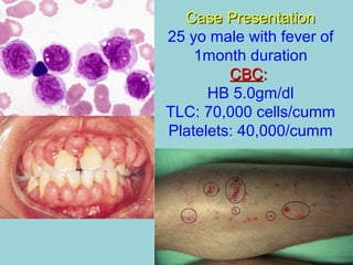
- 1. Case PresentationCase Presentation 25 yo male with fever of 1month duration CBCCBC:: HB 5.0gm/dl TLC: 70,000 cells/cumm Platelets: 40,000/cumm
- 2. Acute LeukemiasAcute Leukemias - AML- AML Dr.CSBR.Prasad, M.D., Professor of Pathology Sri Devaraj Urs Medical College Kolar-563101 Karnataka INDIA csbrprasad@gmail.com
- 4. Differentiation of blood cells In leukemiaIn leukemia there isthere is blockage inblockage in maturationmaturation CSBRP-SDUMC-Oct-2018
- 5. Terms • Dysplasia • Clone • Malignancy • Leukemia • Lineage • CD antigens • Immunophenotyping • Flow cytometry • Acute & Chronic • Gp IIb/IIIa • Oncogenes • Prognosis CSBRP-SDUMC-Oct-2018
- 6. What is the difference between monocyte and macrophage? CSBRP-SDUMC-Oct-2018
- 7. Myeloid neoplasms All have common origin from hemopoietic progenitor cells They primarily involve BM and to a lesser degree the secondary hemopoietic organs (spleen, liver and LNs) and Present with altered hematopoiesis CSBRP-SDUMC-Oct-2018
- 8. Three broad categories: 1. Acute myelogenous leukemia 2. Myelodysplastic syndromes 3. Chronic myeloproliferative disorders Myeloid neoplasms CSBRP-SDUMC-Oct-2018
- 9. Leukemias Main groups: 1-Myeloid (acute / chronic) 2-Lymphoid (acute / chronic) 3-Mixed lineage leukemias CSBRP-SDUMC-Oct-2018
- 10. Leukemias Pathogenesis: Normal hemopoiesis is finely tuned by hemostatic feedback mechanisms involving cytokines and growth factors that modulate the marrow output of red cells, granulocytes and platelets. These mechanisms are deranged in marrows involved by myeloid neoplasms. Loss of control on growth & survival and suppressor fuctions. CSBRP-SDUMC-Oct-2018
- 13. Leukemias – Clinical features • Anemia • Fever • Fatigue • Bleeding • Gum hypertrophy • Hepatosplenomegaly • Lymphadenopathy • Mediastinal mass • CNS symptoms CSBRP-SDUMC-Oct-2018
- 15. Petechiae on the shin of the left leg CSBRP-SDUMC-Oct-2018
- 20. What do you call this? CSBRP-SDUMC-Oct-2018
- 21. Acute Myeloid Leukemia Definition: Acute myeloid leukemia (AML) is a clonal expansion of myeloblasts in bone marrow, blood or other tissue. AML is a quite heterogeneous disease, reflecting the complexities of myeloid cell differentiation CSBRP-SDUMC-Oct-2018
- 24. Classification of AMLClassification of AML Two types of classificationsTwo types of classifications: 1-1- FAB classification - degree of maturation & lineage of blasts - usage of cytochemistry & IHC 2-2- WHO classification - degree of maturation & lineage of blasts - Immunophenotyping - Cytogenetic & molecular features - Clinical outcome CSBRP-SDUMC-Oct-2018
- 25. WHO classificationWHO classification 1. AML with recurrent genetic abnormalities 2. AML with multilineage dysplasia 3. AML and MDS 4. AML - Tx related 5. AML not otherwise categorised 6. AML of ambiguous lineage CSBRP-SDUMC-Oct-2018
- 27. AMLAML GeneticGenetic abnormalityabnormality FAB typeFAB type Out comeOut come AML t(8,21) M2 Favorable AML t(15,17) M3 Intermediate AML t(11q, 23) Poor AML Rx related Very poor CSBRP-SDUMC-Oct-2018
- 28. Very large, immature myeloblasts with many nucleoli. A distincitve feature of these blasts is a linear red "Auer rod" (Blue arrow) composed of crystallized granules. CSBRP-SDUMC-Oct-2018
- 29. Leukemias typically fill up the marrow with abnormal cells, displacing normal hematopoiesis. CSBRP-SDUMC-Oct-2018
- 30. Requisits for diagnosis of AML The diagnostic requisite of 20 percent myeloblasts in the bone marrow or blood cannot be applied uniformly to all types of AML. BLAST Equivalents: 1. In AML (M3), the predominant leukemic cell is promyelocyte 2. In AML (M5A), the predominant proliferating cell is the monoblast 3. In AML (M5B), the predominant cell is the promonocyte. 4. The megakaryoblasts of acute megakaryoblastic leukemia vary in morphology but uniformly lack the cytochemical properties of myeloblasts. Brunning, RD and McKenna, RW. Tumors of the bone marrow. Atlas of Tumor Pathology, 3rd Series, Fascicle 9. Washington D.C.:Armed Forces Institute of Pathology, 1993. pp.23-25. CSBRP-SDUMC-Oct-2018
- 31. CYTOLOGIC FEATURES OF BLASTS IN ACUTE MYELOID & ACUTE LYMPHOBLASTIC LEUKEMIAS AMLAML ALLALL Blast size Medium to large, uniform Variable Small to medium Cytoplasm Pale blue. Fine granules may be present Usually scant and dark blue No granules Auer rods Present in 60-70% of cases Absent Nuclear chromatin Finely dispersed Coarse Nucleoli 2-4, Clearly visible 1-3, Indistinct Other cell types Often dysplastic changes in maturing myeloid cells Myeloid cells are not dysplastic
- 32. CSBRP-SDUMC-Oct-2018 CYTOLOGIC FEATURES OF BLASTS IN ACUTE MYELOID & ACUTE LYMPHOBLASTIC LEUKEMIAS Lymphoblast Myeloblast
- 33. CYTOCHEMICAL PROFILES OF ACUTE LEUKEMIASCYTOCHEMICAL PROFILES OF ACUTE LEUKEMIAS MPOMPO / SBB/ SBB CAECAE NSENSE PASPAS APAP ALL _ _ + / - Focally + 75% + / - Focal in T- ALL AML + + + Monocytic- diffuse - / + + MPO-myeloperoxidase, SBB-Sudan balck B, CAE-chloracetate esterase, NSE- non specific esterase, PAS-periodic acid schiff, AP-acid phosphatase cvcv
- 34. Auer rodsAuer rods Present in the leukemic cells of approximately 60 to 70 percent of cases of AML, The Auer rod is an azurophilic linear structure of varying length and Width Auer rods may be present in numerous blasts or only in rare cells Single or multiple Auer rods may be present. They are generally MPO, SBB, and CAE positive Ultrastructurally, the Auer rod is an alignment and crystallization of azurophilic granules. The presence of an Auer rod in one or more blasts is definitive evidence of AML. The finding is not specific for any one type of AML. Auer rods do not occur in cells of erythroid or megakaryocyte lineage. Brunning, RD and McKenna, RW. Tumors of the bone marrow. Atlas of Tumor Pathology, 3rd Series, Fascicle 9. Washington D.C.:Armed Forces Institute of Pathology, 1993. pp.26-27. CSBRP-SDUMC-Oct-2018
- 37. SBB positivity AML M1 CSBRP-SDUMC-Oct-2018
- 38. AML M0, M1, M2 CSBRP-SDUMC-Oct-2018
- 39. AML M0AML M0 20% blasts < 3% blasts reactive for MPO, SBB or NSE Auer rods are not found Immunophenotyping: –20% blasts express one or more myeloid antigens: CD13, CD14, CD33 –may be TdT positive; –Blasts are negative for lymphocyte antigens CSBRP-SDUMC-Oct-2018
- 40. AML- M1AML- M1 20% blasts >3% blasts reactive for MPO or SBB. <10% of marrow nucleated cells are promyelocytes or more mature neutrophils Immunophenotyping: Blasts express myeloid antigens: CD13, CD14, CD33. CSBRP-SDUMC-Oct-2018
- 41. AML M1 CASE-4 PEROXIDASE 1000X BM CSBRP-SDUMC-Oct-2018
- 42. AML- M2AML- M2 =/>20% Blasts Evidence of maturation to promyelocytes and more mature neutrophils in 10 percent or more of the cells. Immunophenotyping: myeloid antigen positive in blasts. In t(8,21) associated cells: 40-80% are positive for CD19 20% are TdT positive CSBRP-SDUMC-Oct-2018
- 43. AML- M3AML- M3 A special type of leukemia CSBRP-SDUMC-Oct-2018
- 44. AML M3AML M3 A form of AML characterized primarily by a proliferation of abnormal promyelocytes. It is usually accompanied by DIC t(15;17) The disease presents in two morphologic types: -- hypergranular APL in which the predominant cell is an abnormal promyelocyte with markedly increased and coarse azurophilic granules and -- microgranular or hypogranular APL in which the predominant cell is an abnormal promyelocyte with diminished or small azurophilic granules. Brunning, RD and McKenna, RW. Tumors of the bone marrow. Atlas of Tumor Pathology, 3rd Series, Fascicle 9. Washington D.C.:Armed Forces Institute of Pathology, 1993. p.43. CSBRP-SDUMC-Oct-2018
- 45. AML M3AML M3 The most common presenting symptoms, occurring in 90 percent of patients, relate to hemorrhagic manifestations and include easy bruisability, bleeding gums, hemoptysis, epistaxis, petechiae, and symptoms of gastrointestinal bleeding and intracranial hemorrhage. Basic pathology is DIC. Brunning, RD and McKenna, RW. Tumors of the bone marrow. Atlas of Tumor Pathology, 3rd Series, Fascicle 9. Washington D.C.:Armed Forces Institute of Pathology, 1993. p.43. CSBRP-SDUMC-Oct-2018
- 46. AML M3 case-3 BM, MGG x1000 CSBRP-SDUMC-Oct-2018
- 47. AML M3 Case-8 Bone marrow smear, May-Giemsa stain, x1000 CSBRP-SDUMC-Oct-2018
- 48. CSBRP-SDUMC-Oct-2018 Hypogranular variant – AML-M3 may be mistaken for AML-M5
- 49. AML M3 case-3 BM, Proxidase stain, x1000 CSBRP-SDUMC-Oct-2018
- 54. AML M4AML M4 • Acute myelomonocytic leukemia • Proliferation of both neutrophil and monocyte precurssors • BM blast >20% • >3% of blasts MPO + • >20% of the BM cells are monocytes and their precurssors • Monocytes and their precursors are NSE + • Express CD 13, 33 (myeloid Ag), CD64, 36, and Lysozyme (Monocyte differentiation) • A high number of circulating Monocytes (~5000cells/cumm) CSBRP-SDUMC-Oct-2018
- 55. AML M4AML M4 Monoblast: • Monoblasts are large cells with abundant cytoplasm with only moderate basophilia and may show pseudopod formation • Scattered azurophilic granules and vacuoles in the cytoplasm • Round nucleus with delicate lacy chromatin • 0ne or more prominent nucleoli Promonocyte: • More irregular, convoluted nucleus • Granularity and vacuolations are more obvious CSBRP-SDUMC-Oct-2018
- 56. AML M4 Case-1 Bone marrow smear, May-Giemsa stain, x1000 CSBRP-SDUMC-Oct-2018
- 57. AML M4 CASE-1 Bone marrow smear, Peroxidase stain, x1000 CSBRP-SDUMC-Oct-2018
- 58. AML M4 Case-1 Bone marrow smear, alpha-naphthyl butyrate esterase and chloroacetate esterase stains, x1000 CSBRP-SDUMC-Oct-2018
- 60. AML M5AML M5 • Acute monoblastic leukemia • >80% of leukemic cells are monocytic lineage (Mb, PMc, Mc) • Neutrophil component may constitute <20% • Acute monoblastic Vs monocytic leukemias: • Monoblasts >80% in monoblastic leukemia • Promonocytes are predominant in monocytic leukemias Presentation: Extramedullary masses Cutaneous & gingival infiltrations CNS involvement is common CSBRP-SDUMC-Oct-2018
- 63. Granulocytic sarcoma / Myeloid sarcoma CSBRP-SDUMC-Oct-2018
- 64. AML M5 Case-5 Bone marrow smear, May-Giemsa stain, x1000 CSBRP-SDUMC-Oct-2018
- 66. AML M5B Case-2 Bone marrow smear, alpha-naphthyl butyrate esterase and chloroacetate esterase stains, x1000 CSBRP-SDUMC-Oct-2018
- 68. AML M6AML M6 • Def: Erythroleukemia by definition involves both the granulocytes and erythroid cells. – BM shows >50% are erythroid precurssors (of all nucleated cells) – >20% myeloblasts in non-erythroid cell population • PURE erythroleukemia: >80% are erythroid lineage No significant myeloid cells • No myeloid markers in erythroid precurssors • Glycophorin A + • HGB A + • Myeloid precurssors CD13, CD33, CD117, MPO CSBRP-SDUMC-Oct-2018
- 69. AML M6 Case-1 Bone marrow smear, May-Giemsa stain, x1000 CSBRP-SDUMC-Oct-2018
- 70. AML M6 Case-4 Bone marrow smear, PAS stain, x1000 CSBRP-SDUMC-Oct-2018
- 72. AML M7AML M7 • >50% of the blasts are of MKc lineage • Occur in both adults and children • Uncommon (3-5% of all AMLs) • May be associated with mediastinal germ cell tumors CSBRP-SDUMC-Oct-2018
- 73. AML M7AML M7 • MK blast: – 12-18µm – Round nucleus with reticular chromatin – 1-3 nucleoli – Cytoplasm is basophilic and agranular – Cytoplasmic blebs – May resemble Lymphoblast • Circulating MKc fragments, abnormal platelets • Clustering of blasts • Immunophenotyping: CD41, CD61, Gp IIb/IIIa • No lymphoid markers CSBRP-SDUMC-Oct-2018
- 74. AML M7AML M7 • Frequently manifests in neonatal period • Marked leucocytosis • MPO, SBB, TdT are negative • Some scattered PAS positivity CSBRP-SDUMC-Oct-2018
- 75. AML M7 case-1 Bone marrow smear, May-Giemsa stain, x1000 CSBRP-SDUMC-Oct-2018
- 77. Malignant germ cell tumor of anterior mediastinum CSBRP-SDUMC-Oct-2018
- 78. Acute leukemias and Down’s syndrome CSBRP-SDUMC-Oct-2018
- 79. Down’s Syndrome Acute Leukemia – 10 to 20x more common –Most common leukemia: ALL –Most common among AMLs: M7 Transient MyeloproliferativeTransient Myeloproliferative SyndromeSyndrome
- 80. Acute basophilic leukemiaAcute basophilic leukemia CSBRP-SDUMC-Oct-2018
- 81. Acute basophilic leukemia • Myeloid lukemia with differentiation to basophils • No Ph’ • Constitute <1% • Metachromatic positivity with Toludene blue • PAS positivity in blocks • MPO, SBB are negative • Myeloid markers are positive (CD13, 33, 34, 117) CSBRP-SDUMC-Oct-2018
- 82. Specific PresentationsSpecific Presentations • AML M0, M1, M2 : Chloromas • AML M3 : DIC • AML M4, M5 : Gum hypertrophy • AML M7 : Mediastinal mass (germ cell tumors) CSBRP-SDUMC-Oct-2018
- 83. Clinical PresentationsClinical Presentations The arrest in myeloid development leads to marrow failure and complications related to: – Anemia – Thrombocytopenia and – Neutropenia / Infections CSBRP-SDUMC-Oct-2018
- 84. Frequency of AML M2 is the most common leukemia among AMLs (30%) CSBRP-SDUMC-Oct-2018
- 85. Common chromosomalCommon chromosomal abnormalities in AMLabnormalities in AML • t(15,17) in M3 (70-100%) unique to M3 • t(8,21) in M2 (20%) good prognosis • inv 16 in M4 (~25%) good prognosis • del 11q in M5 (30%) CSBRP-SDUMC-Oct-2018
- 87. Summary – Acute leukemiasSummary – Acute leukemias • Leukemias are clonal disorders • Mutations in oncogenes is the most common underlying pathology • Present with: Anemia, Petichiae, infections, hepatosplenomegaly, Lymphadenopathy • There may be normal, low or elevated total white count CSBRP-SDUMC-Oct-2018
- 88. SummarySummary AML is a heterogeneous disease Blast count should be 20% in BM There are blast equivalents The presence of an Auer rod is definitive evidence of AML WHO classification is well accepted Detection of genetic abnormalities dictates Tx and Prognosis CSBRP-SDUMC-Oct-2018
Editor's Notes
- Leukemia is a bone marrow disease. Stem cells gives rise to various elements of blood. In leukemia there is blockage in normal maturation which results in accumulation of immature forms mostly blasts. Blasts proliferate and occupy the marrow cavity and the other elements of the marrow like Erythroid series and megakaryocytes marginalized, hence the patient may suffer form anaemia, bleeding. Another common complaint with which the patients may present with is fever. Though there are many white cells the patients are susceptible for infections, as the neoplastic white cells (Leukemic cells), are inefficient in performing their duties.
- FIGURE 13-1 Differentiation of blood cells. CFU, colony forming unit; SCF, stem cell factor; Flt3L, Flt3 ligand; G-CSF, granulocyte colony-stimulating factor; GM-CSF, granulocyte-macrophage colony-stimulating factor; LIN–, negative for lineage-specific markers; M-CSF, macrophage colony-stimulating factor. Robbin’spath 8th Ed. ======================================= HSCs have two essential properties that are required for the maintenance of hematopoiesis: pluripotency and the capacity for self-renewal. Pluripotency refers to the ability of a single HSC to generate all mature hematopoietic cells. When an HSC divides at least one daughter cell must self-renew to avoid stem cell depletion. Self-renewing divisions are believed to occur within a specialized marrow niche, in which stromal cells and secreted factors nurture and somehow maintain the HSCs.[2] As you may have already surmised from their ability to migrate during embryonic development, HSCs are not sessile. Particularly under conditions of marked stress, such as severe anemia, HSCs are mobilized from the bone marrow and appear in the peripheral blood. In such circumstances, additional HSC niches are sometimes induced or “unveiled” in other tissues, such as the spleen and liver, which can then become sites of extramedullary hematopoiesis. The marrow response to short-term physiologic needs is regulated by hematopoietic growth factors through effects on the committed progenitors. Since mature blood elements are terminally differentiated cells with finite life spans, their numbers must be constantly replenished. In at least some divisions of HSCs, a single daughter cell begins to differentiate. Once past this threshold, these newly committed cells lose the capacity for self-renewal and commence an inexorable journey down a road that leads to terminal differentiation and death. However, as these progenitors differentiate they also begin to express receptors for lineage-specific growth factors, which stimulate their short-term growth and survival. Some growth factors, such as stem cell factor (also called c-KIT ligand) and FLT3-ligand, act on very early committed progenitors. Others, such as erythropoietin, granulocyte-macrophage colonystimulating factor (GM-CSF), granulocyte colony-stimulating factor (G-CSF), and thrombopoietin, act on committed progenitors with more restricted potentials. Feedback loops that are mediated through growth factors tune the marrow output, allowing the numbers of formed blood elements (red cells, white cells, and platelets) to be maintained within appropriate ranges.
- Prognosis (Greek: πρόγνωσις &quot;fore-knowing, foreseeing&quot;) is a medical term for predicting the likely or expected development of a disease, including whether the signs and symptoms will improve or worsen (and how quickly) or remain stable over time; expectations of quality of life, such as the ability to carry out daily activities; the potential for complications and associated health issues; and the likelihood of survival (including life expectancy).[1][2] A prognosis is made on the basis of the normal course of the diagnosed disease, the individual&apos;s physical and mental condition, the available treatments, and additional factors.[2] A complete prognosis includes the expected duration, function, and description of the course of the disease, such as progressive decline, intermittent crisis, or sudden, unpredictable crisis.
- Monocytes occur in blood. Same cells can migrate to tissue and become macrophages. Importance of this knowledge is: When leukemic transformation involves monocytes, the patient may present with tissue infiltration, producing masses such as, CNS masses, Chloromas, Gum hypertrophy.
- The common feature that unites this heterogeneous group of neoplasms is an origin from hemopoietic progenitor cells capable of giving rise to terminally differentiated cells of the myeloid series (erythrocytes, granulocytes, monocytes and platelets) They primarily involve BM and to a lesser degree the secondary hemopoietic organs (spleen, liver and LNs) and present with altered hematopoiesis.
- Acute myeloid leukemias, in which an accumulation of immature myeloid forms (blasts) in the bone marrow suppresses normal hematopoiesis Myelodysplastic syndromes, in which ineffective hematopoiesis leads to cytopenias Myeloproliferative disorders, in which there is usually increased production of one or more types of blood cells ============= Given that all myeloid neoplasms originate from transformed hematopoietic progenitors, it is not surprising that divisions between these neoplasms are sometimes blurred. Myeloid neoplasms, like other malignancies, tend to evolve over time to more aggressive forms of disease. In particular, both myelodysplastic syndromes and myeloproliferative disorders often “transform” to AML. In one of the most important myeloproliferative disorders, chronic myeloid leukemia, transformation to acute lymphoblastic leukemia is also seen, indicating that it originates from a transformed pluripotent hematopoietic stem cell.
- Acute lymphoblastic leukemia (ALL) is a proliferation of lymphoblasts that have their origin in a lymphocyte progenitor cell. Acute myeloid leukemia (AML) originates in the myeloid hematopoietic progenitor cell system which includes myeloblasts, monoblasts, erythroblasts, and megakaryoblasts; the proliferative process may involve one or more of these precursor cells. Brunning, RD and McKenna, RW. Tumors of the bone marrow. Atlas of Tumor Pathology, 3rd Series, Fascicle 9. Washington D.C.:Armed Forces Institute of Pathology, 1993. p.19.
- FIGURE 13-1 Differentiation of blood cells. CFU, colony forming unit; SCF, stem cell factor; Flt3L, Flt3 ligand; G-CSF, granulocyte colony-stimulating factor; GM-CSF, granulocyte-macrophage colony-stimulating factor; LIN–, negative for lineage-specific markers; M-CSF, macrophage colony-stimulating factor. Robbin’spath 8th Ed. ======================================= HSCs have two essential properties that are required for the maintenance of hematopoiesis: pluripotency and the capacity for self-renewal. Pluripotency refers to the ability of a single HSC to generate all mature hematopoietic cells. When an HSC divides at least one daughter cell must self-renew to avoid stem cell depletion. Self-renewing divisions are believed to occur within a specialized marrow niche, in which stromal cells and secreted factors nurture and somehow maintain the HSCs.[2] As you may have already surmised from their ability to migrate during embryonic development, HSCs are not sessile. Particularly under conditions of marked stress, such as severe anemia, HSCs are mobilized from the bone marrow and appear in the peripheral blood. In such circumstances, additional HSC niches are sometimes induced or “unveiled” in other tissues, such as the spleen and liver, which can then become sites of extramedullary hematopoiesis. The marrow response to short-term physiologic needs is regulated by hematopoietic growth factors through effects on the committed progenitors. Since mature blood elements are terminally differentiated cells with finite life spans, their numbers must be constantly replenished. In at least some divisions of HSCs, a single daughter cell begins to differentiate. Once past this threshold, these newly committed cells lose the capacity for self-renewal and commence an inexorable journey down a road that leads to terminal differentiation and death. However, as these progenitors differentiate they also begin to express receptors for lineage-specific growth factors, which stimulate their short-term growth and survival. Some growth factors, such as stem cell factor (also called c-KIT ligand) and FLT3-ligand, act on very early committed progenitors. Others, such as erythropoietin, granulocyte-macrophage colonystimulating factor (GM-CSF), granulocyte colony-stimulating factor (G-CSF), and thrombopoietin, act on committed progenitors with more restricted potentials. Feedback loops that are mediated through growth factors tune the marrow output, allowing the numbers of formed blood elements (red cells, white cells, and platelets) to be maintained within appropriate ranges.
- Seen in monocytic leukemias as they have the tendency to infiltrate the tissue and form masses.
- Generalized lymphadenopathy
- One of the dreaded complication of severe thrombocytopenia is cerebral haemorrhage.
- Ans: Clone (They are all derived from a single cells) (Genetically identical)
- FIGURE 13-1 Differentiation of blood cells. CFU, colony forming unit; SCF, stem cell factor; Flt3L, Flt3 ligand; G-CSF, granulocyte colony-stimulating factor; GM-CSF, granulocyte-macrophage colony-stimulating factor; LIN–, negative for lineage-specific markers; M-CSF, macrophage colony-stimulating factor. Robbin’spath 8th Ed. ======================================= HSCs have two essential properties that are required for the maintenance of hematopoiesis: pluripotency and the capacity for self-renewal. Pluripotency refers to the ability of a single HSC to generate all mature hematopoietic cells. When an HSC divides at least one daughter cell must self-renew to avoid stem cell depletion. Self-renewing divisions are believed to occur within a specialized marrow niche, in which stromal cells and secreted factors nurture and somehow maintain the HSCs.[2] As you may have already surmised from their ability to migrate during embryonic development, HSCs are not sessile. Particularly under conditions of marked stress, such as severe anemia, HSCs are mobilized from the bone marrow and appear in the peripheral blood. In such circumstances, additional HSC niches are sometimes induced or “unveiled” in other tissues, such as the spleen and liver, which can then become sites of extramedullary hematopoiesis. The marrow response to short-term physiologic needs is regulated by hematopoietic growth factors through effects on the committed progenitors. Since mature blood elements are terminally differentiated cells with finite life spans, their numbers must be constantly replenished. In at least some divisions of HSCs, a single daughter cell begins to differentiate. Once past this threshold, these newly committed cells lose the capacity for self-renewal and commence an inexorable journey down a road that leads to terminal differentiation and death. However, as these progenitors differentiate they also begin to express receptors for lineage-specific growth factors, which stimulate their short-term growth and survival. Some growth factors, such as stem cell factor (also called c-KIT ligand) and FLT3-ligand, act on very early committed progenitors. Others, such as erythropoietin, granulocyte-macrophage colonystimulating factor (GM-CSF), granulocyte colony-stimulating factor (G-CSF), and thrombopoietin, act on committed progenitors with more restricted potentials. Feedback loops that are mediated through growth factors tune the marrow output, allowing the numbers of formed blood elements (red cells, white cells, and platelets) to be maintained within appropriate ranges.
- AML is quite heterogeneous, reflecting the complexities of myeloid cell differentiation. A new proposed classification from the WHO subdivides AML into four categories ( Table 13-10 ).[11] The first includes forms of AML that are associated with particular genetic aberrations, which are important because they correlate with prognosis and guide therapy. Also included are categories of AML arising after a myelodysplastic disorder (MDS) or with MDS-like features, and therapy-related AML. AMLs in these two categories have distinct genetic features and respond poorly to therapy. A fourth “wastebasket” category includes AMLs lacking any of these features. These are classified according to the earlier French-American-British (FAB) classification, which divide AMLs into subtypes based on the degree of differentiation and the lineage of the leukemic blasts. Although it has limited utility, the FAB classification is still commonly referred to in practice. In recognition of this, Table 13-10 correlates (to the extent possible) the FAB and WHO classifications. Given the increasing role of cytogenetic and molecular features in directing therapy, a further shift toward genetic classification of AML is both inevitable and desirable.
- AML is quite heterogeneous, reflecting the complexities of myeloid cell differentiation. A new proposed classification from the WHO subdivides AML into four categories ( Table 13-10 ).[11] The first includes forms of AML that are associated with particular genetic aberrations, which are important because they correlate with prognosis and guide therapy. Also included are categories of AML arising after a myelodysplastic disorder (MDS) or with MDS-like features, and therapy-related AML. AMLs in these two categories have distinct genetic features and respond poorly to therapy. A fourth “wastebasket” category includes AMLs lacking any of these features. These are classified according to the earlier French-American-British (FAB) classification, which divide AMLs into subtypes based on the degree of differentiation and the lineage of the leukemic blasts. Although it has limited utility, the FAB classification is still commonly referred to in practice. In recognition of this, Table 13-10 correlates (to the extent possible) the FAB and WHO classifications. Given the increasing role of cytogenetic and molecular features in directing therapy, a further shift toward genetic classification of AML is both inevitable and desirable.
- Identification of myeloblasts is very important to make a diagnosis of AML. Here are very large, myeloblasts with 2-4 nucleoli. A distinctive feature of these blasts is a linear red &quot;Auer rod&quot; composed of crystallized granules. These findings are typical for acute myelogenous leukemia (AML) that is most prevalent in young adults.
- Leukemias typically fill up the marrow with abnormal cells, displacing normal hematopoiesis. The marrow here is essentially 100% cellular, but composed almost exclusively of leukemic cells. Normal hematopoiesis is reduced via replacement (a &quot;myelophthisic&quot; process) or by suppressed stem cell division. Thus, leukemic patients are prone to anemia, thrombocytopenia, and granulocytopenia and all of the complications that ensue, particularly complications of bleeding and infection
- The diagnostic requisite of 20 percent type I and II myeloblasts in the bone marrow or blood cannot be applied uniformly to all types of AML. In acute promyelocytic leukemia (M3), the predominant leukemic cell is an abnormal promyelocyte; the myeloblasts are rarely20 percent or higher (86). In acute monoblastic leukemia (M5A), the predominant proliferating cell is the monoblast, a cell with morphologic and cytochemical features distinct from type I and II myeloblasts. In acute monocytic leukemia, differentiated (M5B), the predominant cell is the promonocyte, which is intermediate in maturation to the monoblast and monocyte. The megakaryoblasts of acute megakaryoblastic leukemia vary in morphology but uniformly lack the cytochemical properties of myeloblasts. For purposes of determining the requisite 20 percent blasts for a diagnosis of AML, the abnormal promyelocytes in M3, the monoblasts in M5A, the monoblasts and promonocytes in M5B and acute myelomonocytic leukemia (M4), and the megakaryoblasts in acute megakaryoblastic leukemia (M7) are considered equivalent to myeloblasts. Brunning, RD and McKenna, RW. Tumors of the bone marrow. Atlas of Tumor Pathology, 3rd Series, Fascicle 9. Washington D.C.:Armed Forces Institute of Pathology, 1993. pp.23-25.
- An important morphologic finding, present in the leukemic cells of approximately 60 to 70 percent of cases of AML, is the Auer rod. The Auer rod is an azurophilic linear structure of varying length and width, that may be found in most types of AML (fig. 26). Auer rods may be present in numerous blasts or only in rare cells; single or multiple Auer rods may be present. Auer rods may uncommonly be detected in more mature cells of the neutrophil series, including segmented forms (fig. 27). They are generally MPO, SBB, and CAE positive and these reactions may aid in their recognition. Ultrastructurally, the Auer rod is an alignment and crystallization of azurophilic granules. In the context of a blast proliferation of 30 percent or more, the presence of an Auer rod in one or more blasts is definitive evidence of AML. The finding is not specific for any one type of AML; it is rarely found in acute monoblastic leukemia. Auer rods do not occur in cells of erythroid or megakaryocyte lineage. Table 10 is a summary of the cytochemical reactivity and incidence of Auer rods for the subtypes of AML. Brunning, RD and McKenna, RW. Tumors of the bone marrow. Atlas of Tumor Pathology, 3rd Series, Fascicle 9. Washington D.C.:Armed Forces Institute of Pathology, 1993. pp.26-27.
- MPO reacts with cells of neutrophil, eosinophil, and monocyte lineage. The reaction in the neutrophils and eosinophils increases in intensity with maturation. Myeloblasts generally have a few MPO-positive granules; segmented neutrophils react intensely (fig. 25). Promyelocyte reactivity is usually intense. Brunning, RD and McKenna, RW. Tumors of the bone marrow. Atlas of Tumor Pathology, 3rd Series, Fascicle 9. Washington D.C.:Armed Forces Institute of Pathology, 1993. p.25.
- http://pathy.med.nagoya-u.ac.jp/atlas
- An AML subtype characterized by 30 percent or more blasts in the blood or bone marrow and evidence of maturation to promyelocytes and more mature neutrophils in 10 percent or more of the cells (20, 22). Brunning, RD and McKenna, RW. Tumors of the bone marrow. Atlas of Tumor Pathology, 3rd Series, Fascicle 9. Washington D.C.:Armed Forces Institute of Pathology, 1993. p.37.
- http://pathy.med.nagoya-u.ac.jp/atlas
- A form of AML characterized primarily by a proliferation of abnormal promyelocytes. It is usually accompanied by DIC (15;17) The disease presents in two morphologic types: -- hypergranular APL in which the predominant cell is an abnormal promyelocyte with markedly increased and coarse azurophilic granules and -- microgranular or hypogranular APL in which the predominant cell is an abnormal promyelocyte with diminished or small azurophilic granules. Brunning, RD and McKenna, RW. Tumors of the bone marrow. Atlas of Tumor Pathology, 3rd Series, Fascicle 9. Washington D.C.:Armed Forces Institute of Pathology, 1993. p.43.
- Ref: NE Oncology Issue - September 2013 Advances in the Management of Acute Promyelocytic Leukemia: Summary of the Presentation by Dr. Lo-Coco at AMHOQ &lt;http://www.newevidence.com/oncology/entries/Advances_in_the_Management_of_Acute_Promyelocytic/&gt; &lt;http://www.newevidence.com/oncology/&gt;
- http://pathy.med.nagoya-u.ac.jp/atlas
- http://pathy.med.nagoya-u.ac.jp/atlas
- Acute monoblastic and monocytic leukemias are distinguished by the relative proportions of monoblasts and promonocytes: Monoblasts &gt;80% in monoblastic leukemia Promonocytes are predominant in monocytic leukemias
- Seen in monocytic leukemias as they have the tendency to infiltrate the tissue and form masses.
- Kidney: Granulocytic sarcoma / Myeloid sarcoma Note the greenish mass. Green color is due to MPO.
- Just as there are many different types of myeloid cells (neutrophils, red cells, monocytes, eosinophils, basophils), there are many different types of acute myeloid leukemia (AML). Two types of AML are composed almost entirely of cells of the monocytic series: acute monoblastic leukemia and acute monocytic leukemia. In both of these types of AML, at least 80% of the leukemic cells are from the monocytic series (monoblasts, promonocytes, and monocytes). In acute monoblastic leukemia, most of these cells are monoblasts, and in acute monocytic leukemia, most of these cells are promonocytes. Promonocytes have a very characteristic appearance, as shown above. They have nuclei that show a delicate folding pattern, almost like a piece of tissue paper that has been crumpled a bit. If you had a case of acute leukemia and most of the cells looked like this, you would think about acute monocytic leukemia – and you’d get an NSE to prove it. &lt;http://www.pathologystudent.com/?p=165&gt;
- http://pathy.med.nagoya-u.ac.jp/atlas
- Erythroleukemia by definition involves both the granulocytes and erythroid cells. Brunning, RD and McKenna, RW. Tumors of the bone marrow. Atlas of Tumor Pathology, 3rd Series, Fascicle 9. Washington D.C.:Armed Forces Institute of Pathology, 1993. p.21.
- http://pathy.med.nagoya-u.ac.jp/atlas
- Heterogeneous mass filling most of the left thoracic cavum and displacing the mediastinum to the right. The tumour contained a single focal calcification and a few spots of fat densities. It invaded the pericardium, the thoracic wall and the diaphragm. CT-guided biopsy was performed. Histopathology report: Non-seminomatous mixed germ cell tumour composed of mature and immature teratoma, AFP-positive yolk sac tumour, and embryonal carcinoma. Growth fraction &gt; 50%. The patient received neoadjuvant chemotherapy before surgical resection.
- Transient Myeoloproliferative Syndrome: Seen in Down’s patients which resembles acute leukemia, but reverses to normalcy on it’s own. With such recurrent events the patient’s may land in acute leukemia – AML. Acute Leukemia – 10 to 20x more risk Most common leukemia: ALL Most common among AMLs: M7
- http://pathy.med.nagoya-u.ac.jp/atlas