Perspectivas Actuales del Cancer
•Descargar como PPTX, PDF•
1 recomendación•188 vistas

Tema: Perspectivas actuales del Cáncer Disertante: Dr. Martin Sangueza Acosta (Médico Patólogo

Recomendados
Recomendados
Más contenido relacionado
La actualidad más candente
La actualidad más candente (20)
Similar a Perspectivas Actuales del Cancer
Similar a Perspectivas Actuales del Cancer (20)
Más de Clínica CEMES
Más de Clínica CEMES (13)
Último
Último (20)
Perspectivas Actuales del Cancer
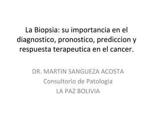
- 1. La Biopsia: su importancia en el diagnostico, pronostico, prediccion y respuesta terapeutica en el cancer. DR. MARTIN SANGUEZA ACOSTA Consultorio de Patologia LA PAZ BOLIVIA
- 2. Que es la Biopsia? Una biopsia es un procedimiento que consiste en la extraccion de una muestra total o parcial de tejido para ser examinada con el fin de establecer un diagnostico.
- 3. Cuando se realiza una biopsia?
- 4. 9-08-17
- 5. Fase Pre-analitica • Toma de la muestra
- 6. El envio a laboratorio Dar una historia clinica minina sobre el paciente y una adecuada descripcion de la(s) lesiones Hipotesis dx Rendimiento de la Bx
- 7. IMPORTANCIA DE LA CLINICA EN LA BIOPSIA • Como se envia una biopsia y en que?
- 8. IMPORTANCIA DE LA CLINICA EN LA BIOPSIA • MITO – Una vez tomanda una biopsia representativa no hay que tener mayor cuidado.
- 9. IMPORTANCIA DE LA CLINICA EN LA BIOPSIA • ES EL PROCESO MEDIANTE EL CUAL SE PRODUCE UNA PARALIZACION INMEDIATA DE LA ACTIVIDAD CELULAR IMPIDIENTO EL PROCESO DE AUTODIGESTION CITOPLASMATICA Y NUCLEAR, PERMITIENDO ASI LA POSIBILIDAD DE SER ESTUDIADA DE UNA MANERA ADECUADA
- 10. IMPORTANCIA DE LA CLINICA EN LA BIOPSIA Caracteristicas de recipiente – trasparente – boca ancha – tapa rosca – de volumen adecuado a la muestra – Identificado – irrompible
- 13. EXPLORANDO LA MITOLOGIA DE LA BIOPSIA • Macroscopia
- 14. – ENCAPSULACION – CORTE – TINCION
- 23. Scanners de lamina completa Hallazgos • Auto-loading mechanism
- 25. Company Confidential – DO NOT DISTRIBUTE4/24/19 26
- 27. MICROSCOPIA CONFOCAL in vivo
- 28. MICROSCOPIA CONFOCAL in vivo
- 29. MICROSCOPIA CONFOCAL in vivo
- 32. A 4 mm thick section is mounted onto an indium-tin oxide coated MALDI target A serial section is placed on a microscope slide for histology staining Many tissues are analyzed simultaneously on one MALDI target A pathologist marks areas of interest using a color code on a photomicrograph of the stained tissue section. Spots placed are 300 µm in diameter, slightly larger than size of the matrix spots to be deposited The spotted photomicrograph and the MALDI plate image are merged and pixel coordinates of the spots determined The coordinates are transferred to an acoustic robotic spotter that deposits trypsin and matrix at the designated locations The matrix spots deposited on the tissue are slightly smaller than the desired location of the colored spots on the photomicrograph Mass spectra are collected from the matrix spots. Peptide profiles show differences depending on the cell type from which they were collected 1 cm 1 cm 1 mm melanoma Spitz nevi
- 37. S - 100
- 38. LA BIOPSIA • ESTUDIOS ESPECIALES – H & E – HISTOQUIMICA – INMUNOHISTOQUIMICA – HIBRIDIZACION IN SITU – INMUNOFLUORESCENCIA – PCR – DNA MICROARRAY – TISSUE MICROARRAY
- 40. Estudio del DNA, GENES y sus alteraciones
- 42. Microdissection • It is a very simple concept • Technical challenge – Amount of material • Several ways to microdissected – Semi-microdissection from frozen tissue blocks – Manual Microdissection • Tumor in a needle • Stroma in a needle – Micromanipulation – Microdissection by LCM
- 47. TMA - Needles Algunas agujas pueden variar de diametro 0,6mm – 1mm – 1,5mm – 2,0mm 500 300 120 80
- 48. TMA
- 50. TMA
- 52. Human Array CGH with ~ 1 Mb Resolution 2500 BACs Triplicate spots 130 mm centers 864 well plates 0.5 mg DNA Random Primed Cy3 and Cy5 16 hr Hybe 12 mm
- 53. TISSUE-ARRAY (MATRICES TISULARES) •Preservación del bloque original • 2 “tissue” de melanomas en fase de crecimiento vertical • 1 “tissue” de Nevus de Spitz
- 60. Importancia de los biomarcadores en la medicina personalizada • Cambio del papel del patólogo • Participación en equipos multidisciplinares: – Her2 neu y cáncer de mama – K-Ras y cáncer de colon – EGFR y carcinoma pulmonar – EML4-ALK y carcinoma de pulmón – Braf y melanoma metastásico
- 61. CANCER DE MAMA • EL CANCER DE MAMA ES UNA NEOPLASIA EPITELIAL MALIGNA CON UNA EVOLUCION Y PRONOSTICO, QUE DEPENDERA DE LA EXTENSION DEL TUMOR AL MOMENTO DEL DIAGNOSTICO.
- 71. Baseline, 3/15/2011 Cycle 4 Day 1, 6/8/2011 PLX4032 = RG7204 = vemurafenib Tumor Response to Vemurafenib
- 72. McDermott U et al. N Engl J Med 2011;364:340-350. Metabolic response to treatment with Vemurafenib
- 73. Vemurafenib treatment: 1.- Flaherty KT, et al. N Engl J Med 2010;363:809–19. Computer tomography revealed regression of lung, liver and bone metastases following 8 weeks Vemurafenib treatment (with each pair of images shown for a different patient) 23
- 86. Biopsia liquida
- 87. Definiciones • cfDNA = cell-free DNA • ctDNA = circulating tumoral DNA • CTCs = Circulating tumor cellsas • Exosomas = vesículas con acidos nucleicos en su interior
- 90. Utilidad de Biopsia liquida Analisis de blancos terapeuticos y de resistencia a drogas conferidos por mutaciones genicas que detectamos en muestras de sangre periferica: • Estimacion del riesgo de metastasis, recurrencia y progression • Permite entender el proceso de desarrollo de metastasis • Prediccion de respuesta a terapias dirigidas • Monitoreo de enfermedad minima residual • Seguimiento de resistencia secundaria (adquirida)
Notas del editor
- Partnered with Philips Family of “machine intelligence” software Assist pathologists in making a diagnosis
- Figure 2. Targeting Treatment to a Specific Variant in the Melanoma Gene. Shown are three-dimensional representations of glucose metabolism in 18F-fluorodeoxyglucose (FDG)–PET scans obtained at baseline and 2 weeks after the initiation of treatment in a patient with melanoma carrying the V600E BRAF mutation. The patient was treated with the BRAF inhibitor PLX4032. Hypermetabolism of injected radioactive glucose is indicated by the red, green, and yellow signals and is a feature of dividing cancer cells, as well as being a normal feature of brain and bladder metabolism or excretion. (Images courtesy of Grant McArthur, Jason Callahan, and Rod Hicks of the Peter MacCallum Cancer Centre.)