SlideShare a Scribd company logo
1 of 9
Download to read offline
Ojvensha E learning Resources-Prepared by Dr.B.B.Gosai
White matter of Cerebrum
Introduction:
 White matter of cerebrum is formed by nerve fibers in the cerebrum.
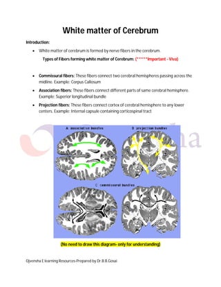
Types of Fibers forming white matter of Cerebrum: (*****Important - Viva)
 Commissural fibers: These fibers connect two cerebral hemispheres passing across the
midline. Example: Corpus Callosum
 Association fibers: These fibers connect different parts of same cerebral hemisphere.
Example: Superior longitudinal bundle
 Projection fibers: These fibers connect cortex of cerebral hemisphere to any lower
centers. Example: Internal capsule containing corticospinal tract
(No need to draw this diagram- only for understanding)
Ojvensha E learning Resources-Prepared by Dr.B.B.Gosai
Commissural Fibers (*****Important - Viva)
 Definition: These fibers connect two cerebral hemispheres passing across the midline.
Examples:
1. Corpus Callosum
2. Anterior commissure
3. Posterior commissure
4. Habenular commissure
5. Commissure of fornix
Corpus Callosum (*****Most Important Short note - Viva)
 Definition: Corpus Callosum is largest commissure of brain which connects all the parts
of two cerebral hemispheres passing across the midline except anteroinferior part of
temporal lobe.
 Parts of Corpus Callosum:
1. Rostrum
2. Genu
3. Trunk (Body)
4. Splenium
Draw this Diagram for the short note)
Ojvensha E learning Resources-Prepared by Dr.B.B.Gosai
 Rostrum: This part runs downwards and backwards from Genu and tapers to continue
with Lamina terminalis.
Relations of Rostrum:
• Inferiorly: Anterior cerebral artery
• Superiorly : Lateral ventricle (Anterior horn)
 Genu: This part is bend between the rostrum and trunk. It is located 4 cms behind the
frontal pole.
Relations of Genu:
• Antriorly: Anterior cerebral artery
• Poseriorly : Lateral ventricle (Anterior horn)
 Trunk (Body): This large part located between the genu and splenium. Its superior
surface is concavo-convex
Relations of Trunk (Body):
• Superiorly : Anterior cerebral artery, Falx cerebri and Inferior sagittal
sinus
• Inferiorly: Lateral ventricle (Anterior horn and central part)
 Splenium: This part is posterior most part of corpus callosum. It is located 6 cms in front
of the occipital pole.
Relations of Splenim:
• Poseriorly : Pineal gland
 Fibers in Corpus Callosum:
1. Forceps minor: These fibers connect two frontal lobes and forms small fork.
2. Forceps major: These fibers connect two occipital lobes and forms larger fork.
3. Tapetum: These fibers connect two temporal lobes and forms strip like fibers
and do not interdigitate with corona radiate fibers.
4. Rest of the fibers connect two parietal lobes and interdigitate with corona
radiate fibers
Ojvensha E learning Resources-Prepared by Dr.B.B.Gosai
(Draw this Diagram for the short note)
 Function of Corpus Callosum:
1. Coordination of activities of two cerebral hemispheres especially for the
steriognosis (Identification of objects with closed eye).
 Applied Anatomy of Corpus Callosum:
1. Corpus callosum can be congenitally absent without causing any abnormal
effects as both hemispheres learn to keep steriognosis
2. Due to this finding many neurological operations are planned by cutting the
corpus callosum.
3. Such operative procedures in blind leads to miserable dependent life as
Steriognosis is lost (Asteriognosis: Inability to identify objects with closed eyes).
Steriognosis is most important for blind for their all day to day activities.
4. Postoperative advice to any person when corpus callosum is cut is to accompany
while moving in the dark location.
Ojvensha E learning Resources-Prepared by Dr.B.B.Gosai
Other commissures
1. Anterior commissure: located in upper part of lamina terminalis connecting
anteroinferior part of temporal lobe
2. Posterior commissure: located in lower stalk of pineal gland connecting
pretectal region
3. Habenular commissure: located in upper stalk of pineal gland connecting
hebenular nuclei
4. Commissure of fornix: located in the posterior part of fornix connecting two
hippocampal nuclei
Association Fibers (***Important - Viva)
 Definition: These fibers connect different parts of same cerebral hemisphere.
Types of Association Fibers:
Short Association Fibers: These fibers connect adjacent gyri of same hemisphere.
Long Association Fibers: These fibers connect distant areas of same hemisphere.
Examples:
1. Superior longitudinal Bundle: Connect Frontal lobe with occipital lobe
2. Inferior longitudinal Bundle: Connect Temporal lobe with occipital lobe
3. Fasciculus Uncinatus: Connect Frontal lobe with temporal lobe
4. Cingulum (Seen on Medial surface within Cingulate gyrus): Connect parts of
limbic system
Ojvensha E learning Resources-Prepared by Dr.B.B.Gosai
Projection fibers: These fibers connect cortex of cerebral hemisphere to any lower centers.
Example: Internal capsule containing corticospinal tract
Internal capsule: (*****Most Important Short note - Viva)
 Definition: Internal capsule consists of projection fibers connecting cortex of cerebrum
to any lower centers.
 Location: Located between Lentiform nucleus (L) (Laterally) and Caudate nucleus (C)
and Thalamus (Th) (Medially).
 Shape: “V” shape in Horizontal section of cerebrum
(Draw this Diagram for the short note)
Ojvensha E learning Resources-Prepared by Dr.B.B.Gosai
 Parts of Internal capsule:
1. Anterior limb
2. Posterior limb
3. Genu
4. Retrolentiform part (Optic radiation)
5. Sublentiform part (Auditory Radiation) (Cannot be seen in Horizontal Section)
(No need to draw this diagram- only for understanding)
Ojvensha E learning Resources-Prepared by Dr.B.B.Gosai
(Draw this Diagram for the short note)
 Anterior limb: located between Lentiform nucleus laterally and Caudate nucleus
medially.
Fibers in the anterior limb:
• Afferent fiber: Thalamocortico radiation
• Efferent fiber : Corticopontine fibers
 Genu: located between anterior and posterior limb.
Fibers in the Genu:
• Efferent fibers:
• Corticonuclear fibers
• Corticospinal tract for upper limb and upper half of trunk
 Posterior limb: located between Lentiform nucleus laterally and Thalamus medially.
Fibers in the posterior limb:
• Afferent fiber: Thalamocortico radiation
• Efferent fiber :
• Corticospinal tract for lower limb and lower part of trunk
• Corticorubral tract
• Corticopontine fibers
Ojvensha E learning Resources-Prepared by Dr.B.B.Gosai
 Retrolentiform part (Optic radiation): located behind Lentiform nucleus.
Fibers in the retrolentiform part:
• Afferent fiber: Optic radiation-part of visual pathway from lateral
geniculate body to visual area of occipital lobe
 Sublentiform part (Auditory radiation): located below Lentiform nucleus.
Fibers in the sublentiform part:
• Afferent fiber: Auditory radiation-part of auditory pathway from medial
geniculate body to auditory area of temporal lobe
 Blood supply of Internal Capsule:
1. Anterior limb (Upper part): Middle cerebral Artery (Internal Carotid Artery).
2. Anterior limb (lower part): Anterior cerebral artery (Internal Carotid Artery).
3. Genu (Upper part): Middle cerebral Artery (Internal Carotid Artery).
4. Genu (lower part): Recurrent branch of Anterior cerebral artery (Heubner’s
Artery)
5. Posterior limb (upper part): Charcot’s artery of cerebral hemorrhage
(Lenticulostriate branch of Middle cerebral artery)
6. Posterior limb (lower part): Anterior choroidal artery (Internal Carotid Artery).
7. Retrolentiform part: Posterior cerebral artery (Basilar artery)
8. Sublentiform part: Anterior choroidal artery (Internal Carotid Artery).
 Applied Anatomy of Internal capsule:
1. Fibers in the internal capsule are compactly arranged. Hence even a small lesion
(damage) can lead to wide spread effects.
2. The common lesion is due to cerebral hemorrhage or cerebral thrombosis. It
results in complete hemiplegia on the opposite side (contralateral hemiplegia).
==================X================

More Related Content

What's hot

What's hot (20)

Sulci,Gyri & Functional areas of cerebrum Dr.N.Mugunthan.M.S
Sulci,Gyri & Functional areas of cerebrum Dr.N.Mugunthan.M.SSulci,Gyri & Functional areas of cerebrum Dr.N.Mugunthan.M.S
Sulci,Gyri & Functional areas of cerebrum Dr.N.Mugunthan.M.S
 
Cerebellum
CerebellumCerebellum
Cerebellum
 
Third ventricle
Third ventricleThird ventricle
Third ventricle
 
The brainstem
The brainstemThe brainstem
The brainstem
 
Posterior triangle of the neck
Posterior triangle of the neckPosterior triangle of the neck
Posterior triangle of the neck
 
Pons Anatomy
Pons AnatomyPons Anatomy
Pons Anatomy
 
Anatomy of brainstem
Anatomy of brainstemAnatomy of brainstem
Anatomy of brainstem
 
White fibers of cerebrum 1
White fibers of cerebrum 1White fibers of cerebrum 1
White fibers of cerebrum 1
 
3rd ventricle n pineal gland
3rd ventricle n pineal gland3rd ventricle n pineal gland
3rd ventricle n pineal gland
 
Anatomy of medulla oblongata
Anatomy of medulla oblongataAnatomy of medulla oblongata
Anatomy of medulla oblongata
 
white fibres of cns modified
white fibres of cns modifiedwhite fibres of cns modified
white fibres of cns modified
 
Anterior triangle of the neck
Anterior triangle of the neckAnterior triangle of the neck
Anterior triangle of the neck
 
Suboccipital triangle
Suboccipital triangleSuboccipital triangle
Suboccipital triangle
 
Basal ganglia
Basal gangliaBasal ganglia
Basal ganglia
 
The cervical plexus
The cervical plexusThe cervical plexus
The cervical plexus
 
Cerebellum ppt
Cerebellum pptCerebellum ppt
Cerebellum ppt
 
Basal ganglia
Basal gangliaBasal ganglia
Basal ganglia
 
Commissural fibres
Commissural fibresCommissural fibres
Commissural fibres
 
Carotid sheath
Carotid sheathCarotid sheath
Carotid sheath
 
Anatomy of midbrain
Anatomy of midbrainAnatomy of midbrain
Anatomy of midbrain
 

Similar to White matter of cerebrum dr.gosai

WHITE FIBERS OF CEREBRUM.pptx
WHITE FIBERS OF CEREBRUM.pptxWHITE FIBERS OF CEREBRUM.pptx
WHITE FIBERS OF CEREBRUM.pptxMODIG1
 
corpus callosum-drroyabasser-AMU.pdf
corpus callosum-drroyabasser-AMU.pdfcorpus callosum-drroyabasser-AMU.pdf
corpus callosum-drroyabasser-AMU.pdfroyabasser
 
Internal structures of hemispheres
Internal structures of hemispheresInternal structures of hemispheres
Internal structures of hemispheresMasooma Alsharakhat
 
THE ANATOMY OF WHITE MATTER AND ITS ANORMALIES.pptx
THE ANATOMY OF WHITE MATTER AND ITS ANORMALIES.pptxTHE ANATOMY OF WHITE MATTER AND ITS ANORMALIES.pptx
THE ANATOMY OF WHITE MATTER AND ITS ANORMALIES.pptxoluwatosinsax
 
Neuroanatomy Cerebellum-5 (Muhadharaty).pdf
Neuroanatomy Cerebellum-5 (Muhadharaty).pdfNeuroanatomy Cerebellum-5 (Muhadharaty).pdf
Neuroanatomy Cerebellum-5 (Muhadharaty).pdfmaktabaalarbaeen
 
15. White matter cerebrum
15. White matter cerebrum15. White matter cerebrum
15. White matter cerebrumRathiNivedhana
 
Disconnection syndromes sharath
Disconnection syndromes sharathDisconnection syndromes sharath
Disconnection syndromes sharathPasham sharath
 
Cerebellum and its connections
Cerebellum and its connectionsCerebellum and its connections
Cerebellum and its connectionsABDUL QADEER MEMON
 
anatomyofcerebellum-181202085620.pdf
anatomyofcerebellum-181202085620.pdfanatomyofcerebellum-181202085620.pdf
anatomyofcerebellum-181202085620.pdfSamarAbdElmonaem
 
gray and white matter of spinal cord.pdf
gray and white matter of spinal cord.pdfgray and white matter of spinal cord.pdf
gray and white matter of spinal cord.pdfnimraadrees2
 
White matter of cerebral hemisphere corpus callosum
White matter of cerebral hemisphere corpus callosum White matter of cerebral hemisphere corpus callosum
White matter of cerebral hemisphere corpus callosum Dr Lovely Jain
 
CEREBELLUM- with clinical and radiology
CEREBELLUM- with clinical and radiologyCEREBELLUM- with clinical and radiology
CEREBELLUM- with clinical and radiologylalit yadav
 
anatomy of cerebellum.pptx
anatomy of cerebellum.pptxanatomy of cerebellum.pptx
anatomy of cerebellum.pptxDipaliTalaviya1
 
Cerebrum internal features by DR.ARSHAD
Cerebrum internal features by DR.ARSHADCerebrum internal features by DR.ARSHAD
Cerebrum internal features by DR.ARSHADSMS_2015
 

Similar to White matter of cerebrum dr.gosai (20)

WHITE FIBERS OF CEREBRUM.pptx
WHITE FIBERS OF CEREBRUM.pptxWHITE FIBERS OF CEREBRUM.pptx
WHITE FIBERS OF CEREBRUM.pptx
 
Cerebellum dr.gosai
Cerebellum dr.gosaiCerebellum dr.gosai
Cerebellum dr.gosai
 
corpus callosum-drroyabasser-AMU.pdf
corpus callosum-drroyabasser-AMU.pdfcorpus callosum-drroyabasser-AMU.pdf
corpus callosum-drroyabasser-AMU.pdf
 
Internal structures of hemispheres
Internal structures of hemispheresInternal structures of hemispheres
Internal structures of hemispheres
 
Brain stem dr.gosai
Brain stem dr.gosaiBrain stem dr.gosai
Brain stem dr.gosai
 
THE ANATOMY OF WHITE MATTER AND ITS ANORMALIES.pptx
THE ANATOMY OF WHITE MATTER AND ITS ANORMALIES.pptxTHE ANATOMY OF WHITE MATTER AND ITS ANORMALIES.pptx
THE ANATOMY OF WHITE MATTER AND ITS ANORMALIES.pptx
 
Neuroanatomy Cerebellum-5 (Muhadharaty).pdf
Neuroanatomy Cerebellum-5 (Muhadharaty).pdfNeuroanatomy Cerebellum-5 (Muhadharaty).pdf
Neuroanatomy Cerebellum-5 (Muhadharaty).pdf
 
15. White matter cerebrum
15. White matter cerebrum15. White matter cerebrum
15. White matter cerebrum
 
Neural circuit of cerebellum,
Neural circuit of cerebellum,Neural circuit of cerebellum,
Neural circuit of cerebellum,
 
Cerebellum
 Cerebellum  Cerebellum
Cerebellum
 
Nervous systems in vertebrates: T.Y.B.Sc. Sem VI Notes
Nervous systems in vertebrates: T.Y.B.Sc. Sem VI NotesNervous systems in vertebrates: T.Y.B.Sc. Sem VI Notes
Nervous systems in vertebrates: T.Y.B.Sc. Sem VI Notes
 
Brain of scoliodon
Brain of scoliodonBrain of scoliodon
Brain of scoliodon
 
Disconnection syndromes sharath
Disconnection syndromes sharathDisconnection syndromes sharath
Disconnection syndromes sharath
 
Cerebellum and its connections
Cerebellum and its connectionsCerebellum and its connections
Cerebellum and its connections
 
anatomyofcerebellum-181202085620.pdf
anatomyofcerebellum-181202085620.pdfanatomyofcerebellum-181202085620.pdf
anatomyofcerebellum-181202085620.pdf
 
gray and white matter of spinal cord.pdf
gray and white matter of spinal cord.pdfgray and white matter of spinal cord.pdf
gray and white matter of spinal cord.pdf
 
White matter of cerebral hemisphere corpus callosum
White matter of cerebral hemisphere corpus callosum White matter of cerebral hemisphere corpus callosum
White matter of cerebral hemisphere corpus callosum
 
CEREBELLUM- with clinical and radiology
CEREBELLUM- with clinical and radiologyCEREBELLUM- with clinical and radiology
CEREBELLUM- with clinical and radiology
 
anatomy of cerebellum.pptx
anatomy of cerebellum.pptxanatomy of cerebellum.pptx
anatomy of cerebellum.pptx
 
Cerebrum internal features by DR.ARSHAD
Cerebrum internal features by DR.ARSHADCerebrum internal features by DR.ARSHAD
Cerebrum internal features by DR.ARSHAD
 

More from Dr.B.B. Gosai

AN 1.1 introduction anatomical terminology-Dr.Gosai
AN 1.1 introduction anatomical terminology-Dr.GosaiAN 1.1 introduction anatomical terminology-Dr.Gosai
AN 1.1 introduction anatomical terminology-Dr.GosaiDr.B.B. Gosai
 
Sexual differentiation-Dr.B.B.Gosai
Sexual differentiation-Dr.B.B.GosaiSexual differentiation-Dr.B.B.Gosai
Sexual differentiation-Dr.B.B.GosaiDr.B.B. Gosai
 
Vestibular and auditory apparatus-Dr.B.B.Gosai
Vestibular and auditory apparatus-Dr.B.B.GosaiVestibular and auditory apparatus-Dr.B.B.Gosai
Vestibular and auditory apparatus-Dr.B.B.GosaiDr.B.B. Gosai
 
Brachial and lumbosacral plexus-Dr.B.B.Gosai
Brachial and lumbosacral plexus-Dr.B.B.GosaiBrachial and lumbosacral plexus-Dr.B.B.Gosai
Brachial and lumbosacral plexus-Dr.B.B.GosaiDr.B.B. Gosai
 
Lower limb bones-joints-muscles-Dr.B.B.Gosai
Lower limb bones-joints-muscles-Dr.B.B.GosaiLower limb bones-joints-muscles-Dr.B.B.Gosai
Lower limb bones-joints-muscles-Dr.B.B.GosaiDr.B.B. Gosai
 
Upper limb bones-joints-muscles-Dr.B.B.Gosai
Upper limb bones-joints-muscles-Dr.B.B.GosaiUpper limb bones-joints-muscles-Dr.B.B.Gosai
Upper limb bones-joints-muscles-Dr.B.B.GosaiDr.B.B. Gosai
 
Introduction human movement apparatus-Dr.B.B.Gosai
Introduction human movement apparatus-Dr.B.B.GosaiIntroduction human movement apparatus-Dr.B.B.Gosai
Introduction human movement apparatus-Dr.B.B.GosaiDr.B.B. Gosai
 
Spinal cord dr.gosai
Spinal cord dr.gosaiSpinal cord dr.gosai
Spinal cord dr.gosaiDr.B.B. Gosai
 
Ventricles of brain dr.gosai
Ventricles of brain dr.gosaiVentricles of brain dr.gosai
Ventricles of brain dr.gosaiDr.B.B. Gosai
 
Classification of joints dr.gosai
Classification of joints dr.gosaiClassification of joints dr.gosai
Classification of joints dr.gosaiDr.B.B. Gosai
 
Anatomy of Breast in clinical perspective-Dr.Gosai
Anatomy of Breast in clinical perspective-Dr.GosaiAnatomy of Breast in clinical perspective-Dr.Gosai
Anatomy of Breast in clinical perspective-Dr.GosaiDr.B.B. Gosai
 
Pericardium and heart
Pericardium and heartPericardium and heart
Pericardium and heartDr.B.B. Gosai
 
Extraocular muscles dr.gosai
Extraocular muscles dr.gosaiExtraocular muscles dr.gosai
Extraocular muscles dr.gosaiDr.B.B. Gosai
 
Blood supply of brain dr.gosai
Blood supply of brain dr.gosaiBlood supply of brain dr.gosai
Blood supply of brain dr.gosaiDr.B.B. Gosai
 
Basal ganglia dr.gosai
Basal ganglia dr.gosaiBasal ganglia dr.gosai
Basal ganglia dr.gosaiDr.B.B. Gosai
 
Introduction abdomen-dr.gosai
Introduction abdomen-dr.gosaiIntroduction abdomen-dr.gosai
Introduction abdomen-dr.gosaiDr.B.B. Gosai
 
Esophagus stomach-dr.gosai
Esophagus stomach-dr.gosaiEsophagus stomach-dr.gosai
Esophagus stomach-dr.gosaiDr.B.B. Gosai
 

More from Dr.B.B. Gosai (20)

AN 1.1 introduction anatomical terminology-Dr.Gosai
AN 1.1 introduction anatomical terminology-Dr.GosaiAN 1.1 introduction anatomical terminology-Dr.Gosai
AN 1.1 introduction anatomical terminology-Dr.Gosai
 
Sexual differentiation-Dr.B.B.Gosai
Sexual differentiation-Dr.B.B.GosaiSexual differentiation-Dr.B.B.Gosai
Sexual differentiation-Dr.B.B.Gosai
 
Vestibular and auditory apparatus-Dr.B.B.Gosai
Vestibular and auditory apparatus-Dr.B.B.GosaiVestibular and auditory apparatus-Dr.B.B.Gosai
Vestibular and auditory apparatus-Dr.B.B.Gosai
 
Brachial and lumbosacral plexus-Dr.B.B.Gosai
Brachial and lumbosacral plexus-Dr.B.B.GosaiBrachial and lumbosacral plexus-Dr.B.B.Gosai
Brachial and lumbosacral plexus-Dr.B.B.Gosai
 
Lower limb bones-joints-muscles-Dr.B.B.Gosai
Lower limb bones-joints-muscles-Dr.B.B.GosaiLower limb bones-joints-muscles-Dr.B.B.Gosai
Lower limb bones-joints-muscles-Dr.B.B.Gosai
 
Upper limb bones-joints-muscles-Dr.B.B.Gosai
Upper limb bones-joints-muscles-Dr.B.B.GosaiUpper limb bones-joints-muscles-Dr.B.B.Gosai
Upper limb bones-joints-muscles-Dr.B.B.Gosai
 
Introduction human movement apparatus-Dr.B.B.Gosai
Introduction human movement apparatus-Dr.B.B.GosaiIntroduction human movement apparatus-Dr.B.B.Gosai
Introduction human movement apparatus-Dr.B.B.Gosai
 
Spinal cord dr.gosai
Spinal cord dr.gosaiSpinal cord dr.gosai
Spinal cord dr.gosai
 
Ventricles of brain dr.gosai
Ventricles of brain dr.gosaiVentricles of brain dr.gosai
Ventricles of brain dr.gosai
 
Classification of joints dr.gosai
Classification of joints dr.gosaiClassification of joints dr.gosai
Classification of joints dr.gosai
 
Anatomy of Breast in clinical perspective-Dr.Gosai
Anatomy of Breast in clinical perspective-Dr.GosaiAnatomy of Breast in clinical perspective-Dr.Gosai
Anatomy of Breast in clinical perspective-Dr.Gosai
 
Pericardium and heart
Pericardium and heartPericardium and heart
Pericardium and heart
 
Larynx
LarynxLarynx
Larynx
 
Pharynx dr.gosai
Pharynx dr.gosaiPharynx dr.gosai
Pharynx dr.gosai
 
Extraocular muscles dr.gosai
Extraocular muscles dr.gosaiExtraocular muscles dr.gosai
Extraocular muscles dr.gosai
 
Blood supply of brain dr.gosai
Blood supply of brain dr.gosaiBlood supply of brain dr.gosai
Blood supply of brain dr.gosai
 
Basal ganglia dr.gosai
Basal ganglia dr.gosaiBasal ganglia dr.gosai
Basal ganglia dr.gosai
 
Cerebrum
CerebrumCerebrum
Cerebrum
 
Introduction abdomen-dr.gosai
Introduction abdomen-dr.gosaiIntroduction abdomen-dr.gosai
Introduction abdomen-dr.gosai
 
Esophagus stomach-dr.gosai
Esophagus stomach-dr.gosaiEsophagus stomach-dr.gosai
Esophagus stomach-dr.gosai
 

Recently uploaded

psychiatric nursing HISTORY COLLECTION .docx
psychiatric  nursing HISTORY  COLLECTION  .docxpsychiatric  nursing HISTORY  COLLECTION  .docx
psychiatric nursing HISTORY COLLECTION .docxPoojaSen20
 
Key note speaker Neum_Admir Softic_ENG.pdf
Key note speaker Neum_Admir Softic_ENG.pdfKey note speaker Neum_Admir Softic_ENG.pdf
Key note speaker Neum_Admir Softic_ENG.pdfAdmir Softic
 
Activity 01 - Artificial Culture (1).pdf
Activity 01 - Artificial Culture (1).pdfActivity 01 - Artificial Culture (1).pdf
Activity 01 - Artificial Culture (1).pdfciinovamais
 
Mixin Classes in Odoo 17 How to Extend Models Using Mixin Classes
Mixin Classes in Odoo 17  How to Extend Models Using Mixin ClassesMixin Classes in Odoo 17  How to Extend Models Using Mixin Classes
Mixin Classes in Odoo 17 How to Extend Models Using Mixin ClassesCeline George
 
Basic Civil Engineering first year Notes- Chapter 4 Building.pptx
Basic Civil Engineering first year Notes- Chapter 4 Building.pptxBasic Civil Engineering first year Notes- Chapter 4 Building.pptx
Basic Civil Engineering first year Notes- Chapter 4 Building.pptxDenish Jangid
 
This PowerPoint helps students to consider the concept of infinity.
This PowerPoint helps students to consider the concept of infinity.This PowerPoint helps students to consider the concept of infinity.
This PowerPoint helps students to consider the concept of infinity.christianmathematics
 
Measures of Dispersion and Variability: Range, QD, AD and SD
Measures of Dispersion and Variability: Range, QD, AD and SDMeasures of Dispersion and Variability: Range, QD, AD and SD
Measures of Dispersion and Variability: Range, QD, AD and SDThiyagu K
 
Holdier Curriculum Vitae (April 2024).pdf
Holdier Curriculum Vitae (April 2024).pdfHoldier Curriculum Vitae (April 2024).pdf
Holdier Curriculum Vitae (April 2024).pdfagholdier
 
Grant Readiness 101 TechSoup and Remy Consulting
Grant Readiness 101 TechSoup and Remy ConsultingGrant Readiness 101 TechSoup and Remy Consulting
Grant Readiness 101 TechSoup and Remy ConsultingTechSoup
 
Gardella_Mateo_IntellectualProperty.pdf.
Gardella_Mateo_IntellectualProperty.pdf.Gardella_Mateo_IntellectualProperty.pdf.
Gardella_Mateo_IntellectualProperty.pdf.MateoGardella
 
Unit-IV- Pharma. Marketing Channels.pptx
Unit-IV- Pharma. Marketing Channels.pptxUnit-IV- Pharma. Marketing Channels.pptx
Unit-IV- Pharma. Marketing Channels.pptxVishalSingh1417
 
1029-Danh muc Sach Giao Khoa khoi 6.pdf
1029-Danh muc Sach Giao Khoa khoi  6.pdf1029-Danh muc Sach Giao Khoa khoi  6.pdf
1029-Danh muc Sach Giao Khoa khoi 6.pdfQucHHunhnh
 
Nutritional Needs Presentation - HLTH 104
Nutritional Needs Presentation - HLTH 104Nutritional Needs Presentation - HLTH 104
Nutritional Needs Presentation - HLTH 104misteraugie
 
SECOND SEMESTER TOPIC COVERAGE SY 2023-2024 Trends, Networks, and Critical Th...
SECOND SEMESTER TOPIC COVERAGE SY 2023-2024 Trends, Networks, and Critical Th...SECOND SEMESTER TOPIC COVERAGE SY 2023-2024 Trends, Networks, and Critical Th...
SECOND SEMESTER TOPIC COVERAGE SY 2023-2024 Trends, Networks, and Critical Th...KokoStevan
 
Unit-IV; Professional Sales Representative (PSR).pptx
Unit-IV; Professional Sales Representative (PSR).pptxUnit-IV; Professional Sales Representative (PSR).pptx
Unit-IV; Professional Sales Representative (PSR).pptxVishalSingh1417
 
How to Give a Domain for a Field in Odoo 17
How to Give a Domain for a Field in Odoo 17How to Give a Domain for a Field in Odoo 17
How to Give a Domain for a Field in Odoo 17Celine George
 

Recently uploaded (20)

Mehran University Newsletter Vol-X, Issue-I, 2024
Mehran University Newsletter Vol-X, Issue-I, 2024Mehran University Newsletter Vol-X, Issue-I, 2024
Mehran University Newsletter Vol-X, Issue-I, 2024
 
Advance Mobile Application Development class 07
Advance Mobile Application Development class 07Advance Mobile Application Development class 07
Advance Mobile Application Development class 07
 
psychiatric nursing HISTORY COLLECTION .docx
psychiatric  nursing HISTORY  COLLECTION  .docxpsychiatric  nursing HISTORY  COLLECTION  .docx
psychiatric nursing HISTORY COLLECTION .docx
 
Key note speaker Neum_Admir Softic_ENG.pdf
Key note speaker Neum_Admir Softic_ENG.pdfKey note speaker Neum_Admir Softic_ENG.pdf
Key note speaker Neum_Admir Softic_ENG.pdf
 
Activity 01 - Artificial Culture (1).pdf
Activity 01 - Artificial Culture (1).pdfActivity 01 - Artificial Culture (1).pdf
Activity 01 - Artificial Culture (1).pdf
 
Mixin Classes in Odoo 17 How to Extend Models Using Mixin Classes
Mixin Classes in Odoo 17  How to Extend Models Using Mixin ClassesMixin Classes in Odoo 17  How to Extend Models Using Mixin Classes
Mixin Classes in Odoo 17 How to Extend Models Using Mixin Classes
 
Basic Civil Engineering first year Notes- Chapter 4 Building.pptx
Basic Civil Engineering first year Notes- Chapter 4 Building.pptxBasic Civil Engineering first year Notes- Chapter 4 Building.pptx
Basic Civil Engineering first year Notes- Chapter 4 Building.pptx
 
This PowerPoint helps students to consider the concept of infinity.
This PowerPoint helps students to consider the concept of infinity.This PowerPoint helps students to consider the concept of infinity.
This PowerPoint helps students to consider the concept of infinity.
 
Measures of Dispersion and Variability: Range, QD, AD and SD
Measures of Dispersion and Variability: Range, QD, AD and SDMeasures of Dispersion and Variability: Range, QD, AD and SD
Measures of Dispersion and Variability: Range, QD, AD and SD
 
Holdier Curriculum Vitae (April 2024).pdf
Holdier Curriculum Vitae (April 2024).pdfHoldier Curriculum Vitae (April 2024).pdf
Holdier Curriculum Vitae (April 2024).pdf
 
Grant Readiness 101 TechSoup and Remy Consulting
Grant Readiness 101 TechSoup and Remy ConsultingGrant Readiness 101 TechSoup and Remy Consulting
Grant Readiness 101 TechSoup and Remy Consulting
 
Mattingly "AI & Prompt Design: Structured Data, Assistants, & RAG"
Mattingly "AI & Prompt Design: Structured Data, Assistants, & RAG"Mattingly "AI & Prompt Design: Structured Data, Assistants, & RAG"
Mattingly "AI & Prompt Design: Structured Data, Assistants, & RAG"
 
Gardella_Mateo_IntellectualProperty.pdf.
Gardella_Mateo_IntellectualProperty.pdf.Gardella_Mateo_IntellectualProperty.pdf.
Gardella_Mateo_IntellectualProperty.pdf.
 
Unit-IV- Pharma. Marketing Channels.pptx
Unit-IV- Pharma. Marketing Channels.pptxUnit-IV- Pharma. Marketing Channels.pptx
Unit-IV- Pharma. Marketing Channels.pptx
 
1029-Danh muc Sach Giao Khoa khoi 6.pdf
1029-Danh muc Sach Giao Khoa khoi  6.pdf1029-Danh muc Sach Giao Khoa khoi  6.pdf
1029-Danh muc Sach Giao Khoa khoi 6.pdf
 
Nutritional Needs Presentation - HLTH 104
Nutritional Needs Presentation - HLTH 104Nutritional Needs Presentation - HLTH 104
Nutritional Needs Presentation - HLTH 104
 
Código Creativo y Arte de Software | Unidad 1
Código Creativo y Arte de Software | Unidad 1Código Creativo y Arte de Software | Unidad 1
Código Creativo y Arte de Software | Unidad 1
 
SECOND SEMESTER TOPIC COVERAGE SY 2023-2024 Trends, Networks, and Critical Th...
SECOND SEMESTER TOPIC COVERAGE SY 2023-2024 Trends, Networks, and Critical Th...SECOND SEMESTER TOPIC COVERAGE SY 2023-2024 Trends, Networks, and Critical Th...
SECOND SEMESTER TOPIC COVERAGE SY 2023-2024 Trends, Networks, and Critical Th...
 
Unit-IV; Professional Sales Representative (PSR).pptx
Unit-IV; Professional Sales Representative (PSR).pptxUnit-IV; Professional Sales Representative (PSR).pptx
Unit-IV; Professional Sales Representative (PSR).pptx
 
How to Give a Domain for a Field in Odoo 17
How to Give a Domain for a Field in Odoo 17How to Give a Domain for a Field in Odoo 17
How to Give a Domain for a Field in Odoo 17
 

White matter of cerebrum dr.gosai

  • 1. Ojvensha E learning Resources-Prepared by Dr.B.B.Gosai White matter of Cerebrum Introduction:  White matter of cerebrum is formed by nerve fibers in the cerebrum. Types of Fibers forming white matter of Cerebrum: (*****Important - Viva)  Commissural fibers: These fibers connect two cerebral hemispheres passing across the midline. Example: Corpus Callosum  Association fibers: These fibers connect different parts of same cerebral hemisphere. Example: Superior longitudinal bundle  Projection fibers: These fibers connect cortex of cerebral hemisphere to any lower centers. Example: Internal capsule containing corticospinal tract (No need to draw this diagram- only for understanding)
  • 2. Ojvensha E learning Resources-Prepared by Dr.B.B.Gosai Commissural Fibers (*****Important - Viva)  Definition: These fibers connect two cerebral hemispheres passing across the midline. Examples: 1. Corpus Callosum 2. Anterior commissure 3. Posterior commissure 4. Habenular commissure 5. Commissure of fornix Corpus Callosum (*****Most Important Short note - Viva)  Definition: Corpus Callosum is largest commissure of brain which connects all the parts of two cerebral hemispheres passing across the midline except anteroinferior part of temporal lobe.  Parts of Corpus Callosum: 1. Rostrum 2. Genu 3. Trunk (Body) 4. Splenium Draw this Diagram for the short note)
  • 3. Ojvensha E learning Resources-Prepared by Dr.B.B.Gosai  Rostrum: This part runs downwards and backwards from Genu and tapers to continue with Lamina terminalis. Relations of Rostrum: • Inferiorly: Anterior cerebral artery • Superiorly : Lateral ventricle (Anterior horn)  Genu: This part is bend between the rostrum and trunk. It is located 4 cms behind the frontal pole. Relations of Genu: • Antriorly: Anterior cerebral artery • Poseriorly : Lateral ventricle (Anterior horn)  Trunk (Body): This large part located between the genu and splenium. Its superior surface is concavo-convex Relations of Trunk (Body): • Superiorly : Anterior cerebral artery, Falx cerebri and Inferior sagittal sinus • Inferiorly: Lateral ventricle (Anterior horn and central part)  Splenium: This part is posterior most part of corpus callosum. It is located 6 cms in front of the occipital pole. Relations of Splenim: • Poseriorly : Pineal gland  Fibers in Corpus Callosum: 1. Forceps minor: These fibers connect two frontal lobes and forms small fork. 2. Forceps major: These fibers connect two occipital lobes and forms larger fork. 3. Tapetum: These fibers connect two temporal lobes and forms strip like fibers and do not interdigitate with corona radiate fibers. 4. Rest of the fibers connect two parietal lobes and interdigitate with corona radiate fibers
  • 4. Ojvensha E learning Resources-Prepared by Dr.B.B.Gosai (Draw this Diagram for the short note)  Function of Corpus Callosum: 1. Coordination of activities of two cerebral hemispheres especially for the steriognosis (Identification of objects with closed eye).  Applied Anatomy of Corpus Callosum: 1. Corpus callosum can be congenitally absent without causing any abnormal effects as both hemispheres learn to keep steriognosis 2. Due to this finding many neurological operations are planned by cutting the corpus callosum. 3. Such operative procedures in blind leads to miserable dependent life as Steriognosis is lost (Asteriognosis: Inability to identify objects with closed eyes). Steriognosis is most important for blind for their all day to day activities. 4. Postoperative advice to any person when corpus callosum is cut is to accompany while moving in the dark location.
  • 5. Ojvensha E learning Resources-Prepared by Dr.B.B.Gosai Other commissures 1. Anterior commissure: located in upper part of lamina terminalis connecting anteroinferior part of temporal lobe 2. Posterior commissure: located in lower stalk of pineal gland connecting pretectal region 3. Habenular commissure: located in upper stalk of pineal gland connecting hebenular nuclei 4. Commissure of fornix: located in the posterior part of fornix connecting two hippocampal nuclei Association Fibers (***Important - Viva)  Definition: These fibers connect different parts of same cerebral hemisphere. Types of Association Fibers: Short Association Fibers: These fibers connect adjacent gyri of same hemisphere. Long Association Fibers: These fibers connect distant areas of same hemisphere. Examples: 1. Superior longitudinal Bundle: Connect Frontal lobe with occipital lobe 2. Inferior longitudinal Bundle: Connect Temporal lobe with occipital lobe 3. Fasciculus Uncinatus: Connect Frontal lobe with temporal lobe 4. Cingulum (Seen on Medial surface within Cingulate gyrus): Connect parts of limbic system
  • 6. Ojvensha E learning Resources-Prepared by Dr.B.B.Gosai Projection fibers: These fibers connect cortex of cerebral hemisphere to any lower centers. Example: Internal capsule containing corticospinal tract Internal capsule: (*****Most Important Short note - Viva)  Definition: Internal capsule consists of projection fibers connecting cortex of cerebrum to any lower centers.  Location: Located between Lentiform nucleus (L) (Laterally) and Caudate nucleus (C) and Thalamus (Th) (Medially).  Shape: “V” shape in Horizontal section of cerebrum (Draw this Diagram for the short note)
  • 7. Ojvensha E learning Resources-Prepared by Dr.B.B.Gosai  Parts of Internal capsule: 1. Anterior limb 2. Posterior limb 3. Genu 4. Retrolentiform part (Optic radiation) 5. Sublentiform part (Auditory Radiation) (Cannot be seen in Horizontal Section) (No need to draw this diagram- only for understanding)
  • 8. Ojvensha E learning Resources-Prepared by Dr.B.B.Gosai (Draw this Diagram for the short note)  Anterior limb: located between Lentiform nucleus laterally and Caudate nucleus medially. Fibers in the anterior limb: • Afferent fiber: Thalamocortico radiation • Efferent fiber : Corticopontine fibers  Genu: located between anterior and posterior limb. Fibers in the Genu: • Efferent fibers: • Corticonuclear fibers • Corticospinal tract for upper limb and upper half of trunk  Posterior limb: located between Lentiform nucleus laterally and Thalamus medially. Fibers in the posterior limb: • Afferent fiber: Thalamocortico radiation • Efferent fiber : • Corticospinal tract for lower limb and lower part of trunk • Corticorubral tract • Corticopontine fibers
  • 9. Ojvensha E learning Resources-Prepared by Dr.B.B.Gosai  Retrolentiform part (Optic radiation): located behind Lentiform nucleus. Fibers in the retrolentiform part: • Afferent fiber: Optic radiation-part of visual pathway from lateral geniculate body to visual area of occipital lobe  Sublentiform part (Auditory radiation): located below Lentiform nucleus. Fibers in the sublentiform part: • Afferent fiber: Auditory radiation-part of auditory pathway from medial geniculate body to auditory area of temporal lobe  Blood supply of Internal Capsule: 1. Anterior limb (Upper part): Middle cerebral Artery (Internal Carotid Artery). 2. Anterior limb (lower part): Anterior cerebral artery (Internal Carotid Artery). 3. Genu (Upper part): Middle cerebral Artery (Internal Carotid Artery). 4. Genu (lower part): Recurrent branch of Anterior cerebral artery (Heubner’s Artery) 5. Posterior limb (upper part): Charcot’s artery of cerebral hemorrhage (Lenticulostriate branch of Middle cerebral artery) 6. Posterior limb (lower part): Anterior choroidal artery (Internal Carotid Artery). 7. Retrolentiform part: Posterior cerebral artery (Basilar artery) 8. Sublentiform part: Anterior choroidal artery (Internal Carotid Artery).  Applied Anatomy of Internal capsule: 1. Fibers in the internal capsule are compactly arranged. Hence even a small lesion (damage) can lead to wide spread effects. 2. The common lesion is due to cerebral hemorrhage or cerebral thrombosis. It results in complete hemiplegia on the opposite side (contralateral hemiplegia). ==================X================