SlideShare ist ein Scribd-Unternehmen logo
1 von 24
Downloaden Sie, um offline zu lesen
Disorders of the heart valves
Lecture 10
Heart valves
238
Disorders of the heart valves
Damage and dysfunction of the heart valves most commonly occurs as a result of :
 Infective endocarditis or
 Rheumatic fever
 Ischemia
 Direct trauma
 Congenital defects or abnormalities of the heart valves
Overview
 Two major types of changes are seen in heart valves:
 Stenosis and Regurgitation (also called insufficiency or incompetence)
 Aortic Stenosis
 Mitral Stenosis
 Aortic Regurgitation
• Acute and Chronic
 Mitral Regurgitation
• Acute and Chronic
Valvular Stenosis
 The valve opening narrows
 The valve leaflets may become fused or thickened that the valve cannot open freely
 obstructs the normal flow of blood
 EFFECTS: the chamber behind the stenotic valve is subject to
o greater stress  must generate more pressure or work
o hard to force blood through the narrowed opening
 initially, the heart compensates for the additional workload by gradual
hypertrophy and dilation of the myocardium  heart failure
239
Valvular Insufficiency or Regurgitation
 Scarring and retraction of valve leaflets or weakening of supporting structures 
incomplete closure of the valve result to leakage or backflow of blood from the previous
chamber
 EFFECTS:
o causes the Heart to pump the same blood twice (as the blood comes back into the
chamber)
o the Heart dilates to accommodate more blood (the usual blood it needs to pump
+ regurgitated blood)
o ventricular dilation and hypertrophy  eventually leads to heart failure
Heart sounds
240
Heart Murmur: is an extra or unusual sound heard during a heartbeat.
Mnemonic: Systolic Murmurs ( ASaMR) Diastolic Murmurs (ARMS)
Aortic Stenosis
Aortic Stenosis Overview:
 Normal Aortic Valve Area: 3-4 cm2
 Symptoms: Occur when valve area is 1/4th
of normal area.
 Types:
o Supravalvular
o Subvalvular
o Valvular
241
 The normal aortic valve consists of three thin and pliable valve leaflets
 Bicuspid aortic valve (BAV) is a congenital condition of the aortic valve where two of
the aortic valvular leaflets fuse during development resulting in a valve that is bicuspid (2
leaflets) instead of the normal tricuspid configuration. Persons born with an abnormal
bicuspid valve are particularly susceptible to calcification later in life.
 Normally the only cardiac valve that is bicuspid is the mitral valve (bicuspid valve).
Aortic Stenosis
242
Aortic Stenosis causes & Consequences
 may be due to rheumatic heart disease, atherosclerosis, congenital valvular disease or
malformations
 narrowing of the aortic valve  flow of blood from the left ventricle to the aorta
  blood volume and pressure in the left ventricle
 Left ventricle hypertrophy develops as a compensatory mechanism to continue pumping
blood through the narrowed opening
 Patients under 70: >50% have a congenital cause
 Patients over 70: 50% due to degenerative
243
Pathophysiology of Aortic Stenosis
Clinical Manifestations
 fatigue & exertional dyspnea – 1st
symptoms – due to  CO and pulmonary congestion
 chest pain (angina) – most common symptom
o occurs during exercise – due to inability of the heart to increase coronary blood
flow to cardiac muscle
 exertional syncope, vertigo, periods of confusion --  CO
 weakness, orthopnea, PND, pulmonary edema (severe cases)
 signs of right-sided heart failure –- end-stage symptoms
- if untreated, survival rate: 1.5-3 years
 Auscultation: harsh, rough, mid-systolic ejection murmur
244
Management of AS
 General- IE prophylaxis in dental procedures with a prosthetic AV or history of
endocarditis.
 Medical:
o limited role since AS is a mechanical problem.
o restrict activity
o digitalis
o Na+ restriction, diuretics
o Nitroglycerin – for chest pain
 Surgical:
o Aortic Balloon Valvotomy- shows little benefit.
o Surgical Replacement: Definitive treatment (even in elderly and CHF)
245
Mitral Stenosis
Mitral stenosis Definition
 Mitral stenosis is characterized by thickening, fibrosis and hardening of the mitral
annulus, such as mitral orifice narrows and blood cannot pass by to the left ventricle.
 obstruct blood flow from left atrium to the left ventricle that prevents LV proper filling
during diastole
 The consequence of this is the accumulation of blood in the left atrium and then to the
lungs (heart failure and pulmonary edema).
 Normal MV Area: 4-6 cm2
, symptoms begin at areas less than 2 cm2
Mitral Stenosis Overview
 Most common valvular disorder in Rheumatic Heart Disease (RHD)
 Considered late complications of rheumatic fever.
 Rheumatic fever is a late sequela to Group A β-hemolytic streptococcal infection of the
throat.
 However, most often strep infection is asymptomatic and thereby properly left untreated
(treatment consist in eradication of infection with penicillin).
246
 14 days after the onset of infection a series of immune complexes are formed with high
affinity for connective tissue (like heart valves – M protein).
 Mitral stenosis occurs after a free interval of 10-20 years from initial rheumatic attack,
but is often asymptomatic in the early stages.
 The incidence of rheumatic mitral stenosis decreased in recent years due to the
identification and proper treatment of beta-hemolytic streptococci infection. However,
the condition is quite common in cold and wet climates.
Mitral Stenosis
247
Mitral Stenosis Pathophysiology
MS Symptoms & Signs
 Progressive Dyspnea (70%): LA dilation  pulmonary congestion (reduced emptying) :
worse with exercise, fever, tachycardia, and pregnancy
 Increased Transmitral Pressures: Leads to left atrial enlargement  Arrhythmia (irregular
heart beats) called atrial fibrillation.
 Atrial fibrillation is the most common arrhythmia in mitral stenosis and can cause serious
complications such as systemic embolisms.
 Anticoagulant medication (Warfarin “Coumadin” : is still used prophylactically, it helps
to prevent thrombus formation at the level of the dilated left atrium.
 Right heart failure symptoms: due to Pulmonary venous HTN
 Hemoptysis (Coughing Blood): due to rupture of bronchial vessels due to elevated
pulmonary pressure
 Mortality: Due to progressive pulmonary congestion, infection, and thromboembolism
248
Physical Exam Findings of MS
 Presence of bilateral peripheral edema, especially in the ankle, with warm skin, slightly
cyanotic;
 The present of hepatojugular reflux ( blood outpouring in the veins of the neck,
secondary to liver stasis); prominent "a" wave in jugular venous pulsations (JVP): Due to
pulmonary hypertension and right ventricular hypertrophy
 Signs of right-sided heart failure: in advanced disease
 Lung auscultation may reveal the present of hyperemia rales, at the level of lung bases
(pulmonary edema);
 Mitral facies:(Malar flush) When MS is severe and the cardiac output is diminished, there
is vasoconstriction, resulting in pinkish-purple patches on the cheeks
Clinical Manifestations
 exertional dyspnea and fatigue (most common)
 orthopnea, paroxysmal nocturnal dyspnea, cough, hemoptysis
 cyanosis
 Right-sided heart failure – distended neck veins, peripheral edema, hepatomegaly,
abdominal discomfort
 Auscultation:
o Mid diastolic murmur (apex)
249
Evaluation of MS
 ECG: may show atrial fibrillation and LA enlargement
 CXR: LA enlargement and pulmonary congestion. Occasionally calcified MV
 ECHO: The GOLD STANDARD for diagnosis. Asses mitral valve mobility, gradient and
mitral valve area
Management of MS
 MS like AS is a mechanical problem and medical therapy does not prevent progression
 Medical Therapy is aimed at preventing the complications of systemic embolization and
bacterial endocarditis as well as atrial fibrillation.
 Patients who have asymptomatic mitral stenosis require only antibiotic prophylaxis.
 IE prophylaxis: also with Patients with prosthetic valves or a Hx of IE for dental
procedures.
 Patients with mild pulmonary congestion can be managed with diuretics alone. β -
Blockers can be used to reduce heart rate and improve diastolic filling time.
250
 When patients have atrial fibrillation, digoxin, β-blockers, or calcium channel blockers
can be used for ventricular response rate control.
 Patients with atrial fibrillation require anticoagulation to prevent thrombus formation in
the atrium.
 Once the patient has symptoms despite adequate medical management, mechanical
correction of mitral stenosis by:
 Balloon valvuloplasty :. It is minimally invasive procedure; done by cardiac
Catheterization.
 Surgery: by Commissurotomy which is an open-heart surgery that repairs a mitral
valve that is narrowed from mitral valve stenosis.
 Mitral valve replacement – when stenosis is severe
MS Surgical Treatment
251
Aortic Regurgitation
 Definition:
o is the leaking of the aortic valve that causes blood to flow in the reverse direction
during ventricular diastole, from the aorta into the left ventricle.
 Compensatory Mechanisms:
o LV dilation, LVH. Progressive dilation leads to heart failure
 Causes
o Rheumatic Fever – most common cause
o Bicuspid aortic valve disease
o Severe hypertension
o Congenital anomaly
252
Symptoms & Signs of AR
 Asymptomatic until 4th
or 5th
decade
 Rate of Progression: 4-6% per year
 Progressive Symptoms include:
o Dyspnea: Shortness of breath
o Orthopnea : is shortness of breath which occurs when lying flat, causing the
person to have to sleep propped up in bed or sitting in a chair
o Paroxsymal Nocturnal Dyspnea (PND): refers to attacks of severe shortness of
breath and coughing that generally occur at night.
o Nocturnal angina: due to slowing of heart rate and reduction of diastolic blood
pressure
o Palpitations: due to increased force of contraction
Physical Findings of AR
 Wide pulse pressure : most sensitive
 Auscultation:
 Diastolic blowing murmur at the left sternal border
 Austin flint murmur (apex) -mid-diastolic or presystolic murmur:
 Due to diastolic displacement of the anterior leaflet of the mitral valve by the
aortic regurgitation stream causing it to vibrate.
 Systolic ejection murmur: due to increased flow across the aortic valve
 Florid pulmonary edema
 Pulmonary edema is accumulation of fluid in the interstitial spaces of the lungs
(Interstitial Edema) due to an imbalance in the oncotic and hydrostatic pressure in
the lungs.
 Florid pulmonary edema: the excess fluid is filled in the space between the
capillary and lung interstitial spaces.
253
Pathophysiology of AR
The Evaluation of AR
 CXR: enlarged cardiac silhouette and aortic root enlargement
 ECHO: Evaluation of the AV and aortic root with measurements of LV dimensions and
function (cornerstone for decision making and follow up evaluation)
 Aortography: Used to confirm the severity of disease
Management of AR
 General: IE prophylaxis in dental procedures with a prosthetic AV or history of
endocarditis.
 Medical: Vasodilators (ACEI’s), Nifedipine improve stroke volume and reduce
regurgitation only if pt symptomatic or HTN.
 Serial Echocardiograms: to monitor progression.
 Surgical Treatment: Definitive Tx
 Simplified Indications for Surgical Treatment of AR
o ANY Symptoms at rest or exercise
o Asymptomatic treatment if:
 EF drops below 50% or LV becomes dilated
254
Mitral Regurgitation
 Definition: Backflow of blood from the LV to the LA during systole
 Mild (physiological) MR is seen in 80% of normal individuals.
MR Causes
 Infective Endocarditis
 Acute MI: papillary muscle rupture or rupture of a chorda tendinea secondary to MI
 Malfunction or disruption of prosthetic valve
 Acute rheumatic fever with carditis is the predominant cause.
The Natural History of MR
 Compensatory phase: 10-15 years
 Patients with asymptomatic severe MR have a 5%/year mortality rate
 Once the patient’s EF becomes <60% and/or becomes symptomatic, mortality rises
sharply
 Mortality: From progressive dyspnea and heart failure
Clinical Manifestations of MR
 Fatigue & weakness – due to  CO – predominant complaint
 exertional dyspnea & cough – pulmonary congestion
 palpitations – due to atrial fibrillation (occur in 75% of pts.)
 Right-sided heart failure – distended neck veins, edema, ascites, hepatomegaly
 Auscultation: holosystolic murmur at the apex radiating to the axilla
255
Pathophysiology of MR
Imaging studies in MR
 ECG: May show, LA enlargement, atrial fibrillation and LV hypertrophy with severe MR
 CXR: LA enlargement, central pulmonary artery enlargement.
 ECHO: Estimation of LA, LV size and function. Valve structure assessment
 TEE (TransEsophageal Echocardiogram) if transthoracic echo is inconclusive.
Management of MR
 The treatment of acute aortic regurgitation is surgery to repair or replace the valve.
 Medical therapy may be used to stabilize the patient en route to surgery; however,
surgery should not be delayed in favor of efforts at medical management.
 Do not attempt to alleviate tachycardia with beta-blockers. Mild-to-moderate tachycardia
is beneficial in these patients because it allows less time for the heart to have backfill,
which lowers regurgitant volume.
 Medications
o Vasodilator such as hydralazine
o Rate control for atrial fibrillation with β-blockers, CCB, digoxin
o Anticoagulation in atrial fibrillation and flutter
256
o Diuretics for fluid overload
 Serial Echocardiography:
o Mild: 2-3 years, Moderate: 1-2 years, Severe: 6-12 months
 IE prophylaxis: Patients with prosthetic valves or a Hx of IE for dental procedures.
 MV Replacement in Severe MR
Mitral Valve Prolapse
 When 1 or both of the valve leaflets bulge into the left atrium during ventricular
contraction
 more common in women
 Cause: due to an inherited connective tissue disorder  enlargement of one or both
valve leaflets
 Elongates/stretches the chordae tendinae & papillary muscles  regurgitation may occur
 usually asymptomatic
 Extra heart sound (Mitral click) – an early sign that a valve leaflet is ballooning into the
left atrium
 fatigue, shortness of breath
 arrhythmias may develop – dizziness, chest pain, dyspnea, palpitations, syncope
 high-pitched late systolic murmur
257
Mitral Valve Prolapse
Interventions:
 antibiotic prophylaxis to prevent endocarditis
 If w/ dysrhythmia – avoid caffeine, alcohol, stop smoking
 anti-arrhythmic drugs
 for chest pain – nitrates, calcium channel blockers, beta blockers
 surgery not indicated
Tricuspid Stenosis
 usually occurs together w/ aortic or mitral stenosis
 may be due to rheumatic heart disease
  blood flow from right atrium to right ventricle
  right ventricular output
  left ventricular filling   CO
 blood accumulates in systemic circulation
  systemic pressure
 S/Sx: symptoms of right-sided heart failure
o hepatomegaly
o peripheral edema
o neck vein engorgement
o  CO – fatigue, hypotension
Tricuspid Regurgitation
 Uncommon
 TR is most commonly secondary, caused by dilation of the right ventricle (RV) with
malfunction of a normal valve, RV dysfunction–induced heart failure (HF), and
pulmonary outflow tract obstruction.
 TR is less commonly primary, due to valvular abnormalities caused by infective
endocarditis in users of illicit IV drugs, blunt chest trauma, rheumatic fever, congenital
defects.
 An insufficient tricuspid valve allows blood to flow back into the right atrium  venous
congestion &  right ventricular output   blood flow towards the lungs
258
Clinical Manifestations of TR
 Symptoms of TR are often nonspecific, and severe TR may be well tolerated, producing
few overt symptoms for a prolonged period.
 With isolated severe TR, Patients may complain of :
o fatigue and decreased exercise tolerance as a result of low cardiac output. Elevated
right atrial (RA) pressure also leads to peripheral edema and hepatic congestion with
decreased appetite and abdominal fullness.
o Long-standing severe TR results in right-heart failure:
o Ascites (accumulation of fluid in the peritoneal cavity, causing abdominal swelling)
o Anasarca (severe generalized, massive edema)
o Decreased urine output
o Active pulsing in the neck veins (Jugular venous distention)
o The murmur of TR is frequently not heard. When evident, it is a holosystolic murmur
259
Pulmonary Stenosis
 rare, usually congenital in origin
  flow of blood to the pulmonary artery due to narrowing

blood flows back to right ventricle and right atrium

right ventricle hypertrophy to compensate for
 blood volume and force blood to the pulmonary artery
 Symptoms & Signs
 harsh systolic murmur
 fatigue, dyspnea on exertion, cyanosis
 poor weight gain or failure to thrive in infants
 hepatomegaly, ascites, edema
Pulmonary Regurgitation
 a rare condition caused by infective endocarditis, tumors or RF
 blood flows back into Right ventricle  Right ventricle and atrium hypertrphy 
symptoms of Right-sided heart failure
Valve Repair
 Valvuloplasty
 Is repair of cardiac valve
 patient does not require continuous anti-coagulant medication
 usually require cardiopulmonary bypass machine
1. Commissurotomy – to separate the fused leaflets
 Balloon Valvuloplasty
 performed in the cardiac cath. lab.
 balloon inflated for 10-30 secs., w/ multiple inflations
 common used for mitral and aortic stenosis
260
 Closed surgical valvuloplasty
 done in the OR under GA
 Midsternal incision, a small hole is cut into the heart, the surgeon’s finger or a
dilator is used to open the commissure
 Open Commissurotomy
 done w/ direct visualization of the valve, thrombus and calcifications may be
identified and removed
2. Annuloplasty
 Is repair of valve annulus (junction of the valve leaflets and the muscular heart
wall)
 narrows the diameter of the valve’s orifice, useful for valvular regurgitation
3. Chordoplasty
 Is repair of chordae tendineae , done for mitral valve regurgitation
 caused by stretched, torn or shortened chordae tendineae
Valve Replacement
 Mechanical valves
 Example: Caged ball valve, Tilting-disk valve
 more durable, used for younger pts.
 risk of thromboembolism – long-term use of anti-coagulants
 Tissue or biological valves:
 xenografts – porcine or bovine heterografts (7-10 yrs viability)
 homografts – from cadaver tissue donations (10-15 yrs)
 autografts – excising the pts.’s own pulmonic valve and portion of pulmonary artery
for use as the aortic valve
 Long-term anticoagulant therapy
 Antibiotic prophylaxis
---------------------------
261

Weitere ähnliche Inhalte

Was ist angesagt? (20)

Aortic regurgitation
Aortic regurgitationAortic regurgitation
Aortic regurgitation
 
Mitral valve prolapse
Mitral valve prolapseMitral valve prolapse
Mitral valve prolapse
 
Aortic stenosis
Aortic stenosisAortic stenosis
Aortic stenosis
 
Mitral stenosis
Mitral stenosisMitral stenosis
Mitral stenosis
 
Cardiomyopathy
CardiomyopathyCardiomyopathy
Cardiomyopathy
 
Infective endocarditis
Infective endocarditisInfective endocarditis
Infective endocarditis
 
Cardiac tumors
Cardiac tumorsCardiac tumors
Cardiac tumors
 
Aortic stenosis
Aortic stenosis Aortic stenosis
Aortic stenosis
 
Aortic regurgitation
Aortic regurgitationAortic regurgitation
Aortic regurgitation
 
Mitral valve stenosis
Mitral valve stenosisMitral valve stenosis
Mitral valve stenosis
 
Infective Endocarditis
Infective EndocarditisInfective Endocarditis
Infective Endocarditis
 
Mitral valve disease
Mitral valve diseaseMitral valve disease
Mitral valve disease
 
Inflammatory Heart Disease
Inflammatory Heart DiseaseInflammatory Heart Disease
Inflammatory Heart Disease
 
Mitral regurgitation
Mitral regurgitationMitral regurgitation
Mitral regurgitation
 
Pericardial effusion
Pericardial effusionPericardial effusion
Pericardial effusion
 
Aortic regurgitation
Aortic regurgitationAortic regurgitation
Aortic regurgitation
 
Rheumatic valvular heart disease pediatrics AG
Rheumatic valvular heart disease pediatrics AGRheumatic valvular heart disease pediatrics AG
Rheumatic valvular heart disease pediatrics AG
 
Myocarditis
MyocarditisMyocarditis
Myocarditis
 
cardiomyopathy
cardiomyopathycardiomyopathy
cardiomyopathy
 
Cardiac tamponade
Cardiac tamponadeCardiac tamponade
Cardiac tamponade
 

Andere mochten auch

VALVULAR HEART DISEASE
VALVULAR HEART DISEASEVALVULAR HEART DISEASE
VALVULAR HEART DISEASEhanisahwarrior
 
5.heart pathology; valvular heart diseases
5.heart pathology; valvular heart diseases5.heart pathology; valvular heart diseases
5.heart pathology; valvular heart diseasesKrishna Tadepalli
 
Congenital heart disease
Congenital heart diseaseCongenital heart disease
Congenital heart diseaseACI Limited
 
Lecture 18 Hepatitis and liver disorders
Lecture 18 Hepatitis and liver disordersLecture 18 Hepatitis and liver disorders
Lecture 18 Hepatitis and liver disordersAreej Abu Hanieh
 
Lecture 15 kidney- pathology
Lecture 15 kidney- pathology Lecture 15 kidney- pathology
Lecture 15 kidney- pathology Areej Abu Hanieh
 
Lecture 22 diabetes mellitus - pathology
Lecture 22 diabetes mellitus - pathologyLecture 22 diabetes mellitus - pathology
Lecture 22 diabetes mellitus - pathologyAreej Abu Hanieh
 
Lecture 20 disorders of hypothalamus, pituitary and thyroid glands - Pathology
Lecture 20 disorders of hypothalamus, pituitary and thyroid glands - PathologyLecture 20 disorders of hypothalamus, pituitary and thyroid glands - Pathology
Lecture 20 disorders of hypothalamus, pituitary and thyroid glands - PathologyAreej Abu Hanieh
 
Lecture 7 diseases of the vascular system - Pathology
Lecture 7 diseases of the vascular system - Pathology Lecture 7 diseases of the vascular system - Pathology
Lecture 7 diseases of the vascular system - Pathology Areej Abu Hanieh
 
Lecture 8 hypertension - Pathology
Lecture 8 hypertension - Pathology Lecture 8 hypertension - Pathology
Lecture 8 hypertension - Pathology Areej Abu Hanieh
 
Rheumatic heart disease
Rheumatic heart diseaseRheumatic heart disease
Rheumatic heart diseaseIndah Syafiqah
 
Lecture 19 pancreatitis - Pathology
Lecture 19 pancreatitis - PathologyLecture 19 pancreatitis - Pathology
Lecture 19 pancreatitis - PathologyAreej Abu Hanieh
 
Valvular heart disease
Valvular heart diseaseValvular heart disease
Valvular heart diseaseraj kumar
 
Lecture 17 Colon Disorders - Pathology
Lecture 17 Colon Disorders - PathologyLecture 17 Colon Disorders - Pathology
Lecture 17 Colon Disorders - PathologyAreej Abu Hanieh
 
Lecture 16 esophagus and stomach disorders - Pathology
Lecture 16 esophagus and stomach disorders - PathologyLecture 16 esophagus and stomach disorders - Pathology
Lecture 16 esophagus and stomach disorders - PathologyAreej Abu Hanieh
 

Andere mochten auch (20)

VALVULAR HEART DISEASE
VALVULAR HEART DISEASEVALVULAR HEART DISEASE
VALVULAR HEART DISEASE
 
5.heart pathology; valvular heart diseases
5.heart pathology; valvular heart diseases5.heart pathology; valvular heart diseases
5.heart pathology; valvular heart diseases
 
Congenital heart disease
Congenital heart diseaseCongenital heart disease
Congenital heart disease
 
Lecture 18 Hepatitis and liver disorders
Lecture 18 Hepatitis and liver disordersLecture 18 Hepatitis and liver disorders
Lecture 18 Hepatitis and liver disorders
 
10 heart failure
10 heart failure10 heart failure
10 heart failure
 
Lecture 15 kidney- pathology
Lecture 15 kidney- pathology Lecture 15 kidney- pathology
Lecture 15 kidney- pathology
 
Pathology - Cancer
Pathology - CancerPathology - Cancer
Pathology - Cancer
 
Lecture 22 diabetes mellitus - pathology
Lecture 22 diabetes mellitus - pathologyLecture 22 diabetes mellitus - pathology
Lecture 22 diabetes mellitus - pathology
 
Lecture 20 disorders of hypothalamus, pituitary and thyroid glands - Pathology
Lecture 20 disorders of hypothalamus, pituitary and thyroid glands - PathologyLecture 20 disorders of hypothalamus, pituitary and thyroid glands - Pathology
Lecture 20 disorders of hypothalamus, pituitary and thyroid glands - Pathology
 
Pathology - Cell Injury
Pathology - Cell Injury Pathology - Cell Injury
Pathology - Cell Injury
 
Lecture 7 diseases of the vascular system - Pathology
Lecture 7 diseases of the vascular system - Pathology Lecture 7 diseases of the vascular system - Pathology
Lecture 7 diseases of the vascular system - Pathology
 
Lecture 8 hypertension - Pathology
Lecture 8 hypertension - Pathology Lecture 8 hypertension - Pathology
Lecture 8 hypertension - Pathology
 
Rheumatic heart disease
Rheumatic heart diseaseRheumatic heart disease
Rheumatic heart disease
 
Lecture 19 pancreatitis - Pathology
Lecture 19 pancreatitis - PathologyLecture 19 pancreatitis - Pathology
Lecture 19 pancreatitis - Pathology
 
Pathology - HIV (AIDS)
Pathology - HIV (AIDS)Pathology - HIV (AIDS)
Pathology - HIV (AIDS)
 
pathology - Hematology
pathology - Hematology pathology - Hematology
pathology - Hematology
 
Valvular heart disease
Valvular heart diseaseValvular heart disease
Valvular heart disease
 
Lecture 17 Colon Disorders - Pathology
Lecture 17 Colon Disorders - PathologyLecture 17 Colon Disorders - Pathology
Lecture 17 Colon Disorders - Pathology
 
Pathology - Hemostasis
Pathology - HemostasisPathology - Hemostasis
Pathology - Hemostasis
 
Lecture 16 esophagus and stomach disorders - Pathology
Lecture 16 esophagus and stomach disorders - PathologyLecture 16 esophagus and stomach disorders - Pathology
Lecture 16 esophagus and stomach disorders - Pathology
 

Ähnlich wie Lecture 10 valvular heart disease - Pathology

Coronary vascular disorder
Coronary vascular disorderCoronary vascular disorder
Coronary vascular disorderMa Icban
 
VALVULAR HEART DISEASES.pptx
VALVULAR HEART DISEASES.pptxVALVULAR HEART DISEASES.pptx
VALVULAR HEART DISEASES.pptxMaina64
 
Aortic valve disorders
Aortic valve disordersAortic valve disorders
Aortic valve disordersHizbullah Khan
 
Mitral valve disorders
Mitral valve disordersMitral valve disorders
Mitral valve disordersHizbullah Khan
 
INDICATIONS FOR VALVULAR SURGERIES
INDICATIONS FOR VALVULAR SURGERIESINDICATIONS FOR VALVULAR SURGERIES
INDICATIONS FOR VALVULAR SURGERIESRevathy Ambikadevi
 
CHRONIC RHEUMATIC FEVER.pptx
CHRONIC RHEUMATIC FEVER.pptxCHRONIC RHEUMATIC FEVER.pptx
CHRONIC RHEUMATIC FEVER.pptxmiroofafrika
 
3. ARRHYTHMIAS.pptx
3. ARRHYTHMIAS.pptx3. ARRHYTHMIAS.pptx
3. ARRHYTHMIAS.pptxmariaidrees3
 
Mitral stenosis and regurgitation sushila
Mitral stenosis and regurgitation sushilaMitral stenosis and regurgitation sushila
Mitral stenosis and regurgitation sushilaSushilaHamal
 
Acquired valvular heart diseases with x ray findings
Acquired valvular heart diseases with x ray findingsAcquired valvular heart diseases with x ray findings
Acquired valvular heart diseases with x ray findingsairwave12
 
Valvular Heart Disease
Valvular Heart DiseaseValvular Heart Disease
Valvular Heart DiseaseJessie Madz
 
Valvular heart disease
Valvular heart diseaseValvular heart disease
Valvular heart diseasehamid-miyanaji
 
Valvular Heart Disease, Esther
Valvular Heart Disease, EstherValvular Heart Disease, Esther
Valvular Heart Disease, EstherEsther Mary Mathew
 

Ähnlich wie Lecture 10 valvular heart disease - Pathology (20)

Coronary vascular disorder
Coronary vascular disorderCoronary vascular disorder
Coronary vascular disorder
 
VALVULAR HEART DISEASES.pptx
VALVULAR HEART DISEASES.pptxVALVULAR HEART DISEASES.pptx
VALVULAR HEART DISEASES.pptx
 
Chronic constrictive pericarditis
Chronic constrictive pericarditisChronic constrictive pericarditis
Chronic constrictive pericarditis
 
Chronic constrictive pericarditis
Chronic constrictive pericarditisChronic constrictive pericarditis
Chronic constrictive pericarditis
 
Aortic valve disorders
Aortic valve disordersAortic valve disorders
Aortic valve disorders
 
Mitral valve disorders
Mitral valve disordersMitral valve disorders
Mitral valve disorders
 
Aortic Regurgitation - Rivin
Aortic Regurgitation - RivinAortic Regurgitation - Rivin
Aortic Regurgitation - Rivin
 
Valvular heart disease
Valvular heart disease Valvular heart disease
Valvular heart disease
 
INDICATIONS FOR VALVULAR SURGERIES
INDICATIONS FOR VALVULAR SURGERIESINDICATIONS FOR VALVULAR SURGERIES
INDICATIONS FOR VALVULAR SURGERIES
 
Cardio3
Cardio3Cardio3
Cardio3
 
CHRONIC RHEUMATIC FEVER.pptx
CHRONIC RHEUMATIC FEVER.pptxCHRONIC RHEUMATIC FEVER.pptx
CHRONIC RHEUMATIC FEVER.pptx
 
3. ARRHYTHMIAS.pptx
3. ARRHYTHMIAS.pptx3. ARRHYTHMIAS.pptx
3. ARRHYTHMIAS.pptx
 
Mitral stenosis and regurgitation sushila
Mitral stenosis and regurgitation sushilaMitral stenosis and regurgitation sushila
Mitral stenosis and regurgitation sushila
 
Acquired valvular heart diseases with x ray findings
Acquired valvular heart diseases with x ray findingsAcquired valvular heart diseases with x ray findings
Acquired valvular heart diseases with x ray findings
 
Valvular heart disease2 . samir rafla
Valvular heart disease2 . samir raflaValvular heart disease2 . samir rafla
Valvular heart disease2 . samir rafla
 
Valvular heart disease
Valvular heart diseaseValvular heart disease
Valvular heart disease
 
Valvular Heart Disease
Valvular Heart DiseaseValvular Heart Disease
Valvular Heart Disease
 
Aortic Regurgitation
Aortic RegurgitationAortic Regurgitation
Aortic Regurgitation
 
Valvular heart disease
Valvular heart diseaseValvular heart disease
Valvular heart disease
 
Valvular Heart Disease, Esther
Valvular Heart Disease, EstherValvular Heart Disease, Esther
Valvular Heart Disease, Esther
 

Mehr von Areej Abu Hanieh

Announcement about my previous presentations - Thank you
Announcement about my previous presentations - Thank youAnnouncement about my previous presentations - Thank you
Announcement about my previous presentations - Thank youAreej Abu Hanieh
 
Hospital acquired pneumonia
Hospital acquired pneumoniaHospital acquired pneumonia
Hospital acquired pneumoniaAreej Abu Hanieh
 
catheter related blood stream infection
catheter related blood stream infection catheter related blood stream infection
catheter related blood stream infection Areej Abu Hanieh
 
Community acquired pneumonia - Pharmacotherapy
Community acquired pneumonia - Pharmacotherapy Community acquired pneumonia - Pharmacotherapy
Community acquired pneumonia - Pharmacotherapy Areej Abu Hanieh
 
Carbapenems - Pharmacology
Carbapenems - PharmacologyCarbapenems - Pharmacology
Carbapenems - PharmacologyAreej Abu Hanieh
 
Cephalosporins - Pharmacology
Cephalosporins - Pharmacology Cephalosporins - Pharmacology
Cephalosporins - Pharmacology Areej Abu Hanieh
 
Hypertensive urgencies and emergencies
Hypertensive urgencies and emergenciesHypertensive urgencies and emergencies
Hypertensive urgencies and emergenciesAreej Abu Hanieh
 
Asthma and COPD exacerbation - Emergency
Asthma and COPD exacerbation - Emergency  Asthma and COPD exacerbation - Emergency
Asthma and COPD exacerbation - Emergency Areej Abu Hanieh
 
Acute decompensated heart failure
Acute decompensated heart failureAcute decompensated heart failure
Acute decompensated heart failureAreej Abu Hanieh
 
Glycemic Control - Diabetes Mellitus
Glycemic Control - Diabetes Mellitus Glycemic Control - Diabetes Mellitus
Glycemic Control - Diabetes Mellitus Areej Abu Hanieh
 
Deep Vein Thrombosis - DVT
Deep Vein Thrombosis  - DVTDeep Vein Thrombosis  - DVT
Deep Vein Thrombosis - DVTAreej Abu Hanieh
 

Mehr von Areej Abu Hanieh (20)

Announcement about my previous presentations - Thank you
Announcement about my previous presentations - Thank youAnnouncement about my previous presentations - Thank you
Announcement about my previous presentations - Thank you
 
Infection - penicillins
Infection - penicillinsInfection - penicillins
Infection - penicillins
 
Hospital acquired pneumonia
Hospital acquired pneumoniaHospital acquired pneumonia
Hospital acquired pneumonia
 
catheter related blood stream infection
catheter related blood stream infection catheter related blood stream infection
catheter related blood stream infection
 
Community acquired pneumonia - Pharmacotherapy
Community acquired pneumonia - Pharmacotherapy Community acquired pneumonia - Pharmacotherapy
Community acquired pneumonia - Pharmacotherapy
 
Cellulitis - Treatment
Cellulitis - TreatmentCellulitis - Treatment
Cellulitis - Treatment
 
Carbapenems - Pharmacology
Carbapenems - PharmacologyCarbapenems - Pharmacology
Carbapenems - Pharmacology
 
Cephalosporins - Pharmacology
Cephalosporins - Pharmacology Cephalosporins - Pharmacology
Cephalosporins - Pharmacology
 
Sickle cell anemia
Sickle cell anemia Sickle cell anemia
Sickle cell anemia
 
Poisoning - Treatment
Poisoning - TreatmentPoisoning - Treatment
Poisoning - Treatment
 
Hypertensive urgencies and emergencies
Hypertensive urgencies and emergenciesHypertensive urgencies and emergencies
Hypertensive urgencies and emergencies
 
Diabetic ketoacidosis DKA
Diabetic ketoacidosis DKADiabetic ketoacidosis DKA
Diabetic ketoacidosis DKA
 
Asthma and COPD exacerbation - Emergency
Asthma and COPD exacerbation - Emergency  Asthma and COPD exacerbation - Emergency
Asthma and COPD exacerbation - Emergency
 
Acute decompensated heart failure
Acute decompensated heart failureAcute decompensated heart failure
Acute decompensated heart failure
 
Acute Coronary syndrome
Acute Coronary syndrome Acute Coronary syndrome
Acute Coronary syndrome
 
Glycemic Control - Diabetes Mellitus
Glycemic Control - Diabetes Mellitus Glycemic Control - Diabetes Mellitus
Glycemic Control - Diabetes Mellitus
 
Stress ulcer prophylaxis
Stress ulcer prophylaxis Stress ulcer prophylaxis
Stress ulcer prophylaxis
 
Pain in the ICU
Pain in the ICUPain in the ICU
Pain in the ICU
 
Deep Vein Thrombosis - DVT
Deep Vein Thrombosis  - DVTDeep Vein Thrombosis  - DVT
Deep Vein Thrombosis - DVT
 
Anti - Coagulants agents
Anti - Coagulants agentsAnti - Coagulants agents
Anti - Coagulants agents
 

Kürzlich hochgeladen

Big Data Analysis Suggests COVID Vaccination Increases Excess Mortality Of ...
Big Data Analysis Suggests COVID  Vaccination Increases Excess Mortality Of  ...Big Data Analysis Suggests COVID  Vaccination Increases Excess Mortality Of  ...
Big Data Analysis Suggests COVID Vaccination Increases Excess Mortality Of ...sdateam0
 
Wessex Health Partners Wessex Integrated Care, Population Health, Research & ...
Wessex Health Partners Wessex Integrated Care, Population Health, Research & ...Wessex Health Partners Wessex Integrated Care, Population Health, Research & ...
Wessex Health Partners Wessex Integrated Care, Population Health, Research & ...Wessex Health Partners
 
LUNG TUMORS AND ITS CLASSIFICATIONS.pdf
LUNG TUMORS AND ITS  CLASSIFICATIONS.pdfLUNG TUMORS AND ITS  CLASSIFICATIONS.pdf
LUNG TUMORS AND ITS CLASSIFICATIONS.pdfDolisha Warbi
 
PULMONARY EDEMA AND ITS MANAGEMENT.pdf
PULMONARY EDEMA AND  ITS  MANAGEMENT.pdfPULMONARY EDEMA AND  ITS  MANAGEMENT.pdf
PULMONARY EDEMA AND ITS MANAGEMENT.pdfDolisha Warbi
 
SWD (Short wave diathermy)- Physiotherapy.ppt
SWD (Short wave diathermy)- Physiotherapy.pptSWD (Short wave diathermy)- Physiotherapy.ppt
SWD (Short wave diathermy)- Physiotherapy.pptMumux Mirani
 
Apiculture Chapter 1. Introduction 2.ppt
Apiculture Chapter 1. Introduction 2.pptApiculture Chapter 1. Introduction 2.ppt
Apiculture Chapter 1. Introduction 2.pptkedirjemalharun
 
Culture and Health Disorders Social change.pptx
Culture and Health Disorders Social change.pptxCulture and Health Disorders Social change.pptx
Culture and Health Disorders Social change.pptxDr. Dheeraj Kumar
 
systemic bacteriology (7)............pptx
systemic bacteriology (7)............pptxsystemic bacteriology (7)............pptx
systemic bacteriology (7)............pptxEyobAlemu11
 
world health day presentation ppt download
world health day presentation ppt downloadworld health day presentation ppt download
world health day presentation ppt downloadAnkitKumar311566
 
COVID-19 (NOVEL CORONA VIRUS DISEASE PANDEMIC ).pptx
COVID-19  (NOVEL CORONA  VIRUS DISEASE PANDEMIC ).pptxCOVID-19  (NOVEL CORONA  VIRUS DISEASE PANDEMIC ).pptx
COVID-19 (NOVEL CORONA VIRUS DISEASE PANDEMIC ).pptxBibekananda shah
 
97111 47426 Call Girls In Delhi MUNIRKAA
97111 47426 Call Girls In Delhi MUNIRKAA97111 47426 Call Girls In Delhi MUNIRKAA
97111 47426 Call Girls In Delhi MUNIRKAAjennyeacort
 
Biomechanics- Shoulder Joint!!!!!!!!!!!!
Biomechanics- Shoulder Joint!!!!!!!!!!!!Biomechanics- Shoulder Joint!!!!!!!!!!!!
Biomechanics- Shoulder Joint!!!!!!!!!!!!ibtesaam huma
 
Informed Consent Empowering Healthcare Decision-Making.pptx
Informed Consent Empowering Healthcare Decision-Making.pptxInformed Consent Empowering Healthcare Decision-Making.pptx
Informed Consent Empowering Healthcare Decision-Making.pptxSasikiranMarri
 
Basic principles involved in the traditional systems of medicine PDF.pdf
Basic principles involved in the traditional systems of medicine PDF.pdfBasic principles involved in the traditional systems of medicine PDF.pdf
Basic principles involved in the traditional systems of medicine PDF.pdfDivya Kanojiya
 
METHODS OF ACQUIRING KNOWLEDGE IN NURSING.pptx by navdeep kaur
METHODS OF ACQUIRING KNOWLEDGE IN NURSING.pptx by navdeep kaurMETHODS OF ACQUIRING KNOWLEDGE IN NURSING.pptx by navdeep kaur
METHODS OF ACQUIRING KNOWLEDGE IN NURSING.pptx by navdeep kaurNavdeep Kaur
 
maternal mortality and its causes and how to reduce maternal mortality
maternal mortality and its causes and how to reduce maternal mortalitymaternal mortality and its causes and how to reduce maternal mortality
maternal mortality and its causes and how to reduce maternal mortalityhardikdabas3
 
Statistical modeling in pharmaceutical research and development.
Statistical modeling in pharmaceutical research and development.Statistical modeling in pharmaceutical research and development.
Statistical modeling in pharmaceutical research and development.ANJALI
 
Presentation for Bella Mahl 2024-03-28-24-MW-Overview-Bella.pptx
Presentation for Bella Mahl 2024-03-28-24-MW-Overview-Bella.pptxPresentation for Bella Mahl 2024-03-28-24-MW-Overview-Bella.pptx
Presentation for Bella Mahl 2024-03-28-24-MW-Overview-Bella.pptxpdamico1
 
Music Therapy's Impact in Palliative Care| IAPCON2024| Dr. Tara Rajendran
Music Therapy's Impact in Palliative Care| IAPCON2024| Dr. Tara RajendranMusic Therapy's Impact in Palliative Care| IAPCON2024| Dr. Tara Rajendran
Music Therapy's Impact in Palliative Care| IAPCON2024| Dr. Tara RajendranTara Rajendran
 

Kürzlich hochgeladen (20)

Epilepsy
EpilepsyEpilepsy
Epilepsy
 
Big Data Analysis Suggests COVID Vaccination Increases Excess Mortality Of ...
Big Data Analysis Suggests COVID  Vaccination Increases Excess Mortality Of  ...Big Data Analysis Suggests COVID  Vaccination Increases Excess Mortality Of  ...
Big Data Analysis Suggests COVID Vaccination Increases Excess Mortality Of ...
 
Wessex Health Partners Wessex Integrated Care, Population Health, Research & ...
Wessex Health Partners Wessex Integrated Care, Population Health, Research & ...Wessex Health Partners Wessex Integrated Care, Population Health, Research & ...
Wessex Health Partners Wessex Integrated Care, Population Health, Research & ...
 
LUNG TUMORS AND ITS CLASSIFICATIONS.pdf
LUNG TUMORS AND ITS  CLASSIFICATIONS.pdfLUNG TUMORS AND ITS  CLASSIFICATIONS.pdf
LUNG TUMORS AND ITS CLASSIFICATIONS.pdf
 
PULMONARY EDEMA AND ITS MANAGEMENT.pdf
PULMONARY EDEMA AND  ITS  MANAGEMENT.pdfPULMONARY EDEMA AND  ITS  MANAGEMENT.pdf
PULMONARY EDEMA AND ITS MANAGEMENT.pdf
 
SWD (Short wave diathermy)- Physiotherapy.ppt
SWD (Short wave diathermy)- Physiotherapy.pptSWD (Short wave diathermy)- Physiotherapy.ppt
SWD (Short wave diathermy)- Physiotherapy.ppt
 
Apiculture Chapter 1. Introduction 2.ppt
Apiculture Chapter 1. Introduction 2.pptApiculture Chapter 1. Introduction 2.ppt
Apiculture Chapter 1. Introduction 2.ppt
 
Culture and Health Disorders Social change.pptx
Culture and Health Disorders Social change.pptxCulture and Health Disorders Social change.pptx
Culture and Health Disorders Social change.pptx
 
systemic bacteriology (7)............pptx
systemic bacteriology (7)............pptxsystemic bacteriology (7)............pptx
systemic bacteriology (7)............pptx
 
world health day presentation ppt download
world health day presentation ppt downloadworld health day presentation ppt download
world health day presentation ppt download
 
COVID-19 (NOVEL CORONA VIRUS DISEASE PANDEMIC ).pptx
COVID-19  (NOVEL CORONA  VIRUS DISEASE PANDEMIC ).pptxCOVID-19  (NOVEL CORONA  VIRUS DISEASE PANDEMIC ).pptx
COVID-19 (NOVEL CORONA VIRUS DISEASE PANDEMIC ).pptx
 
97111 47426 Call Girls In Delhi MUNIRKAA
97111 47426 Call Girls In Delhi MUNIRKAA97111 47426 Call Girls In Delhi MUNIRKAA
97111 47426 Call Girls In Delhi MUNIRKAA
 
Biomechanics- Shoulder Joint!!!!!!!!!!!!
Biomechanics- Shoulder Joint!!!!!!!!!!!!Biomechanics- Shoulder Joint!!!!!!!!!!!!
Biomechanics- Shoulder Joint!!!!!!!!!!!!
 
Informed Consent Empowering Healthcare Decision-Making.pptx
Informed Consent Empowering Healthcare Decision-Making.pptxInformed Consent Empowering Healthcare Decision-Making.pptx
Informed Consent Empowering Healthcare Decision-Making.pptx
 
Basic principles involved in the traditional systems of medicine PDF.pdf
Basic principles involved in the traditional systems of medicine PDF.pdfBasic principles involved in the traditional systems of medicine PDF.pdf
Basic principles involved in the traditional systems of medicine PDF.pdf
 
METHODS OF ACQUIRING KNOWLEDGE IN NURSING.pptx by navdeep kaur
METHODS OF ACQUIRING KNOWLEDGE IN NURSING.pptx by navdeep kaurMETHODS OF ACQUIRING KNOWLEDGE IN NURSING.pptx by navdeep kaur
METHODS OF ACQUIRING KNOWLEDGE IN NURSING.pptx by navdeep kaur
 
maternal mortality and its causes and how to reduce maternal mortality
maternal mortality and its causes and how to reduce maternal mortalitymaternal mortality and its causes and how to reduce maternal mortality
maternal mortality and its causes and how to reduce maternal mortality
 
Statistical modeling in pharmaceutical research and development.
Statistical modeling in pharmaceutical research and development.Statistical modeling in pharmaceutical research and development.
Statistical modeling in pharmaceutical research and development.
 
Presentation for Bella Mahl 2024-03-28-24-MW-Overview-Bella.pptx
Presentation for Bella Mahl 2024-03-28-24-MW-Overview-Bella.pptxPresentation for Bella Mahl 2024-03-28-24-MW-Overview-Bella.pptx
Presentation for Bella Mahl 2024-03-28-24-MW-Overview-Bella.pptx
 
Music Therapy's Impact in Palliative Care| IAPCON2024| Dr. Tara Rajendran
Music Therapy's Impact in Palliative Care| IAPCON2024| Dr. Tara RajendranMusic Therapy's Impact in Palliative Care| IAPCON2024| Dr. Tara Rajendran
Music Therapy's Impact in Palliative Care| IAPCON2024| Dr. Tara Rajendran
 

Lecture 10 valvular heart disease - Pathology

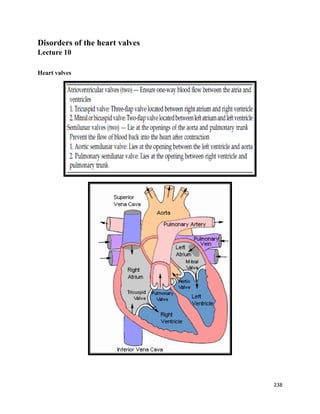
  • 1. Disorders of the heart valves Lecture 10 Heart valves 238
  • 2. Disorders of the heart valves Damage and dysfunction of the heart valves most commonly occurs as a result of :  Infective endocarditis or  Rheumatic fever  Ischemia  Direct trauma  Congenital defects or abnormalities of the heart valves Overview  Two major types of changes are seen in heart valves:  Stenosis and Regurgitation (also called insufficiency or incompetence)  Aortic Stenosis  Mitral Stenosis  Aortic Regurgitation • Acute and Chronic  Mitral Regurgitation • Acute and Chronic Valvular Stenosis  The valve opening narrows  The valve leaflets may become fused or thickened that the valve cannot open freely  obstructs the normal flow of blood  EFFECTS: the chamber behind the stenotic valve is subject to o greater stress  must generate more pressure or work o hard to force blood through the narrowed opening  initially, the heart compensates for the additional workload by gradual hypertrophy and dilation of the myocardium  heart failure 239
  • 3. Valvular Insufficiency or Regurgitation  Scarring and retraction of valve leaflets or weakening of supporting structures  incomplete closure of the valve result to leakage or backflow of blood from the previous chamber  EFFECTS: o causes the Heart to pump the same blood twice (as the blood comes back into the chamber) o the Heart dilates to accommodate more blood (the usual blood it needs to pump + regurgitated blood) o ventricular dilation and hypertrophy  eventually leads to heart failure Heart sounds 240
  • 4. Heart Murmur: is an extra or unusual sound heard during a heartbeat. Mnemonic: Systolic Murmurs ( ASaMR) Diastolic Murmurs (ARMS) Aortic Stenosis Aortic Stenosis Overview:  Normal Aortic Valve Area: 3-4 cm2  Symptoms: Occur when valve area is 1/4th of normal area.  Types: o Supravalvular o Subvalvular o Valvular 241
  • 5.  The normal aortic valve consists of three thin and pliable valve leaflets  Bicuspid aortic valve (BAV) is a congenital condition of the aortic valve where two of the aortic valvular leaflets fuse during development resulting in a valve that is bicuspid (2 leaflets) instead of the normal tricuspid configuration. Persons born with an abnormal bicuspid valve are particularly susceptible to calcification later in life.  Normally the only cardiac valve that is bicuspid is the mitral valve (bicuspid valve). Aortic Stenosis 242
  • 6. Aortic Stenosis causes & Consequences  may be due to rheumatic heart disease, atherosclerosis, congenital valvular disease or malformations  narrowing of the aortic valve  flow of blood from the left ventricle to the aorta   blood volume and pressure in the left ventricle  Left ventricle hypertrophy develops as a compensatory mechanism to continue pumping blood through the narrowed opening  Patients under 70: >50% have a congenital cause  Patients over 70: 50% due to degenerative 243
  • 7. Pathophysiology of Aortic Stenosis Clinical Manifestations  fatigue & exertional dyspnea – 1st symptoms – due to  CO and pulmonary congestion  chest pain (angina) – most common symptom o occurs during exercise – due to inability of the heart to increase coronary blood flow to cardiac muscle  exertional syncope, vertigo, periods of confusion --  CO  weakness, orthopnea, PND, pulmonary edema (severe cases)  signs of right-sided heart failure –- end-stage symptoms - if untreated, survival rate: 1.5-3 years  Auscultation: harsh, rough, mid-systolic ejection murmur 244
  • 8. Management of AS  General- IE prophylaxis in dental procedures with a prosthetic AV or history of endocarditis.  Medical: o limited role since AS is a mechanical problem. o restrict activity o digitalis o Na+ restriction, diuretics o Nitroglycerin – for chest pain  Surgical: o Aortic Balloon Valvotomy- shows little benefit. o Surgical Replacement: Definitive treatment (even in elderly and CHF) 245
  • 9. Mitral Stenosis Mitral stenosis Definition  Mitral stenosis is characterized by thickening, fibrosis and hardening of the mitral annulus, such as mitral orifice narrows and blood cannot pass by to the left ventricle.  obstruct blood flow from left atrium to the left ventricle that prevents LV proper filling during diastole  The consequence of this is the accumulation of blood in the left atrium and then to the lungs (heart failure and pulmonary edema).  Normal MV Area: 4-6 cm2 , symptoms begin at areas less than 2 cm2 Mitral Stenosis Overview  Most common valvular disorder in Rheumatic Heart Disease (RHD)  Considered late complications of rheumatic fever.  Rheumatic fever is a late sequela to Group A β-hemolytic streptococcal infection of the throat.  However, most often strep infection is asymptomatic and thereby properly left untreated (treatment consist in eradication of infection with penicillin). 246
  • 10.  14 days after the onset of infection a series of immune complexes are formed with high affinity for connective tissue (like heart valves – M protein).  Mitral stenosis occurs after a free interval of 10-20 years from initial rheumatic attack, but is often asymptomatic in the early stages.  The incidence of rheumatic mitral stenosis decreased in recent years due to the identification and proper treatment of beta-hemolytic streptococci infection. However, the condition is quite common in cold and wet climates. Mitral Stenosis 247
  • 11. Mitral Stenosis Pathophysiology MS Symptoms & Signs  Progressive Dyspnea (70%): LA dilation  pulmonary congestion (reduced emptying) : worse with exercise, fever, tachycardia, and pregnancy  Increased Transmitral Pressures: Leads to left atrial enlargement  Arrhythmia (irregular heart beats) called atrial fibrillation.  Atrial fibrillation is the most common arrhythmia in mitral stenosis and can cause serious complications such as systemic embolisms.  Anticoagulant medication (Warfarin “Coumadin” : is still used prophylactically, it helps to prevent thrombus formation at the level of the dilated left atrium.  Right heart failure symptoms: due to Pulmonary venous HTN  Hemoptysis (Coughing Blood): due to rupture of bronchial vessels due to elevated pulmonary pressure  Mortality: Due to progressive pulmonary congestion, infection, and thromboembolism 248
  • 12. Physical Exam Findings of MS  Presence of bilateral peripheral edema, especially in the ankle, with warm skin, slightly cyanotic;  The present of hepatojugular reflux ( blood outpouring in the veins of the neck, secondary to liver stasis); prominent "a" wave in jugular venous pulsations (JVP): Due to pulmonary hypertension and right ventricular hypertrophy  Signs of right-sided heart failure: in advanced disease  Lung auscultation may reveal the present of hyperemia rales, at the level of lung bases (pulmonary edema);  Mitral facies:(Malar flush) When MS is severe and the cardiac output is diminished, there is vasoconstriction, resulting in pinkish-purple patches on the cheeks Clinical Manifestations  exertional dyspnea and fatigue (most common)  orthopnea, paroxysmal nocturnal dyspnea, cough, hemoptysis  cyanosis  Right-sided heart failure – distended neck veins, peripheral edema, hepatomegaly, abdominal discomfort  Auscultation: o Mid diastolic murmur (apex) 249
  • 13. Evaluation of MS  ECG: may show atrial fibrillation and LA enlargement  CXR: LA enlargement and pulmonary congestion. Occasionally calcified MV  ECHO: The GOLD STANDARD for diagnosis. Asses mitral valve mobility, gradient and mitral valve area Management of MS  MS like AS is a mechanical problem and medical therapy does not prevent progression  Medical Therapy is aimed at preventing the complications of systemic embolization and bacterial endocarditis as well as atrial fibrillation.  Patients who have asymptomatic mitral stenosis require only antibiotic prophylaxis.  IE prophylaxis: also with Patients with prosthetic valves or a Hx of IE for dental procedures.  Patients with mild pulmonary congestion can be managed with diuretics alone. β - Blockers can be used to reduce heart rate and improve diastolic filling time. 250
  • 14.  When patients have atrial fibrillation, digoxin, β-blockers, or calcium channel blockers can be used for ventricular response rate control.  Patients with atrial fibrillation require anticoagulation to prevent thrombus formation in the atrium.  Once the patient has symptoms despite adequate medical management, mechanical correction of mitral stenosis by:  Balloon valvuloplasty :. It is minimally invasive procedure; done by cardiac Catheterization.  Surgery: by Commissurotomy which is an open-heart surgery that repairs a mitral valve that is narrowed from mitral valve stenosis.  Mitral valve replacement – when stenosis is severe MS Surgical Treatment 251
  • 15. Aortic Regurgitation  Definition: o is the leaking of the aortic valve that causes blood to flow in the reverse direction during ventricular diastole, from the aorta into the left ventricle.  Compensatory Mechanisms: o LV dilation, LVH. Progressive dilation leads to heart failure  Causes o Rheumatic Fever – most common cause o Bicuspid aortic valve disease o Severe hypertension o Congenital anomaly 252
  • 16. Symptoms & Signs of AR  Asymptomatic until 4th or 5th decade  Rate of Progression: 4-6% per year  Progressive Symptoms include: o Dyspnea: Shortness of breath o Orthopnea : is shortness of breath which occurs when lying flat, causing the person to have to sleep propped up in bed or sitting in a chair o Paroxsymal Nocturnal Dyspnea (PND): refers to attacks of severe shortness of breath and coughing that generally occur at night. o Nocturnal angina: due to slowing of heart rate and reduction of diastolic blood pressure o Palpitations: due to increased force of contraction Physical Findings of AR  Wide pulse pressure : most sensitive  Auscultation:  Diastolic blowing murmur at the left sternal border  Austin flint murmur (apex) -mid-diastolic or presystolic murmur:  Due to diastolic displacement of the anterior leaflet of the mitral valve by the aortic regurgitation stream causing it to vibrate.  Systolic ejection murmur: due to increased flow across the aortic valve  Florid pulmonary edema  Pulmonary edema is accumulation of fluid in the interstitial spaces of the lungs (Interstitial Edema) due to an imbalance in the oncotic and hydrostatic pressure in the lungs.  Florid pulmonary edema: the excess fluid is filled in the space between the capillary and lung interstitial spaces. 253
  • 17. Pathophysiology of AR The Evaluation of AR  CXR: enlarged cardiac silhouette and aortic root enlargement  ECHO: Evaluation of the AV and aortic root with measurements of LV dimensions and function (cornerstone for decision making and follow up evaluation)  Aortography: Used to confirm the severity of disease Management of AR  General: IE prophylaxis in dental procedures with a prosthetic AV or history of endocarditis.  Medical: Vasodilators (ACEI’s), Nifedipine improve stroke volume and reduce regurgitation only if pt symptomatic or HTN.  Serial Echocardiograms: to monitor progression.  Surgical Treatment: Definitive Tx  Simplified Indications for Surgical Treatment of AR o ANY Symptoms at rest or exercise o Asymptomatic treatment if:  EF drops below 50% or LV becomes dilated 254
  • 18. Mitral Regurgitation  Definition: Backflow of blood from the LV to the LA during systole  Mild (physiological) MR is seen in 80% of normal individuals. MR Causes  Infective Endocarditis  Acute MI: papillary muscle rupture or rupture of a chorda tendinea secondary to MI  Malfunction or disruption of prosthetic valve  Acute rheumatic fever with carditis is the predominant cause. The Natural History of MR  Compensatory phase: 10-15 years  Patients with asymptomatic severe MR have a 5%/year mortality rate  Once the patient’s EF becomes <60% and/or becomes symptomatic, mortality rises sharply  Mortality: From progressive dyspnea and heart failure Clinical Manifestations of MR  Fatigue & weakness – due to  CO – predominant complaint  exertional dyspnea & cough – pulmonary congestion  palpitations – due to atrial fibrillation (occur in 75% of pts.)  Right-sided heart failure – distended neck veins, edema, ascites, hepatomegaly  Auscultation: holosystolic murmur at the apex radiating to the axilla 255
  • 19. Pathophysiology of MR Imaging studies in MR  ECG: May show, LA enlargement, atrial fibrillation and LV hypertrophy with severe MR  CXR: LA enlargement, central pulmonary artery enlargement.  ECHO: Estimation of LA, LV size and function. Valve structure assessment  TEE (TransEsophageal Echocardiogram) if transthoracic echo is inconclusive. Management of MR  The treatment of acute aortic regurgitation is surgery to repair or replace the valve.  Medical therapy may be used to stabilize the patient en route to surgery; however, surgery should not be delayed in favor of efforts at medical management.  Do not attempt to alleviate tachycardia with beta-blockers. Mild-to-moderate tachycardia is beneficial in these patients because it allows less time for the heart to have backfill, which lowers regurgitant volume.  Medications o Vasodilator such as hydralazine o Rate control for atrial fibrillation with β-blockers, CCB, digoxin o Anticoagulation in atrial fibrillation and flutter 256
  • 20. o Diuretics for fluid overload  Serial Echocardiography: o Mild: 2-3 years, Moderate: 1-2 years, Severe: 6-12 months  IE prophylaxis: Patients with prosthetic valves or a Hx of IE for dental procedures.  MV Replacement in Severe MR Mitral Valve Prolapse  When 1 or both of the valve leaflets bulge into the left atrium during ventricular contraction  more common in women  Cause: due to an inherited connective tissue disorder  enlargement of one or both valve leaflets  Elongates/stretches the chordae tendinae & papillary muscles  regurgitation may occur  usually asymptomatic  Extra heart sound (Mitral click) – an early sign that a valve leaflet is ballooning into the left atrium  fatigue, shortness of breath  arrhythmias may develop – dizziness, chest pain, dyspnea, palpitations, syncope  high-pitched late systolic murmur 257
  • 21. Mitral Valve Prolapse Interventions:  antibiotic prophylaxis to prevent endocarditis  If w/ dysrhythmia – avoid caffeine, alcohol, stop smoking  anti-arrhythmic drugs  for chest pain – nitrates, calcium channel blockers, beta blockers  surgery not indicated Tricuspid Stenosis  usually occurs together w/ aortic or mitral stenosis  may be due to rheumatic heart disease   blood flow from right atrium to right ventricle   right ventricular output   left ventricular filling   CO  blood accumulates in systemic circulation   systemic pressure  S/Sx: symptoms of right-sided heart failure o hepatomegaly o peripheral edema o neck vein engorgement o  CO – fatigue, hypotension Tricuspid Regurgitation  Uncommon  TR is most commonly secondary, caused by dilation of the right ventricle (RV) with malfunction of a normal valve, RV dysfunction–induced heart failure (HF), and pulmonary outflow tract obstruction.  TR is less commonly primary, due to valvular abnormalities caused by infective endocarditis in users of illicit IV drugs, blunt chest trauma, rheumatic fever, congenital defects.  An insufficient tricuspid valve allows blood to flow back into the right atrium  venous congestion &  right ventricular output   blood flow towards the lungs 258
  • 22. Clinical Manifestations of TR  Symptoms of TR are often nonspecific, and severe TR may be well tolerated, producing few overt symptoms for a prolonged period.  With isolated severe TR, Patients may complain of : o fatigue and decreased exercise tolerance as a result of low cardiac output. Elevated right atrial (RA) pressure also leads to peripheral edema and hepatic congestion with decreased appetite and abdominal fullness. o Long-standing severe TR results in right-heart failure: o Ascites (accumulation of fluid in the peritoneal cavity, causing abdominal swelling) o Anasarca (severe generalized, massive edema) o Decreased urine output o Active pulsing in the neck veins (Jugular venous distention) o The murmur of TR is frequently not heard. When evident, it is a holosystolic murmur 259
  • 23. Pulmonary Stenosis  rare, usually congenital in origin   flow of blood to the pulmonary artery due to narrowing  blood flows back to right ventricle and right atrium  right ventricle hypertrophy to compensate for  blood volume and force blood to the pulmonary artery  Symptoms & Signs  harsh systolic murmur  fatigue, dyspnea on exertion, cyanosis  poor weight gain or failure to thrive in infants  hepatomegaly, ascites, edema Pulmonary Regurgitation  a rare condition caused by infective endocarditis, tumors or RF  blood flows back into Right ventricle  Right ventricle and atrium hypertrphy  symptoms of Right-sided heart failure Valve Repair  Valvuloplasty  Is repair of cardiac valve  patient does not require continuous anti-coagulant medication  usually require cardiopulmonary bypass machine 1. Commissurotomy – to separate the fused leaflets  Balloon Valvuloplasty  performed in the cardiac cath. lab.  balloon inflated for 10-30 secs., w/ multiple inflations  common used for mitral and aortic stenosis 260
  • 24.  Closed surgical valvuloplasty  done in the OR under GA  Midsternal incision, a small hole is cut into the heart, the surgeon’s finger or a dilator is used to open the commissure  Open Commissurotomy  done w/ direct visualization of the valve, thrombus and calcifications may be identified and removed 2. Annuloplasty  Is repair of valve annulus (junction of the valve leaflets and the muscular heart wall)  narrows the diameter of the valve’s orifice, useful for valvular regurgitation 3. Chordoplasty  Is repair of chordae tendineae , done for mitral valve regurgitation  caused by stretched, torn or shortened chordae tendineae Valve Replacement  Mechanical valves  Example: Caged ball valve, Tilting-disk valve  more durable, used for younger pts.  risk of thromboembolism – long-term use of anti-coagulants  Tissue or biological valves:  xenografts – porcine or bovine heterografts (7-10 yrs viability)  homografts – from cadaver tissue donations (10-15 yrs)  autografts – excising the pts.’s own pulmonic valve and portion of pulmonary artery for use as the aortic valve  Long-term anticoagulant therapy  Antibiotic prophylaxis --------------------------- 261