PROTEIN STRUCTURE PREDICTION USING SUPPORT VECTOR MACHINE
PaulZRXie_Nucl. Acids Res.-2013
1. LISE: a server using ligand-interacting and
site-enriched protein triangles for prediction
of ligand-binding sites
Zhong-Ru Xie1
, Chuan-Kun Liu2
, Fang-Chih Hsiao2
, Adam Yao2
and Ming-Jing Hwang1,
*
1
Institute of Biomedical Sciences, Academia Sinica, Taipei 115, Taiwan and 2
National Center for Genome
Medicine, Academia Sinica, Taipei 115, Taiwan
Received January 28, 2013; Revised March 19, 2013; Accepted March 31, 2013
ABSTRACT
LISE is a web server for a novel method for predict-
ing small molecule binding sites on proteins. It
differs from a number of servers currently available
for such predictions in two aspects. First, rather
than relying on knowledge of similar protein struc-
tures, identification of surface cavities or estimation
of binding energy, LISE computes a score by
counting geometric motifs extracted from sub-
structures of interaction networks connecting
protein and ligand atoms. These network motifs
take into account spatial and physicochemical
properties of ligand-interacting protein surface
atoms. Second, LISE has now been more thoroughly
tested, as, in addition to the evaluation we previ-
ously reported using two commonly used small
benchmark test sets and targets of two commu-
nity-based experiments on ligand-binding site pre-
dictions, we now report an evaluation using a large
non-redundant data set containing >2000 protein–
ligand complexes. This unprecedented test, the
largest ever reported to our knowledge, demon-
strates LISE’s overall accuracy and robustness.
Furthermore, we have identified some hard to
predict protein classes and provided an estimate
of the performance that can be expected from a
state-of-the-art binding site prediction server, such
as LISE, on a proteome scale. The server is freely
available at http://lise.ibms.sinica.edu.tw.
INTRODUCTION
Once a protein’s 3D structure becomes available, our
knowledge about the protein and our ability to use the
knowledge gained can be greatly enhanced. One
approach for obtaining knowledge from structures is the
prediction of binding sites for small molecule ligands that
is needed in a variety of structure-based investigations,
notably virtual docking in computer-aided drug design.
Mainly because of this need, an increasing number of
web servers and databases have been established for pre-
dicting/archiving small molecule-binding sites. They
include those identifying the largest cavities on protein
surfaces, those computing energetically favoured binding
regions, and those using other computational
methodologies (1–10). However, although these servers/
databases have been useful in structural and functional
genomics studies, their performance on a large set of
protein structures remains largely unknown. This is
because most of the binding site prediction methods
have been tested on two small benchmark data sets con-
taining, respectively, 210 and 48 pairs of ligand-bound and
-unbound protein structures (6–10).
We recently reported a new ligand-binding site predic-
tion method, LISE (11), in which the prediction is made
based on triplets of protein surface atoms, called protein
triangles, that are statistically enriched at ligand-binding
sites (12). A brief description of the methodology is
provided later in the text; details of the methodology
and all its parameters and how they were determined
can be found in our previous publications (11,12). We
modelled potential ligand-binding sites as spheres of a
fixed size (5.5 A˚ radius) containing regularly spaced grid
points on protein surface. For each grid point, we
extracted its surrounding protein triangles, which are tri-
angles formed by three non-hydrogen protein surface
atoms that are confined in certain triangular geometries
determined from a statistical analysis of 6276 protein–
ligand complex structures. These triangles were classified
into various types based on chemical nature of their con-
stituent atoms, and each type had a statistically derived
propensity value to be present at ligand-binding sites. The
geometric/network motifs mentioned in abstract refer to
these triangles. By summing the propensity values of its
surrounding triangles, each grid point received a grid
score; by summing the grid scores of its grid points,
each sphere received a sphere score. The spheres
*To whom correspondence should be addressed. Tel: +866 2 2788 9033; Fax: +866 2 2788 7641; Email: mjhwang@ibms.sinica.edu.tw
W292–W296 Nucleic Acids Research, 2013, Vol. 41, Web Server issue Published online 22 April 2013
doi:10.1093/nar/gkt300
ß The Author(s) 2013. Published by Oxford University Press.
This is an Open Access article distributed under the terms of the Creative Commons Attribution License (http://creativecommons.org/licenses/by/3.0/), which
permits unrestricted reuse, distribution, and reproduction in any medium, provided the original work is properly cited.
atYeshivaGottesmanonDecember12,2014http://nar.oxfordjournals.org/Downloadedfrom
2. (i.e. predicted ligand-binding sites) were then ranked by
sphere scores.
We now present the LISE server, which allows users to
retrieve LISE’s pre-computed results for 63783 protein
structures or request a new prediction on structures
provided in PDB (13) format. LISE’s performance using
the two commonly used small benchmark data sets and
targets of two critical assessment of protein structure
prediction experiments (14,15) has been previously
reported (11). The evaluation has now been extended to a
large set of non-redundant protein–ligand complex struc-
tures compiled by Brylinski and Skolnick (3). To our know-
ledge, this is the largest test (2073 structures evaluated) ever
reported evaluating ligand-binding site predictions.
MATERIALS AND METHODS
Data sets of protein structures
We downloaded the biological units of protein structures
from the ftp site of the PDB (13) on 14 March 2012. The
number of entries (i.e. PDB format files) downloaded was
72106. For those with multiple biological unit files (i.e.
pdb1, .pdb2 and so forth), the first file was used. Those
containing nucleic acid fragments or not containing coord-
inates of protein atoms were ignored, leaving 63783 struc-
tures for LISE to predict their ligand-binding sites regardless
of whether the structure was a complex containing a bound
ligand molecule. The results were stored as pre-computed
predictions, which can be retrieved when an input PDB ID
matches that of any of the 63783 structures computed.
To further extend the evaluation of LISE beyond that
described in our previous study (11), we also downloaded
a non-redundant data set of protein–ligand complex struc-
tures from the FINDSITE website (3). In this data set, a
PDB entry may be separated into several different files,
each containing only a single protein chain and, as a
result, the predictions can contain many duplications.
Furthermore, as biological units should be used to avoid
missing the opportunity of predicting binding sites located
at the interface between multiple protein chains (11), the
same PDB IDs of these FINDSITE entries were used to
download their biological units from the PDB website to
evaluate LISE’s performance. The exclusion of $60
entries in which the ligands were small solvent molecules,
such as SOÀ2
3 or SOÀ2
4 , left 2073 ligand-bound protein
structures to be used for evaluating LISE.
Input and output
The LISE server was written in Java language. A Java
script was used to take the input, extract protein sequences
and atom coordinates and execute PSI-BLAST (16) (to
generate residue conservation scores to be used by LISE)
and other routines of the LISE method (11). The resulting
files were parsed and combined, and the predictions were
displayed on two windows of the interactive browser
plugin, Jmol (17). One window displayed the Top3 pre-
dicted sites, each represented by a cluster of tiny grid
points, and the other the Top10 predicted sites, each rep-
resented by a sphere (Figure 1). There is an option to
download the coordinates of these grid points and
sphere centres in a PDB format output file in which
these binding sites coordinates are added to the original
structure coordinates file.
All computations and file handlings have been auto-
mated and users need only provide an input, which can
be either a PDB ID or the uploading of a protein structure
in PDB format. When the input is a PDB ID matching one
of the 63 783 pre-computed structures, prediction results
will be immediately displayed (see Figure 1 for an
example). When the input is an upload of a PDB structure
file, the server will carry out a new LISE prediction, which
may take several minutes or longer to complete, and a web
link will be provided for users to check the progress of the
computation. The server does not require a login.
On a PC with four 2 GHz CPUs, the LISE algorithm
took $7 min to complete the computation for PDB 1a6w
(229 residues) and $47 min for PDB 3dao (539 residues).
The algorithm has a time complexity of n2
(n refers to the
number of protein surface atoms) because it computes dis-
tances between any two surface atoms of the target
protein. When the size of the protein or its biological
unit becomes very large, such as that of a virus capsid, it
could take a week for LISE to finish the prediction, and
longer if there are other jobs running on the server at the
same time. Users are, therefore, advised to input PDB ID
for pre-computed results or structure files of a single
protein chain for large proteins. A stand-alone version
of LISE is also available for download from the server.
RESULTS AND DISCUSSION
Evaluation of the prediction results
LISE has been shown to achieve significantly better Top1
(>80%) and Top3 (>90%) success rates than many other
methods when tested on two commonly used small bench-
mark data sets (11). For the much larger non-redundant
set of 2073 protein–ligand complexes retrieved from the
FINDSITE website (3) (see ‘Materials and Methods’
section), LISE’s Top1 and Top3 success rates were 72.7
and 85.6%, respectively (Table 1), both showing a
decrease of $10% compared with those using the small
data sets. However, they are still significantly better than
the reported Top5 success rates for FINDSITE (70.9%)
and LIGSITEcsc
($50%) on a subset (901 complexes) of
the large non-redundant data set (3).
To examine possible factors contributing to the lower
success rates of LISE using the large data set, proteins
were grouped into functional categories based on the mo-
lecular description included in the HEADER section of
each PDB entry. As shown in Table 1, LISE’s success
rates for several functional categories, namely, hormone-
binding proteins, kinases and transcription-related
proteins, were significantly worse than for others, espe-
cially in the case of the Top1 success rates. Two possible
explanations are described later in the text.
Kinases
Except for kinases, it is relatively easier for LISE to
predict ligand-binding sites of enzymes than those of
other types of proteins, as clearly shown in Table 1.
Nucleic Acids Research, 2013, Vol. 41, Web Server issue W293
atYeshivaGottesmanonDecember12,2014http://nar.oxfordjournals.org/Downloadedfrom
3. Most kinases belong to a family of proteins that, although
having similar 3D structures, bind a variety of ligand mol-
ecules in overlapping regions, which, when taken together,
form a long, narrow binding site pocket (18) (Figure 2). As
we have discussed previously (11), the binding site pre-
dicted by LISE, which is represented by a rather small
sphere, does not cover the entire pocket and, as a result,
often fails to ‘hit’ a specific ligand molecule within the
distance required to be determined as a successful predic-
tion (Figure 2). Nevertheless, at least in the case of
kinases, the low success rate of LISE’s prediction should
not diminish its usefulness in most applications, such as
docking computations, as the predicted binding site sphere
is usually located within the large binding site pocket. For
example, visual inspection of the 112 kinase structures
computed (Table 1) showed that for 101 (90%) of them,
LISE’s Top1 sphere was inside the binding site pocket.
However, there is room for improvement in LISE’s pre-
diction specificity for protein families with a large binding
site pocket, such as kinases. A more informative and
useful way to compute binding site prediction success
rates taking into account multiple protein structures and
diverse ligands is also desirable.
Data set composition
The binding site scoring of LISE was developed from a
statistical analysis of >6000 protein–ligand complex struc-
tures from the PDB (11,12). About 70% of these struc-
tures are enzymes, reflecting a bias in the experimental
determination of protein–ligand complex structures for
the purpose of, for example, drug development. Such a
bias towards enzymes is also evident in the non-redundant
test data set in which transferases, oxidoreductases and
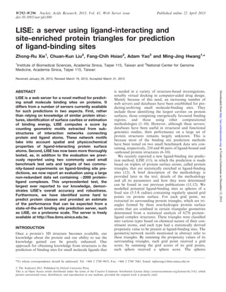
Figure 1. Display of LISE-predicted ligand-binding sites. LISE’s prediction results are displayed as (left) the Top3 predicted sites with their grid
points colour-coded according to binding site-enrichment score (11) and (right) the Top10 predicted sites represented by spheres colour-coded
according to their predicted rank. Users can also view individual site(s) separately by ticking their rank box.
Table 1. LISE success rates for different functional categories of
proteinsa
Protein categoryb
Number of
proteins
Top1 success
rate (%)
Top3 success
rate (%)
Hormone-binding protein 23 17 48
Kinase 112 49 67
Membrane protein 22 64 73
Transcription-related
protein
38 37 75
Unknown function 46 65 78
Specific molecule
(e.g. sugar/lipid/odorant)
binding protein
99 66 81
Transport protein 110 73 82
Chaperone 22 75 82
Transferase 311 69 85
Oxidoreductase 230 77 90
Immune system 30 60 90
Hydrolase 615 84 91
Signalling protein 32 72 91
Lyase 85 80 94
Isomerase 66 88 95
Ligase 41 83 95
All other categories
(<20 structures
in each category)
191 62 78
Overall 2073 72.7 85.6
a
Success rates were computed as the percentage of the query structures
in a category for which the best (Top1) or any one of the best three
(Top3) predicted binding sites satisfied the distance criterion (shortest
distance between the centre of the predicted site and the bound ligand’s
non-hydrogen atoms <4 A˚ ).
b
Classified according to the molecular description included in the
HEADER section of each PDB file.
W294 Nucleic Acids Research, 2013, Vol. 41, Web Server issue
atYeshivaGottesmanonDecember12,2014http://nar.oxfordjournals.org/Downloadedfrom
4. hydrolases were even more abundant than kinases
(Table 1). In fact, 80% of the 210 structures commonly
used as a benchmark data set for ligand-binding site
predictions (8) are enzymes, and some of these structures
are similar in proteins belonging to the same family (e.g.
kinases), even though their amino acid sequences have
diverged. With such a bias and as a result of the situation
with kinases described earlier in the text, it is not
surprising that LISE performed particularly well for
enzymes. It remains to be determined whether other
prediction methods suffer the same data set bias, but
our evaluation points out the need for caution in inter-
preting the success rates of ligand-binding site predictions.
SUMMARY
In this article, we introduced the LISE web server for a
novel ligand-binding site prediction method for any given
protein 3D structure. The server takes a PDB ID or a PDB
format structure file and, by an automated process,
displays/outputs Top3 and Top10 predicted sites. Tests
on commonly used benchmark data sets and on a very
large non-redundant data set indicated that the new
server provides accurate and reliable predictions of
protein ligand-binding sites. Therefore, the new server
can help in virtual drug design and other structure-based
studies, although considerably lower success rates can be
expected for some particular types of proteins, such as
hormone carriers or hormone-binding proteins.
ACKNOWLEDGEMENTS
The authors acknowledge the personnel who compiled the
benchmark data sets and made them available for the
community to use and for us to test the LISE server:
these are Dr B. Huang and Dr M. Schroeder (Technical
University of Dresden, Germany) and Dr Z. Zhang
(Zhejiang University, China) for the LIGSITEcsc
data
sets and Dr M. Brylinski and Dr J. Skolnick (Georgia
Institute of Technology, USA) for the FINDSITE data
set. They thank Dr T. Barkas for English editing.
FUNDING
National Science Council of Taiwan [NSC 96-2627-B-001-
004 and NSC 100-2811-B-001-067 to Z.R.X.]. Funding for
open access charge: Institute of Biomedical Sciences
Academia of Sinica, Taiwan.
Conflict of interest statement. None declared.
REFERENCES
1. Wass,M.N., Kelley,L.A. and Sternberg,M.J. (2010) 3DLigandSite:
predicting ligand-binding sites using similar structures. Nucleic
Acids Res., 38, W469–W473.
2. Roy,A., Kucukural,A. and Zhang,Y. (2010) I-TASSER: a unified
platform for automated protein structure and function prediction.
Nat. Protoc., 5, 725–738.
3. Brylinski,M. and Skolnick,J. (2008) A threading-based method
(FINDSITE) for ligand-binding site prediction and functional
annotation. Proc. Natl Acad. Sci. USA, 105, 129–134.
4. Tseng,Y.Y. and Li,W.H. (2011) Evolutionary approach to
predicting the binding site residues of a protein from its primary
sequence. Proc. Natl Acad. Sci. USA, 108, 5313–5318.
5. Hernandez,M., Ghersi,D. and Sanchez,R. (2009) SITEHOUND-
web: a server for ligand binding site identification in protein
structures. Nucleic Acids Res., 37, W413–W416.
6. Zhang,Z., Li,Y., Lin,B., Schroeder,M. and Huang,B. (2011)
Identification of cavities on protein surface using multiple
computational approaches for drug binding site prediction.
Bioinformatics, 27, 2083–2088.
7. Laurie,A.T. and Jackson,R.M. (2005) Q-SiteFinder: an energy-
based method for the prediction of protein-ligand binding sites.
Bioinformatics, 21, 1908–1916.
8. Huang,B. and Schroeder,M. (2006) LIGSITEcsc: predicting ligand
binding sites using the Connolly surface and degree of
conservation. BMC Struct. Biol., 6, 19.
9. Yu,J., Zhou,Y., Tanaka,I. and Yao,M. (2010) Roll: a new
algorithm for the detection of protein pockets and cavities with a
rolling probe sphere. Bioinformatics, 26, 46–52.
10. Zhu,H. and Pisabarro,M.T. (2011) MSPocket: an orientation-
independent algorithm for the detection of ligand binding
pockets. Bioinformatics, 27, 351–358.
11. Xie,Z.R. and Hwang,M.J. (2012) Ligand-binding site prediction
using ligand-interacting and binding site-enriched protein
triangles. Bioinformatics, 28, 1579–1585.
12. Xie,Z.R. and Hwang,M.J. (2010) An interaction-motif-based
scoring function for protein-ligand docking. BMC Bioinformatics,
11, 298.
13. Berman,H.M., Westbrook,J., Feng,Z., Gilliland,G., Bhat,T.N.,
Weissig,H., Shindyalov,I.N. and Bourne,P.E. (2000) The Protein
Data Bank. Nucleic Acids Res., 28, 235–242.
14. Lopez,G., Ezkurdia,I. and Tress,M.L. (2009) Assessment of ligand
binding residue predictions in CASP8. Proteins, 77(Suppl. 9),
138–146.
15. Schmidt,T., Haas,J., Gallo Cassarino,T. and Schwede,T. (2011)
Assessment of ligand-binding residue predictions in CASP9.
Proteins, 79(Suppl. 10), 126–136.
16. Altschul,S.F., Madden,T.L., Schaffer,A.A., Zhang,J., Zhang,Z.,
Miller,W. and Lipman,D.J. (1997) Gapped BLAST and
PSI-BLAST: a new generation of protein database search
programs. Nucleic Acids Res., 25, 3389–3402.
17. Jmol: an open-source Java viewer for chemical structures in 3D.
http://www.jmol.lorg.
Figure 2. The binding of ligands in the binding site pocket of various
kinases. The Figure shows a superimposition of 81 kinase–ligand
complex structures downloaded from the Pocketome server (19), but,
for clarity, only one protein structure, MAP kinase p38 (PDB ID:
1a9u), is shown (green ribbons). Ligand molecules are shown as
sticks; that for MAP kinase p38 is shown in red and all others in
yellow. LISE’s Top1 predicted site (purple sphere) for 1a9u is close
to its ligand (red stick), but not close enough to be determined as a
successful prediction by the <4 A˚ distance criterion. This figure was
created using the ICM browser (20).
Nucleic Acids Research, 2013, Vol. 41, Web Server issue W295
atYeshivaGottesmanonDecember12,2014http://nar.oxfordjournals.org/Downloadedfrom
5. 18. Endicott,J.A., Noble,M.E. and Johnson,L.N. (2012) The
structural basis for control of eukaryotic protein kinases. Annu.
Rev. Biochem., 81, 587–613.
19. Kufareva,I., Ilatovskiy,A.V. and Abagyan,R. (2012) Pocketome:
an encyclopedia of small-molecule binding sites in 4D. Nucleic
Acids Res., 40, D535–D540.
20. Abagyan,R., Lee,W.H., Raush,E., Budagyan,L., Totrov,M.,
Sundstrom,M. and Marsden,B.D. (2006) Disseminating structural
genomics data to the public: from a data dump to an animated
story. Trends Biochem. Sci., 31, 76–78.
W296 Nucleic Acids Research, 2013, Vol. 41, Web Server issue
atYeshivaGottesmanonDecember12,2014http://nar.oxfordjournals.org/Downloadedfrom