2. A Tribute to Dr. Herbert Schilder
The two most popular solid cores are gutta percha and silver Some criticism has been directed toward the solvent techniques in
cones. Silver cones are used always with a suitable cement. Gutta percha spite of the excellent results which have been obtained by experienced
cones may be used with a cementing material, or they may be rendered endodontists. Since all of the solvents which have been used are highly
plastic with various solvents and used without root canal cement. A brief volatile, a measurable amount of shrinkage takes place gradually as the
review of the more popular methods of using gutta percha and silver root canal filling hardens. The amount of shrinkage varies directly with
cones is helpful in the evolution of a technique which may most closely the amount of solvent used, and, if shrinkage is excessive, failure may
fulfill the requirements of a permanent three-dimensional root canal result from the inadequate sealing of the root canal. Likewise, when
filling. excess solvent has been employed, it may be difficult to confine the
plastic material to the root canals, and gross excess may be forced into
the periodontal tissues. While this surplus in no way prejudices the final
Gutta Percha Techniques
outcome of the case, the solvents are irritants, and considerable dis-
Solvent Techniques
comfort may occasionally occur until the chloroform has been carried
Gutta percha may be dissolved in various solvents, such as chlo- away from the apical periodontium. The excess material is resorbed
roform, oil of eucalyptus, or xylol. When dissolved in chloroform, a over a period of years with complete bone regeneration at the root apex.
paste of chloropercha is formed, the thickness of which is determined The Nygaard–Ostby technique is a variation of the Callahan–John-
by the amount of solvent used and by the amount which evaporates in the son method, where finely ground specially prepared gutta percha par-
course of the filling procedure. ticles are spatulated with chloroform to produce the necessary paste
At the beginning of the filling visit, several pieces of gutta percha which will be used in conjunction with the master gutta percha cone.
are cut into a dappen dish and stirred with a small amount of chloro- This method is reported to reduce greatly both apical excess and the
form. The dappen dish should be covered with a glass slab to minimize shrinkage in the final filling.
evaporation of the chloroform as the gutta percha dissolves into it while
the dentist is selecting his master gutta percha cone. Lateral Condensation with Nonsolvent Cements
Gutta percha cones are available in many widths as well as in the Gutta percha also has been used with nonsolvent cements. Gross-
standardized sizes which purport to conform to the standardized sizes of man has set forth the properties of a good root canal cement as follows:
root canal reamers and files. Except insofar as the standardized gutta
percha cones provide a greater variety of sizes available for selection, 1. The cement should be tacky when mixed so as to provide good
they are clinically of no special value. For example, a #40 gutta percha adhesion, when set, between it and the canal wall.
cone may not reach deeply into a curved root canal which has been 2. It should make an hermetic seal.
prepared with a #40 file or reamer, since the flexibility and softness of 3. It should be radiopaque so that it can be visualized in the
the gutta percha may result in its binding and collapsing on its long axis roentgenogram.
at the first suggestion of lateral resistance, long before the apical end of 4. The particles of powder should be very fine so that they can mix
the root canal has been reached. Good clinical judgment will quickly easily with the cement liquid.
lead to the selection of a gutta percha cone which fits the canal reason- 5. It should not shrink upon setting.
ably well and is 1 or 2 mm short of the end of the root. 6. It should not stain tooth structure.
A small amount of chloropercha is streaked onto the walls of the 7. It should be bacteriostatic, or at least should not encourage
dry root canal with a fine root canal spreader or other suitable instru- bacterial growth.
ment. The apical third of the master cone is dipped into the chloroper- 8. It should set slowly.
cha paste, and the entire master cone is gently repositioned into the 9. It should be insoluble in tissue fluids.
canal. The material in the canal is now forced laterally with root canal 10. It should be tissue tolerant, i.e., nonirritating to periapical
spreaders, making room for additional gutta percha cones which are tissue.
added repeatedly in sufficient number to provide a dense root canal 11. It should be soluble in a common solvent if it be necessary to
filling. Each piece of gutta percha blends with the gutta percha and remove the root canal filling.
chloropercha already in the canal to form a homogeneous mass which These properties are incorporated in the Grossman nonstaining root
conforms quite adequately to the configuration of the root canal system. canal cement, which is available as Procosol Nonstaining Sealer or Kerr
The lateral pressure on the plastic gutta percha– chloropercha mixture Tubli-seal. The original Richert formula (Kerr sealer) and the Gross-
automatically imparts a small vertical component of pressure, owing to man silver cement containing precipitated silver, zinc oxide, and stabil-
the shape of most prepared canals. The entire mass moves apically ite resin are equally useful and will not stain tooth structure when
during lateral condensation with any solvent technique. adequate access cavities and coronal restorations are employed (see
Many variations of solvent techniques have been used successfully the papers by Dr. Levin and Dr. Baraban).
in addition to the basic chloropercha or eucapercha methods. In the In lateral condensation with a nonsolvent cement, the original
Callahan–Johnson diffusion technique, the root canal system is flooded gutta percha cone is selected to coincide as closely as possible to the
with 95 percent ethyl alcohol and then dried with paper points. The shape and length of the root canal, and this is confirmed with a radio-
alcohol flooding and drying removes some organic and most aqueous graph. The master gutta percha cone is then rolled in the cement and
material from the walls of the canals. The canals are flooded again, now placed in position in the dry canal. This cone is now pressed laterally
with a chlororosin solution. The chlororosin solution spreads exten- repeatedly with a root canal spreader, and a series of fine gutta percha
sively into inaccessible eccentricities in the root canal and into acces- cones are added until a dense filling has been obtained. The advantage
sory canals as well. This solution acts as a solvent both for a previously of this method over the solvent techniques is that positive dimensional
prepared master cone and for fatty organic material, so that when the stability of the root canal filling can be anticipated, and there is less
master cone and additional cones are added in the condensation pro- likelihood of carrying filling material beyond the root apex.
cedures, dissolved gutta percha will diffuse effectively into otherwise One disadvantage is that at no time is a homogeneous mass devel-
inaccessible portions of the root canal system. Very good results have oped. The final filling consists of a large number of separate gutta
been obtained with these methods. percha cones tightly pressed together and joined by frictional grip and
282 Schilder JOE — Volume 32, Number 4, April 2006
3. A Tribute to Dr. Herbert Schilder
the cementing substance only. It is the gradual setting of the root canal which the cone and root canal walls are in contact. This extra effort is
cement which gives the digital illusion of homogeneity as the lateral well worthwhile, and will be rewarded by greatly increased success
condensation procedure continues. Only at the point where the coronal rates where silver cones are used.
excess is removed with a hot instrument is true homogeneity estab- The nature of the “locking,” “binding,” or “fitting” of silver cones
lished. By the nature of lateral condensation procedures with nonsol- in root canals must be understood if consistent success is to be
vent cements and the shape of most root canals, lateral condensation achieved. While both silver and dentin are slightly compressible, they
produces the densest filling in the middle and coronal thirds of the are both substances of considerable firmness. It is possible, therefore,
tooth, and the apical seal is little improved as additional cones are for a silver cone to bind in an eccentric root canal, although it contacts
added. It should be clear, however, that in spite of criticism directed the walls at only two points. Indeed, this happens not infrequently.
toward the value of this type of lateral condensation in establishing Although elliptical root canals tend to become round in their apical
positive apical seal, the method does insure a thorough, dimensionally thirds, they are never geometrically round, and it is doubtful if the best
stable obturation of the major volume of the root canal. It offers the directed efforts of any endodontist will make them geometrically round.
distinct advantages of greater control and greater patient comfort, and It has been observed that in inexperienced hands root canal instrumen-
has been successfully employed by large numbers of dentists for many tation often results in canals with elliptical or teardrop shaped foramina.
years. Great attention must be paid to maintaining or developing roundness at
the apical foramen if success is to be achieved with silver cones. To the
Silver Cone Techniques extent that the apical portion of the root canal is eccentric, the seal at the
Silver cones were introduced into endodontics about 40 years ago apex will depend on root canal cement, or the seal will be deficient when
and have had a rather checkered career as a root canal filling material. silver cones are used. To the extent that the apical portion of the canal
They are always used in conjunction with a root canal cement. They are approaches roundness, the compressibility of silver and dentin will
decidedly inert, dimensionally stable, and, except in the rarest instances provide a permanent apical seal.
of manufacturing defect, they cannot be resorbed. Their semi-rigidity A correctly fitted silver cone is one which reaches to the end of the
permits them to be wedged forcibly toward the critical root apex, where cleaned shaped canal, which cannot be forced farther with strong pres-
they are expected to provide a hermetic seal for the root canal. As in the sure, and which offers resistance to removal before cementation. A
case of gutta percha, silver cones are available in a variety of widths and silver cone which cannot be pushed deeper, but which can be with-
in standardized sizes which are intended to conform to the shapes of drawn without frictional resistance is sitting on a ledge in the root or
standardized reamers and files. Ideally, the silver cone should pass against bone, but it does not seal the foramen. This is a very common
loosely through the coronal and middle third of a root canal and bind in error in the buccal roots of upper molars, where many silver cones are
the apical portion only. The shape and rigidity of silver cones make this fitted against the zygoma without sealing the root canals. This condition
possible in most instances. can be diagnosed and corrected if small portions of the apical end of the
cone are cut off and it is found that the silver cone still appears to stop
Proper Fitting at the root-end radiographically. Clearly, then, dense bone and not
A silver cone is not properly fitted unless it reaches very close to the frictional grip has prevented the silver cone from being pushed farther.
root apex and cannot be pushed farther with any amount of pressure. By repeated reduction at the end of the silver cone, the cone will be
For this reason, silver cones should be manipulated with silver cone made to fit so that it can neither be forced deeper nor be removed from
pliers specifically designed for this purpose, such as the Anteos silver the canal without considerable effort. It is now ready for cementation.
cone pliers. Hemostats cannot provide the kind of pressure necessary Another point must be borne in mind. Silver cones were some-
for the forcible seating of silver cones. times recommended for use especially in extremely narrow and curved
Great care must be taken in the proper selection and fitting of silver canals. Nothing could be more incorrect unless the canals were first
cones. It is fantasy to expect a canal that has been cleaned and shaped shaped and enlarged considerably. The #3 silver cone is the minimum
with a certain number reamer or file to be obturated by a silver cone of sized one that can possibly seal an apex. A #2 silver cone may be
the same size. Should a dentist complete the shaping of a root canal with threaded or teased into a narrow, underprepared root canal, but it
a #70 file, for example, the very fact that the #70 file can be placed to the defies logic to expect such a slender wire introduced at the coronal end
root apex and removed again indicates that the apical portion of the of the canal to offer a frictional seal at the apical foramen. It was the
canal must be wider than the apical end of the file. Usually, the differ- improper use of silver cones in this way, as well as the occasional
ence in the respective diameters of the root canal and the final reamer defective cementation of silver cones, which led to objections to the use
or file is considerable, and a silver cone of the same size as the last of silver cones from time to time. Silver cones almost always provide
cutting instrument will fit too loosely at the apical end of the canal. Two esthetic radiographs, but it is only the dentist who checks the case
possibilities exist: namely, that the tip of a silver cone which fits too several years later who can determine the thoroughness of the obtura-
loosely may be cut off step by step until by trial and error a tight fit at the tion.
apex is obtained, or that a silver cone somewhat wider than the last
cutting instrument can be selected and its apical end narrowed some- Cementation of Silver Cones
what until it binds tightly at the root apex. Any of the above-mentioned root canal cements may be used with
When using silver cones, I prefer the latter method of gradually silver cones. A small amount of cement should be introduced into the
shaving down a slightly oversized cone. The apical 3 or 4 mm of the root canal first, and then the silver cone may be rolled in the cement and
silver cone are placed within the abrasive surfaces of a folded-over gradually replaced into the canal. Gradual seating of the silver cone will
medium grit -inch sandpaper disk. By pressing the folds of the disk prevent excess cement from extruding past the root apex as well as
tightly over the silver cone with the thumb and index finger of the left obviate pressure discomfort to the patient caused by air being com-
hand, and rotating the silver cone back and forth rapidly with the thumb pressed against the periodontal tissues. The seating of a silver cone
and index finger of the right hand, a gentle taper is imparted to the silver should be as certain as the seating of a well-made gold inlay. As it takes
cone. This taper permits the silver cone to be locked into position at the its position at the root apex, it may be rotated slightly to increase the
end of the root canal while importantly increasing the surface area over wedging effect near the foramen. Some men, fearful that cement may be
JOE — Volume 32, Number 4, April 2006 Filling Root Canals in Three Dimensions 283
4. A Tribute to Dr. Herbert Schilder
condensed laterally beside the mail silver cone. In all silver cone cases,
I prefer to place warm pellets of gutta percha in the floor of the pulp
chamber adjacent to the silver cone or cones, and to press the gutta
percha apically around the silver cones with narrow root canal pluggers
(Fig. 1). The gutta percha may be rewarmed from time to time with a
heated spreader and the softened mass forced repeatedly into the mouth
of the root canal. Many lateral canals are filled in this manner.
Split Cone Technique
A variation of the above technique may be employed where a cast
post is needed to support a coronal restoration. This variation has been
presented under many names, but it is usually recognized as the split
cone technique. The silver cone is fitted carefully as above. Before
cementation, however, the cone is scored deeply with a disk or bur at
some distance from its apical end. Cementation proceeds as indicated,
but after the cone has been seated well, its coronal end is rotated
repeatedly along its long axis. While rotating, firm apical pressure is
applied through the pliers to prevent unseating at the root apex. As the
rotary motion continues, the cone will be severed at the score line, and
all but the apical portion removed. The middle and coronal parts of the
canal are now available for impressions for a post, or the deeper portion
of the canal may be filled with warm gutta percha condensed vertically
against the apical segment of the silver cone and cement (Fig. 2). In-
terestingly, this condensation provides a severe test for the apical fit of
the silver cone. If the apical segment cannot be moved beyond the
foramen under the tremendous pressure that is imparted to it through
the warm gutta percha, the dentist can be assured that some form of
apical wedging has been effected. If, on the other hand, cement or gutta
percha can be extruded outside the canal beyond the stationary silver
cone, then the apical wedging was to some extent incomplete. Fortu-
Figure 1. Silver cones are used always in conjunction with a cement and often
with lateral gutta percha as well. A, A lower molar where the mesiolingual and
mesiobuccal canals shared a common foramen. Both the distal and mesiolin-
gual canals were obturated with silver cones, while warm gutta percha was
plugged into the mesiobuccal canal. The rather typical puff of cement at the apex
of the mesial root resulted from the initial cementation of the mesiolingual cone
and not from the pressure developed subsequently in the buccal canal. This
verified the fit of the mesiolingual silver cone. B, An upper premolar, where a
lateral canal was filled while gutta percha was forcibly plugged alongside a
recently cemented silver cone. In this case, too, the apical puff is typical, and is
looked for as an indication that the silver cone has been cemented along its
entire length. The apical cement was not increased by the vertical pressure
which forced cement into the lateral canal. Note the bone resorption that had
occurred opposite the unfilled accessory foramen.
deposited outside the canal, only roll the cone in cement and do not
introduce cement into the canal prior to the final placement of the cone.
If no cement is placed in the root canal ahead of the silver cone, the
cement on the cone quite often wipes off halfway up the canal, so that the
apical portion of the cone is placed in position without any cement at all.
The basic problem of sealing potentially eccentric foramina with rela-
tively round silver cones requires that a film of cement be provided
along the entire length of the cone. A properly fitted and cemented silver
cone cannot be removed after the cement has set without the use of
much solvent (alcohol, chloroform, xylol, etc.) and much mechanical
effort. Some can never be removed. Silver cones which can be removed
easily were not properly fitted and cemented. Figure 2. The split silver cone technique in an upper second premolar. The
Since silver cones rarely fill the root canals laterally, the remaining apical plug of silver, perhaps 4 to 5 mm long, is seen wedged at the root apex.
space also must be obturated in some manner. In narrow canals, the A cast crown has been built upon a gold post placed in the coronal third of the
lateral space sometimes is filled with root canal sealer only. In wider, root canal, and the tooth helps to support a partial denture. The apical puff of
more tapering canals, gutta percha or additional silver cones may be cement has been resorbed almost fully from this old silver cone case.
284 Schilder JOE — Volume 32, Number 4, April 2006
5. A Tribute to Dr. Herbert Schilder
Figure 3. A, This case was specially selected to illustrate the problems that may occur when cold gutta percha is used with inadequate lateral condensation. It is not
typical of good lateral condensation cases. Both the apical fit of the gutta percha and its lateral adaptation to the walls of the root canal are defective. A fistular opening
has erupted on the labial alveolus. B, A typical example of a dense homogeneous root canal filling obtained with warm gutta percha. The solidity of the apical obturation
is most important. C, Another fairly routine maxillary anterior tooth filled with vertical condensation of warm gutta percha. Three accessory foramina as well as an
additional one through the plane of the film have been filled.
nately, most silver cones will remain stationary under this test; but, The Master Gutta Percha Cone
unfortunately, extrusion can be produced in almost half the cases. The original piece of gutta percha must be carefully selected so that
Hundreds of thousands of cases have been treated successfully its taper is more gradual than the taper of the root canal. For this reason,
with well managed silver cones, but extrusion past stationary apical it is important to reject gutta percha cones which are too pointed, since
segments makes one wonder if a more effective manner of root canal they will bind somewhere within the body of the canal and not near the
obturation cannot be employed. apex. It is preferable to cut off the apical end of a less heavy gutta percha
cone than to select a heavier gutta percha cone with a fine tip. In any
Vertical Condensation with Warm Gutta Percha event, the apical end of the master gutta percha cone must be wider than
It would seem desirable to fill root canals with a homogeneous, the apical end of the root canal. The task, then, is to insure the seal by
inert, dimensionally stable, physiologically acceptable material which forcibly wedging the gutta percha into the narrower portion of the
could be manipulated with sufficient plasticity to form a permanent cast shaped canal.
of the internal configuration of the root canal system. Borrowing heavily A small amount of root canal cement is introduced into the root
from the experience gained by others in the development and use of the canal by means of a lentulo, which is rotated clockwise between the
above techniques, and from my own clinical and laboratory experience thumb and index finger. Care should be taken to use as little cement as
with them, I now favor warm gutta percha as the filling material of possible, and only enough to streak the walls in several places is nec-
choice for most root canals. The value of gutta percha in endodontics essary. On the one hand, it is undesirable to have any large portion of the
has long been established, and its use in a plastic state, without depen- final filling composed of the potentially resorbable cement; on the other
dence on solvents, imparts to the filling the dimensional stability which hand, a small amount of cement is fully adequate for spreading over all
sometimes was lacking in the past. Of equal importance, the use of the surfaces of the canals after condensing pressure has been applied to
warm gutta percha with vertical condensation permits great density to the warm gutta percha.
be created in the apical portion of the filling. Last, no other technique The apical end of the properly sterilized master cone is dipped in
provides for the filling of accessory canals and foramina with such the cement, and the gutta percha is introduced into the dry root canal.
frequency as warm gutta percha used in conjunction with vertical con- Care should be taken to deliver the gutta percha cone gently back to its
densation (Fig. 3). original position so as not to force cement apically. This should be
In order to employ the warm gutta percha technique, the root checked radiographically. With the use of rapid developers, in less than
canal must be shaped so that a continuously tapering funnel is created a minute the dentist is prepared to begin the vertical condensation.
with its narrowest diameter at the periodontal ligament and its widest
diameter at the coronal opening or access cavity (see the paper by Dr.
Levin). However this funnel may curve, it must be progressively wider Vertical Condensation
coronally. This continuous taper permits the introduction into the canal The Heat Carrier
of a graded series of pluggers with which the gutta percha is wedged The coronal end of the gutta percha cone is seared off with a hot
apically. spreader, and the warm end which remains in the tooth is folded into the
The materials to be employed are gutta percha and an acceptable pulp chamber with a broad plugger. It is well to remember that pluggers
root canal cement. The only mechanical instruments necessary are a differ from spreaders in that, regardless of their width, pluggers have
lentulo or past filler, one root canal spreader or heat carrier, and the blunt ends, whereas the ends of spreaders are pointed. In this tech-
graded series of pluggers. nique, the pluggers are always used cold, whereas the spreader is only
JOE — Volume 32, Number 4, April 2006 Filling Root Canals in Three Dimensions 285
6. A Tribute to Dr. Herbert Schilder
Figure 4. A few steps in the development of a dense gutta percha filling in the apical third of a badly broken down maxillary premolar. A, The pulpless tooth at the time
of examination, with the suggestion of bone breakdown mesially below the root apex. This radiolucency was not always apparent in subsequent working films. B, A
file at the radiographic apex. C, The master gutta percha cone in position. The canal has been shaped and the gutta percha selected so that the cone binds apically
only. Still, the cross-sectional diameter of the gutta percha is too great to fit into the apical end of the canal. D, The master cone is shown cemented into the root canal.
Care has been taken to prevent root canal cement from preceding the master cone. Vertical condensation has been started only in the coronal third of the canal. E,
Here can be seen what has happened after the gutta percha was repeatedly heat softened, vertically condensed, and partially removed. A wave of condensation moved
apically, and finally the gutta percha was delivered to the root apex, as a lateral canal leading to the mesial radiolucent area was filled. No reverse wave of condensation
took place here, but a pellet of cotton was placed in the canal to prevent temporary cement from blocking the space that will be used for a cast post.
used hot. The spreader is not used to “spread” gutta percha, as is dimensional vertical condensation in the cervical region is achieved
common in lateral condensation techniques, but only to carry heat first, while the middle and apical thirds of the canal remain relatively
which softens the gutta percha sufficiently to allow vertical condensation unaffected.
with the pluggers. The pluggers must never be heated, since heat will After the canal has been filled laterally and in depth in the cervical
make them stick to the gutta percha and will soften the metal, rendering region, the heat carrier can be used not only to soften the gutta percha
the instrument useless for the application of strong vertical pressure. A but to remove portions of it as it is withdrawn from the main mass of
change in term from “spreader” to “heat carrier” is suggested, since at material. By this means, the 3 or 4 mm plunges of the heated instrument
no time should this hot instrument be directed laterally in this tech- will progressively soften deeper portions of the gutta percha, while
nique. removing portions of the gutta percha at the working end of the canal.
The heat carrier is now heated to cherry redness, and it is brought Sequential strong vertical condensation with the graded series of plug-
quickly to the root canal, where it pierces the coronal 3 or 4 mm of the gers will slowly carry a wave of condensed gutta percha down into the
gutta percha. Immediately after the gutta percha is softened, the heat middle portion of the root canal, which in its turn will be filled laterally
carrier is removed and a suitable plugger is applied to the plastic mass, and in depth. This wavelike condensation with warm gutta percha ex-
forcing it apically. It must be understood clearly that gutta percha is an tending to a depth of 3, 4, or 5 mm at a time will effectively seal lateral
excellent insulating material, and that only the gutta percha near the canals which are large enough to receive filling material. At this time, the
heat carrier is softened. Gutta percha several millimeters beyond the apical portion of the gutta percha cone is still unaffected. A radiograph
cervical region is completely unaffected by any condensation at this should be taken when the pluggers are brought to bear approximately
point. As the process of reheating and plugging continues, a three- halfway into the depth of the root. In this way, the dentist can prepare
286 Schilder JOE — Volume 32, Number 4, April 2006
7. A Tribute to Dr. Herbert Schilder
himself for the movement of the apical plug of gutta percha up to the
root surface.
In order to simplify the depth perception which is necessary in
delivering the apical plug of gutta percha tightly to the apex, 5-mm
indications have been scored into the pluggers. The dentist can readily
compare the length of his last reamer or file with the score lines on the
pluggers and judge his progress toward the root apex.
Apical Filling and Lateral Canals
It is only after a series of heatings, condensations, and gutta percha
removals that the hot spreader can soften the apical portion of the gutta
percha. With experience, the dentist can now deliver this portion of
gutta percha progressively into cross-sectional portions of the apical
end of the canal, into which it could not fit in its original solid state. The
gutta percha fully adapts itself to the infinite anatomic complexities of
the root canal, forming a cast of its interior while filling the major apical
accessory canals (Fig. 4). The root canal is now essentially empty,
except in its apical end. The remaining portion of the canal is filled with
warmed segments of gutta percha, 2, 3, or 4 mm in length which have
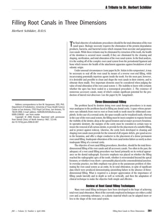
been previously selected to conform to the diameters of the root canal Figure 6. Lines have been scored into the pluggers at 5-mm intervals to indicate
pluggers and the canal. One by one these plugs of gutta percha are the effective level of condensation at all times. The measurements also permit
heated and condensed vertically as described above, but with no attempt the intelligent prefitting of the pluggers to be used at each level before the actual
filling procedure commences. By the addition of half sizes, a more useful series
to remove material with the heat carrier. No cement at all is used in this
of pluggers has been developed.
phase, and a new wave of vertically condensed gutta percha passes
through the root, reversing the direction of the original movement. In
this manner, the root canal is filled three-dimensionally with a solid
core of inert gutta percha and a minimum amount of potentially resorb-
able paste.
Should it be deemed desirable to use a cast core in restoring the
tooth, the repacking of the canal cervically can be stopped at any dis-
tance and the depth of the post space approximated accurately by the
score lines on the pluggers.
It must be recalled that only a minimum amount of cement is to be
used, since the firm vertical condensation of the gutta percha mass will
spread the cement uniformly under enormous hydrostatic pressure.
Indeed, lateral canals which are too fine to receive the warm gutta
percha will be filled by the cement. Although this is less desirable than
filling with gutta percha, it is preferable to leaving them unobturated.
Fears that the gutta percha will shrink on cooling are groundless.
While molten gutta percha does contract on solidifying, at no time in this
technique is the apical gutta percha in contact with the heat carrier. It is
softened only by proximity to the heat source, and actually moves only
under firm pressure. In no part of the filling is the gutta percha liquefied,
and considerable attention must be placed on the prompt delivery of the
heat carrier to the gutta percha lest the heat be inadequate to soften the
gutta percha at all. It is control of the amount of heat in the spreader
which determines whether the gutta percha is to be softened or is to be
removed. If the instrument is not heated sufficiently, the gutta percha
will harden on it, and excessive portions of the filling will be inadver-
tently carried away. Also, the repetitious nature of the heating and con-
densation in this technique is similar to the paint-on technique with
acrylic filling materials. The application and reapplication of pressure
on the gutta percha as it cools with the continuous augmentation of
material eliminates the significance of dimensional change as the tem-
perature drops (Fig. 5).
Figure 5. Canals of posterior teeth also may be filled with warm gutta percha
and vertical condensation. A, A maxillary second molar with three canals obtu- This technique incorporates many elements of the sectional
rated with warm gutta percha. The apical puffs are somewhat typical and indi- method of filling root canals. In the sectional method, segments of gutta
cate the completion of the filling. B, A mandibular second molar where all percha too wide to fit the apical end of the root canal are forcibly
canals share a common foramen. The root canal system has been filled three- wedged into position piece by piece until a complete root canal filling
dimensionally as well as a fine network of accessory canals near the apex. has been achieved. The pieces may be warmed or pressed in cold, in
JOE — Volume 32, Number 4, April 2006 Filling Root Canals in Three Dimensions 287
8. A Tribute to Dr. Herbert Schilder
Figure 7. This tooth presented a history of a silver cone having been cemented in the distal canal many years previous to the more recent inadvertent perforation of
the mesial root. The mesial root perforation apparently had led to a severe lateral abscess. A, A large file passed easily through the perforation, giving some indication
of its size and the extent of the associated bone destruction. B, The periapical condition immediately after the sealing of the true foramen and the overfilling of the false
canal with surplus gutta percha in the periradicular bone. C, The case one year later. Very little resorption of the excess gutta percha has taken place as yet, but the
alveolar bone has regenerated tightly against the surplus material. The slight void above the apical plug of gutta percha in the mesial root shows evidence of the
segmented nature of the technique.
which case solely the compressibility of the gutta percha is relied upon dentin, instead of vertical pressure of metal against gutta percha. It is
to insure the seal. The sectional technique lacks the first wave of con- decidedly possible to split a longstanding pulpless tooth by needless
densation from the cervical to apical portions of the root, and does not wedging with an oversized plugger. Prefitting will prevent unnecessary
take advantage of the continuous resoftening of the gutta percha to root splitting, will insure that proper instruments are available to reach
improve its adaptation to the walls of the root canal system. the apical third of the canal, will help the dentist plan the sequence of
pluggers he will use in any given case, and may indicate areas where the
Vertical Condensation Pluggers shaping of the root canal should be improved.
The pluggers for the warm gutta percha technique are not dissim-
ilar to the pluggers used in lateral condensation. As has been men-
tioned, it was found convenient to place score lines on the pluggers at Vertical Extent of Root Canal Fillings
5-mm intervals. Also, both to simplify and to perfect the filling, a more It should be clear that the totality of the three-dimensional filling of
evenly graded series of pluggers was required, and half sizes have been the root canal is more important than its vertical extent alone. What
added to the regular series. A complete series of these instruments is significance should be given, then, to so-called “underfilled” and “over-
made by the Star Dental Manufacturing Company, and includes num- filled” root canals?
bers 8, 9, 9½, 10, 10½, 11, 11½, 12, although most vertical condensa-
tion is found to be done with numbers 8 through 11 (Fig. 6). Where a Cementodentinal Junction
plugger must be bent in order to conform to a curved canal, it should be Many endodontists rely on filling root canals to the cementoden-
left in its new shape and used again only for a similarly curved canal. tinal junction. The cementodentinal junction is, theoretically, the point
Rebending the instrument will soften it or lead to breakage, and in either which divides the pulp tissue from the tissue of the periodontal ligament.
case it will be rendered useless. This junction is usually found some small distance within the apical end
of the root canal and at a somewhat constricted portion of the apical
Prefitting of Pluggers opening. Filling to this point, it is assumed, fills the root canal without
Two additional points must be made about the use of the pluggers. impingement on the periapical tissues and encourages the eventual
One is that a small amount of dry cement powder may be used as a physiologic closure of the root canal with cementum. In order to fill to
separating medium on the end of the pluggers to keep warm gutta the cementodentinal junction, the endodontist relies either on his sense
percha from adhering to them. The other is that, to plan the root filling of feel or he fills all canals .5 to 1 mm short in the hope of ending the root
procedure most intelligently, the pluggers should be prefitted into the canal filling properly. Countless successful cases have been obturated in
canal. A plugger which cannot fit to within a few millimeters of the end this manner.
of a canal will be valueless in obturating the apical end of the canal. Filling to the theoretical cementodentinal junction is not an un-
Attempting to force a plugger deeper into a canal than its own cross- mixed blessing. Studies of the anatomy of root canals indicate clearly
sectional diameter will permit results in lateral pressure of metal against that the position of this line is highly variable from one tooth to another,
288 Schilder JOE — Volume 32, Number 4, April 2006
9. A Tribute to Dr. Herbert Schilder
canal system, but are probably overextended beyond the root canal by a
fraction of a millimeter in most cases, owing to the physics of dental
radiography. Countless teeth have been treated successfully to this point
as well, indicating the high degree of tolerance of the periodontal tissues
for most root canal filling materials. It should be observed that some
markedly curved canals exit their roots at a point which is invisible
radiographically, and that filling of root canals to the radiographic lim-
itation of the root is to be avoided in such cases.
The enormous success which has followed filling root canals to
their radiographic apices or beyond has led some to theorize that heal-
ing of large periapical radiolucencies may be stimulated by root canal
filling material outside the confines of the canal. While this may be
possible, it is more likely that, as in the case of filling to the cemento-
dentinal junction, continued success results from the thoroughness of
the three-dimensional filling along the major extent of the root canal
and not on fractional overextension or underextension of the filling. The
wise old suggestion to slightly underextend root canal fillings in cases of
vital extirpation and to fill to the radiographic apex or slightly beyond in
cases of pulpal necrosis and gangrene is probably more meaningful in
terms of patient comfort than in terms of the ultimate result.
Overfilling Versus Overextension
An important distinction must be made between overfilling and
underfilling, and overextension and underextension. Over and under-
extension refer solely to the vertical dimension of the root canal filling,
beyond or short of the root apex. Underfilling refers to a tooth whose
root canal system has been inadequately obturated in any dimension,
leaving large reservoirs for recontamination and infection. An overfilled
tooth is one whose root canal system has been filled in three dimen-
sions, and where a surplus of material extrudes beyond the foramina. In
spite of a long and conscientious search, I have never encountered a
Figure 8. The filling of complex root canal systems with warm gutta percha and
vertical condensation. Studies of root canal systems show them to be often highly case of endodontic failure due to overfilling, where one means by “over-
complex, with multiple foramina. A, An upper first premolar with three filled filling” that the root canal has been obturated in its entirety and surplus
foramina in the buccal and lingual canals. B, A lower second molar with five material has been intruded into the apical periodontium (Fig. 7). On the
filled foramina as well as a lateral canal filled through the plane of the film in the other hand, as have other endodontists, I have encountered numerous
mesial portion of the distal root. cases of failure of vertical overextensions of underfilled root canals.
In the latter cases, gutta percha or silver cones which never did seal the
and even from one wall to the opposite wall of the same root. The circumference of the apical foramen were carelessly forced into the
cementum may join the dentin .5 mm inside the root on one surface, and apical periodontium, where their presence added additional insult to
3 or 4 mm inside on the opposite surface; or, owing to a variety of the primary problem, namely the underfilled root canal.
causes, the cementodentinal junction may occur outside the root canal The benign nature of most root canal filling materials, if the root
completely. Feeling for the apical constriction, while not an infallible canal system is properly sealed, has been demonstrated over decades of
guide in the hands of the experienced endodontist, can be very difficult clinical practice. One must recall only the extrusions of the diffusion
for less experienced operators. Also, the closure of the root end with techniques, the classic Callahan button, the chloropercha and eucaper-
cementum, while both possible and desirable, is demonstrable much cha surplusses of the solvent techniques, and, more recently, the inten-
more readily in animal experimentation than in human patients, and is tional overfillings with metal posts in severe periodontal cases (see the
unnecessary for the health and function of the apical periodontium. The paper by Dr. Frank) to realize that the vertical extent of the acceptable
success which has been encountered in filling to this point has resulted root canal filling within a 1-mm range cannot be considered of major
more likely from the care with which the root canals were instrumented importance. Excess material beyond the cementodentinal junction
and by the lateral completeness of the fillings than from apical closure probably plays no part in healing and should be avoided solely on the
with cementum. A root canal system filled in three dimensions to within basis of its needlessness and its possible potential for discomforting the
.5 or 1 mm of its radiographic apex is essentially filled to its entirety. The patient at the time of the filling procedure.
shortness of such a filling should in no way be quated with the massive
underfilling produced by a laterally incomplete filling procedure, re-
gardless of the vertical extent of the filling material. Summary
The ultimate objective of endodontic technique is the elimination
Radiographic Apex of the root system as a source of infection and inflammation to the apical
Other endodontists prefer to fill to the radiographic apex or, more periodontium after irreversible pulp pathosis. The most desirable way
precisely, to the point where the root canal appears to join the peri- to render root canals innocuous is to clean and shape them, to eliminate
odontal ligament as viewed in a roentgenogram. Fillings carried to this bacteria and tissue debris from within them, and then to obliterate them
point may more closely approach the 100 percent total filling of the root by means of a dense three-dimensional root canal filling.
JOE — Volume 32, Number 4, April 2006 Filling Root Canals in Three Dimensions 289
10. A Tribute to Dr. Herbert Schilder
Accessory canals are present in practically all teeth. Many acces- The final test of a root canal filling is its capacity to seal off the root
sory canals are very small and calcify spontaneously during chronic canal system from the periapical tissues. The tissue compatibility of
pulp irritation, and others contain too little tissue to be clinically signif- almost all commonly used root canal filling materials is very high, and
icant. Often, however, accessory canals are of considerable size, and, for decades bone has been demonstrated to be laid down in close
where the tissue within them becomes necrotic or infected, they may proximity to all of them. Overfilling, while not necessarily beneficial, will
contribute to lateral root abscesses unless sealed off from the periodon- not prejudice the outcome of a case or prevent healing. Overfilling must
tal ligament. Root canal filling procedures should be directed toward be distinguished from overextension of underfilled cases.1–10
the filling of significant lateral canals as well as the filling of main root
canals (Fig. 8).
Many techniques have been used to obturate root canals success- References
fully. Most of these techniques employ either silver cones or gutta per- 1. Berg B. Endodontic management of multirooted teeth. Oral Surg, Oral Med, Oral Path
cha in some form. When used well, all of these techniques are valuable; 1953;6:3.
when abused, no technique can succeed. The difficulty of adapting a 2. Coolidge ED, Kesel, RG. Endodontology. Philadelphia, Lea & Febiger, 1956.
silver cone to a less than geometrical round foramen sets certain po- 3. Dow P, Ingle, JI. Isotope determination of root canal failure. Oral Surg, Oral Med,
tential limitations upon the use of silver cones in all cases. Likewise, Oral Path 1955;8:1100.
4. Grossman LI. Endodontic Practice. Philadelphia, Lea & Febiger, 1965.
small dimensional changes inherent in the use of gutta percha and a 5. Grossman LI. Present status of plastic root canal filling materials. Transactions of the
solvent, as well as certain problems of apical adaptation of the gutta Third International Conference on Endodontics, Univ. of Penna., 1963.
percha when no solvent is used, encourages the evolvement of a tech- 6. Healey HJ. Endodontics. St. Louis, The C. V. Mosby Co., 1960.
nique by means of which gutta percha is rendered plastic without the 7. Ingle JI. Endodontics. Philadelphia, Lea & Febiger, 1965.
use of solvents. 8. Schilder H. Endodontic therapy. In: Current Therapy in Dentistry, GoldmanH. , et al.,
ed. St. Louis, The C. V. Mosby Co., 1964.
Vertical condensation of warm gutta percha produces consistently
9. Schilder H. The value of culturing in endodontic treatment. Dent Clin North America
dense, dimensionally stable, three-dimensional root canal fillings. Lat- 1966;March:127–138.
eral canals are filled with extraordinary frequency, often with gutta 10. Sommer RF, Ostrander FD, Crowley MC. Clinical Endodontics. Philadelphia, W. B.
percha, sometimes with cement. Saunders Co., 1966.
290 Schilder JOE — Volume 32, Number 4, April 2006