Use of mutants in understanding seedling development.pptx
Pteridophytes
1. 1
MenuPSILOPSIDA
PSILOPSIDA
TYPE PSILOTUM
Occurrence
Psilotum is distributed in tropical and subtropical regions. It may grow as an epiphyte on the bark of
trees.It also grows on soil where humus isabundantlyavailable.
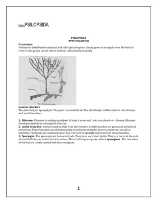
General structure
The plant body is sporophyte. The plant is a small shrub. The plant body is differentiated intorhizome
and aerial branches.
1. Rhizome: Rhizome is underground part of stem. Leavesand roots are absent on rhizome. Rhizome
develops rhizoids for absorption of water.
2. Aerial branches: Aerial branchesarise from the rhizome. Aerial branchesare green and cylindrical
at the base. These branchesare dichotomously branched repeatedly. Leavesare present on aerial
branches.The leavesare small and scale-like. They are irregularlyscattered over these branches.
3. Sporangia: The sporangia are borne in triads. They have very short stalks. They are borne in the axils
of small bifid leaveson the aerial branches. This triad of sporangia is called a synangium. The two lobes
of the leaf are closely united with the synangium.
2. 2
Internal Structure
Aerial branches: In transverse section, the aerial brancheshave central stele and outer cortex.
1. Cortex: The cortex is covered by a single layered epidermis. Stomata are present in the epidermis.
The inner part of the cortex is formed of parenchymatouscells. Outer to thisparenchyma are few layers
of sclerenchymatouscells.The cells in outer most part of the cortex are rich in chloroplasts. Cambium is
absent in the stem.
2. Stele: There is a well developed endodermisbetween the stele and the cortex. The xylem is
actinostelic. It hassix rays. A core of thick walled sclerenchymatousfibers(pith) is present in the centre
of the xylem. Phloem is present between the endodermisand xylem.
Rhizome: The structure of the rhizome is similar to that of aerial branches. But pith or
sclerenchymatoustissuesare not present in the centre of the xylem core. The phloem is poorly
developed. The cortex is composed of thin walled parenchyma. A mycorrhizal fungus livesin it. The cells
of lower epidermiscontain rhizoids.
Leaves: The leaveshave simple structure. The epidermisis formed of cutinized cells and is without any
stomata. The internal tissue is formed of photosynthetictissue. The leavesare without a vein.
Reproduction
3. 3
Vegetative reproduction:
Vegetative reproduction takesplace by the death of the older partsof the rhizome. The younger partsof
rhizome separate from the dead rhizome. They grow as long as independent plants. Sometimes, the
upper cell of the rhizoids dividesand producesa small gemma. The gemma develops into a new rhizome
after detachment.
Sporangium:
Psilotum is homosporous. Sporangia form groups of three on short stalks. This stalk is present in the
axilsof small bifid leaf. The group of three fused sporangia is called a synangium. It is believed that
synangium is sporangiophore. It hasbifid bract at its base. The sporangia develop independentlyfrom
each other. The sporangiophore dividesearlyin a dichotomous manner. One branch terminatesin a
sporangium. But the other branch again divides into two branches. Each of which terminatesin a
sporangium. Thus it produces closely united three sporangia.
Fig: Stages of development of sporangium, A- VS of stem bearing leaf and sporangiophore. B-C-section of
sporangiophore at later development, D-Transverse section of mature sporangium.
Development of Sporangia
1. Each sporangium developsfrom a superficial cell of the sporangiophore. This cell divides transversely
into an outer jacket initial and an inner archesporial initial.
‘2. The jacket initial dividesto produce wall. This wall is four to five cells thick. The archesporial initial
dividesto produce a mass of archesporial cells. Tapetum is not produced in Psilotum.
3. In the mature sporangium some of the archesporial cells become elongated. They are filled with dense
cytoplasmic contents.These cells act as spore mother cells. Each spore mother cell undergoes meiosis
and producesfour spores. The rest of the archesporial cells disintegrate toform protoplasmic mass
or tapetal fluid. It nourishes the developingspores.
----------------------------
----------------------------
The epidermal cellsof the sporangial wall become thick walled. But a single vertical line from the base of
the sporangium to the apex remainsthin walled. The mature sporangium dehiscesalong this line and
the spores are liberated.
4. 4
Gametophyte:
Each spore germinatesto producesa small thallose gametophyte or prothallus. The gametophyte is
colourless and subterranean (underground). It hasone two or more short dichotomous branches.
Gametophyte isinfested with mycorrhizal fungi. There are no vascular strandsin the gametophyte. It
bearsnumerousunicellular rhizoids. The gametophyte does not have much internal differentiation of
tissues.It is monoecious. The sex organs are produced near the growing apex.
Antheridia:
Antheridia are produced earlier than archegonia. The mature antheridium isglobular structures. It
project out on the surface of the gametophyte.
Development of antheridium:
Each antheridium developsfrom a single superficial cell. It dividesinto an outer jacket initial and an
inner primaryandrogonial cell. The jacket initial dividesto produce a single layered wall. The primary
androgonial cell dividesto producesa mass of androcytes or antherozoid mother cells. Each androcyte
gives rise to a single, coiled and multiflagellate antherozoid. The antheridial wall rupturesto release the
antherozoid.
Archegonium:
The mature archegonium consists of a neck and basal part. The neck contains one or two neck canal
cells. The basal part is embedded in the gametophytictissue. It is without any well defined venter. It
contains a single large oosphere.
5. 5
Development of archegonium
Each archegonium developsfrom a single superficial cell. It dividestransverselyinto an upper primary
cover cell and a lower central cell. The primarycover cell divides to produce a group of four neck
initials.These neck initial dividesto produce neck. The central cell dividestransverselyinto a primary
neck canal cell and a primaryventral cell. Primaryventral cell functions as an egg directly.
Fertilization:
The neck canal cellsof mature archegonium disintegrate. It producesa pore through which antherozoids
enter the archegonium. Only one antherozoid fuses with the oosphere to produce oospore.
Development of Sporophyte:
I. The oospore dividestransverselyinto an upper and a lower cell.
2. The lower cell by further divisions producesa foot. Foot buried into the tissue of the prothallus.It
absorbs nourishment for the developingembryo.
3. The upper cell divides to produce a mass of cells. Its one or two peripheral cellsact as apical cells.The
apical cell dividesand increasesthe size of embryo. The gametophytictissue completelysurrounds the
young embryo like calyptra in early stages. But later, it comes out of the calyptra. Some of itssurface
cells produce rhizoids. Other cellsare infested with the mycorrhizal fungi and the embryobecomes
independent. The embryoby further growth becomesthe rhizome. Rhizome develops aerial
dichotomous branches.
Alternation of Generation
Psilotum shows regular alternation generations. The vegetative plant is sporophyte. It produceshaploid
spores by meioses.Spores germinate to give rise to the prothallusor gametophyte. The prothallus
6. 6
produces antheridia and archegonia. Fertilization producesdiploid oospore. Oospore givesrise to the
sporophyte. Thussporophyte and gametophyte alternateswith each other.
LYCOPSIDA
Botany 1 Comment
LYCOPSIDA
TYPE SELAGINELLA
Occurrence
Selaginella isa tropical plant. It has world wide distribution. It grows in damp forests. Some species
occur in temperate regions.They grow in moist shadyplaces.
General structure
The plant body is sporophyte. The body is divided into root, stern and leaves.
Stem: The main stein is prostrate. Some erect brachesarise form the main stem.
Rhizophore: Main stem developsleafless structurescalled rhizophore. Rhizophore grows downward. It
develops adventitiousroots at itstip. The rhizophore are intermediate in structure between the root and
the stem. It is without nodes and intemodes.
Leaves: The main stem and the branchesare covered by green leaves. Each leave hasa ligule. The leaves
are of two sizes, large and small. The leaves are arranged in four vertical rows. Leaves present in pairs.
The larger leaf of each pair is attached toward, die ventral side of the stem and the smaller leaf towards
the dorsal side. The leavesbearingsporangia in their axils are called sporophylls. Many sporophylls
form cones or strobili.
7. 7
Internal structure of the stem
In cross section, the stem is composed of epidermis, cortex and central stele.
1. Epidermis: It is outermost layer. It is without stomata.
2. Cortex: Cortex is present inner to the epidermis. It has manylayered. It composed of
parenchymatouscells.The cortex surroundscentral stele. Cell of peripheral region of cortex contain
chloroplasts. In mature regionsof stem, the cortex form sclerenchymatoushypodermis.
3. Stele: Their stele is from monostelic to polystelic condition. Each
stele is protostelic in nature. The metaxylem forms the solid central core. The protoxylem groups on the
periphery. The xylem core is surrounded by the phloem. Outside the phloem is the pericycle. It is
composed of single layer of parenchymatouscells. The stele is separated from the cortex by a wide,air
space. These spaces have long radiatingcellscalled trabeculea. Trabeculea connect the stele with the
cortex.
Internal Structure of the Root:
The root has a single layered epidermis.Inner to the epidermisis a manylayered cortex. A well
developed single layered endodermisseparatesthe cortex from the stele. There is no air space
surroundingthe stele. The stele is protostelicand monarch. There is a single layered pericycle between
the phloem and the endodermis.The internal structure of the rhizophore is similar to that of the root
Internal Structure of the Leaf:
The leaf is covered by a single layered epidermis. The cells of epidermiscontain chloroplasts. Stomata
are present on the upper, or on the lower, or on both sides of the leaf. The mesophyll is formed of
parenchymatouscells.These cells are loosely arranged and theyhave numerousintercellular spaces.
Each cell containsone or more chloroplasts. Each chloroplast contains several pyrenoid-like bodies.The
mesophyll is traversed by a single vein.
Sporangia
Selaginella is heterosporous. The larger spores are megaspores and the smaller spores
are microspores. Megaspores are produced in megasporangia and microspores are produced in
microsporangia. Both sporangia are borne in the axilsof leavescalled microsporophyll and
megasporophylls.This condition is called stachyosporous. The sporophylls form definite cones or
strobili. Both kinds of sporangia are found in the same strobilus. Megasporangia are present in the basal
portion and the microsporangia are present in the upper part of the cone.
8. 8
Each microsporangium contains several microspores. But them are only four megasporesin each
sporangium. The mature sporesare pyramidal in shape. The sporangial wall consists of three layers.The
inner most layer is tapetum. They provide nourishment to the developingspores. A slit is produced in
mature sporangia.The spores come out of thisslit.
The spores germinate to develop gametophytes. Microspore give rise to male gametophytesand the
megasporesproduces female gametophytes. Both male and female gametophytesremain within the
wallsof the spores.The young embryo develops in the megaspore. This is an approach towards the seed
habit.
Development of Sporangia
The development of micro and megasporangia is similar uptothe formation of spore mother cells.
1. The sporangia initialsare present in the axil of the leaf. The sporangial initialsdivide to form outer
cells called the jacket initials, and an inner group of cells called archesporial initials.
2. The archesporial initialsdividesto form mass of sporogenous tissue. The outer most layer of the
sporogenous tissue forms tapetum. The jacket initials by further divisions give rise to a jacket.
3. All the sporogenous cells in the microsporangia become spore mother cells. The spore mother cells
separate from each other. They undergo meiosisto form microspores. Several spore mother cells are
produced in the megasporangium. But only one spore mother cell is functional. All othersdisintegrate.
9. 9
The spore mother cells divide meioticallyto produce four megaspores. The development of the
megasporesstarted before their sheddingfrom the sporangia.
Gametophytes
Development of the Male Gametophyte:
I. The development of the male gametophyte started within the microsporangia. Microspore dividesinto
two unequal cells.The smaller cell is called prothalial cell. The larger cell is called the antheridial cell.
2. The prothalial cell does not divide further. Antheridial cell dividesto produce 12 cells. Four cells
occupy the centre. They become primary androgonial cells. These cells are surrounded by the
remainingeight peripheral cells.The microspores are liberated from the sporangia at this12-cell stage.
----------------------------
----------------------------
3. The outer eight cells form the jacket of the antheridium. The androgonial cellsdivide to produce a
mass of 128-256 androcytesor antherozoid mother cells. Each androcyte developsinto biflagellate
antherozoid. The prothalial cell and jacket cells disintegrate and liberate the antherozoidsin the
surroundingwater.
Development of the Female Gametophyte:
The germination of the megasporesstarted in the megasporangium. Spore increasesin size. Nucleusof
the spore undergoes several divisions. It makes the spore multinucleate. A large central vacuole
develops in the spore. It pushesthe whole of cytoplasm towardsthe pointed end of the spore. The
vacuole graduallydisappears.Two or three layers of cells are formed towards the pointed end of the
spore. A clear membrane diaphragm separatesthe cellular layers from the rest of the cytoplasm. The
spore wall rupturesat the pointed end exposes the cellular layers. The exposed cellsdevelop
chloroplasts.
Some cells produce rhizoids.
1. Several superficial cells of exposed tissuesbecome archegonial initials. The archegonial initial
dividesinto an upper primary cover cell and a lower central cell.
10. 10
2. The primarycover cell dividesto form the neck of the archegonium. The central cell dividesto
produce an upper primary canal cell and a lower primary ventral cell. The primarycanal cell
functions as single neck canal cell:
3. The primaryventral cell divides to producesa lower egg or oospbere and an upper ventral canal cell.
The surroundingvegetative tissue forms the wall of the venter. The ventral canal cell and the neck canal
cells of mature archegonia disintegrate. Theyform a passage for the entryof antherozoids.
Fertilization
Fertilization alwaystakes place in moisture. Antherozoids swim in water. One antherozoid entersinto
archegonium. It fuses with oosphere to produce oospore.
Development of the Embryo:
1. The oospore divides into two cells.The upper cell enlarges. It is cilled suspensor. The lower cell is
called the embryonal cell. It develops into the embryo. The suspensor pushesthe developingembryo
into the tissue of the gametophyte.
2. The embryonal cell divides to form eight cells or octants. Two cells of the octants divide more
rapidly. They produce an outgrowth called foot on one side. Foot is the chief food absorbingorgan of the
developingembryo.
3. The remainingcells of octant form a mass of cells. The cel tral group of cells in this missforms
the apical meristem. The remainingcellsof these massproduce rudimentsof the first leuves
or cotyledonary leaves.
4. Root primordium arisesas a protuberance between the foot and the suspensor. The root
primordium forms rhizcrphore.
5. Further growth of the apical meristem pushesthe embryo out of the gametophytictissue. Stem grows
upward takingwith it the cotyledonary leaves. The rhizophore grows downward and produces
adventitiousroots.
Alternation of Generation
Selaginella shows a regular alternation of sporophytic and gametophyticgenerations. The vegetative
plant is diploid sperophyte. It produceshaploid micro and mega spores bymeiosis. These spores give
11. 11
rise to male and female gametophytes.Gantetophytesproduce male and female gametes. The gametes
fuse to form diploid oospore. This oospore develops into the sporophyte.
Evolutionary advancement of Selaginella:
Approach to seed habit:
Selaginella shows an evolutionary advancement over the other Pteridophyta. It has an approach
towards seed habit due to following advanced characteristics.
1. The production of gametes,fertilization and the development of the embryo, take place on the
sporophyte. Megaspore is never released from the sporophyte.
2. Selaginella isheterosporous. The microspore producesthe male gametophyte: It completesits
development within the wall of the spore.
3. Megaspore containsa large amount of reserve food material. The female gametophyte completesits
whole development within the megaspore wall. Fertilization and the development of the embryoalso
take place within spore wall. The developinggametophyte arid the embryo use the reserve food.
4. In many cases the megaspore is not released from the megasporangium. The development of the
gametophyte, fertilization of the oosphere and the earlydevelopment of the embryo take place while the
spore is still in the sporangium.
12. 12
Sphenopsida– Occurrence & Structure
Botany No Comments
SPHENOPSIDAF
TYPE EQUISETUNI
Occurrence
The genusEquisetum has25 species. It is world wide in distribution. Theyare most common in
temperate regions.It generallygrows in moist places.
General structure
The plant body is sporophyte. It is composed of rhizome, aerial branches, scale leaves and roots.
1. Rhizome: Plant body has horizontal underground rhizome. Rhizoine gives off erect aerial brandies.
Rhizome and aerial brancheshave nodes and intemodes. Intemodeshave ridges and furrows. Lateral
branchesarise from the nodes. Some budsproduce short branchescalled tubers. Tubersgive rise to new
plants.
2. Leaves: Each node has a whorl of small scale leaves. These leavesform sheathingat the base of node.
Leavesperform little photosynthesis.
3. Roots: Roots are adventitious.Roots arise in whorlsat the nodes of the rhizome.
4. Aerial branches: The aerial branchesare green. Thus aerial branchesperform photosynthesis. The
aerial branchesare differentiated intonodes and internodes. Each aerial branch bears a whorl of lateral
branchesat each node. These lateral brancheshave whorls of tertiarybrancheson their nodes. Some
species of equisetum have two types of aerial branches: fertile and sterile.
(a) Fertile branches: Fertile branchesare short and brownish in colour. They are without lateral
branches.Each fertile branch produces a cone or strobilusat the apeqrhe fertile branchesare produced
in the spring. These branchesdie after the production of cones
(b) Sterile branches: The sterile branchesare green. These brancheshave numerouslateral branches.
Sterile branchespersist throughout the year.
Internal Structure of the Stem (aerial branches & rhizome)
13. 13
Internallythe stem is differentiated intoepidermis, cortex, and central stele.
1. Epidermis: It is the outermost layer. Epidermisconsists of a single layer of cells. Cell wall of these
cells is highlysilicified. Stomata are present in the epidermis. Stomata are absent in the underground
portion or rhizome.
2. Cortex: Cortex is present between epidermisand endodermis. Cortex haslong canals
called vallecular canals. Cortex is composed of parenchymatouscells. There are large bandsof
sclerenchyma in the peripheral portion of the cortex. These sclerenchyma bandsform the main
supportingtissue. Loosely arranged parenchymatouscellsof cortex contain chloroplasts. They are
called chlorenchyma. They are the main photosynthetictissue of the plant.
3. Endodermis: Endodermisis present inner to cortex. Endodermis is formed of a single layer. These
cells have the characteristiccasparian bandson the radial walls.
4. Pericycle: Pericycle is present inner to the endodermis. It consists of a single layer of
parenchymatouscells.
5. Stele: Parenchymatouscells form the ground tissue of the stele. Pith is present in the centre. In the
primaryaerial branch this pith has central canal. But these canals are absent in the rhizome and lateral
branches.
Equisetum has siphonostele. The. vascular bundlesare arranged in a ringaround pith. Each vascular
bundle is collateral. In this case, xylem is inner and phloem is outer. The xylem is in the form of a V. The
protoxylem is present at the basal position and the metaxylem on the tipsof the arms. Phloem is found
in a massbetween the metaxylem groups. Mature vascular bundleshave a cavity called carinal canal.
Internal structure of the rhizome is similar to aerial branches. But it hasno central canal in the pith. Its
epidermisis without stomata. It also hasno chlorenchyma in the cortex.
14. 14
Internal Structure of the Root
Each mature root has a single layered epidermis. Cortex is composed of parenchymatouscells. Two
layersof endodermisare present below the cortex. Cells of the inner layer of endodermisgive rise to the
secondary branchesof the root. A definite pericycle is absent in roots. Stele is present in the centre. It is
without pith.
Internal Structure of Leaf
Each leaf hasa single vein. Its vascular bundle is collateral. The xylem is formed of only protoxylem
elements.There is no carinal canal. Vein is surrounded by the endodermis. Parenchyma is present
outside the endodermis.Parenchyma of the adjacent leavesis continuous in the region of the sheath.
Leaveshave small bandsof the functionless assimilatorytissue.
Sporangia
Equisetum ishomosporous. The cones or strobili are produced at the apex of branches. Each cone has an
elongated central axis. Sporophylls or sporangiophores are attached on it. Sporophylls have a stalk and
15. 15
flattened head. Sporangia are attached to the underside of the head of the sporophyll. Sporangium fills
the whole space between the head and the central axis. The headsthe sporophylls are closely attached
with each. They become hexagonal in outline. A ring like outgrowth annulus is present at the base of the
axis.The number of sporangia on each sporophyll variesfrom 5-10.
Development of the Sporangium
1. A single cull initiatesdie development of each sporangium. It dividesinto an inner and an outer cell.
2. The inner cell further dividesto produce the sporogenous tissue or archesporium. Outer cell takes
part in formation the sporangial wall. Wall of the sporangium consists of several layersof cells. The
inner most layer is the tapetum. The cellsof the outer layer develop spiral thickenings on their walls.
----------------------------
----------------------------
3. One third archesporial cellsenlarge and become spore mother cells. The remainingcells
disintegrate toa mucilaginousliquid. This liquid provides the nourishment to the developingspores.
4. Each spore mother cell dividesmeioticallyto form four spores. Wall of spore becomesfour layered.
The outer most layer epispore splits to form four bands. These bands separate from the spore wall on
drying. These bandsare called elatersor Hapetra. They coil round the spore under moist conditions.
These elatershelp in the splittingof the sporangial wall.
Germination of spore and formation of Prothallus
The spore germinateson suitable substratum. It dividesintotwo unequal cells. Smaller cell forms the
first rhizoid. It forms many rhizoids on the underside of prothellus. The larger cell dividesto produces
prothallus.Peripheral cellsof prothellusare meristematic. Theydivide to increase the size of prothellus.
It produces circular prothallusor gametophyte. Many short, multicellular lobesare produced on the
upper side.
Sex organs are produced on the upper side. The upper portion of mature prothallushas green cells and a
lower portion hascolourless cells.Spores of Equisetum produce two typesof prothalli. Half of the spores
produce smaller male prothellus.The remaininghalfproduces large female prothellus. But sometimes,
female prothellusproduces antheridia. SoEquisetum is not perfectlydioecious.
16. 16
Fig: A–Vertical section of gametophyte, B-D-stages of development of antheridia
Sex Organs:
Sex organs are produced at the margin of the prothallus. But later theyare embedded in the
thallus. Antheridium
1. Each antheridium isproduced from a superficial cell. This cell divides to produce outer jacket
initial and an inner androgonial initial.
2. The jacket initial divides to produce wall of the antheridium. The wall has a triangular opercular
cell at the top. The androgonial initial dividesto produce a mass of androgonial cells.
3. Each androgonial cell dividesto produce two androcytes or antherozoid mother cells. Each
androcyte is changed into an antherozoid. Antherozoid is spirallycoiled. It has a row of cilia near the
upper end. The mature antherozoids come out by the lifting of the opercular cell of the wall.
Fig: C-development of arehegonia, D-G-old archegonium, E-F-young end,‘ vo, G-endnyo at advance stage
Archegonium
1. Archegonium also develops from a superficial cell of the thallus. This cell dividesto
produce an upper neck initial and a lower central cell.
2. The neck initial dividesto produce a neck. Neck is composed of four vertical rows of cells. The central
cell dividestransverselyto produce an upper neck canal initial and lower ventral cell.
3. The neck canal initial produces a row of two or three neck canal cells. The ventral cell dividesto
produce a large egg or oosphere at the base and a smaller ventral canal cell. The surrounding tissue of
the thallusforms the wall of the venter. The mature archegonia lie on the dorsal side of the thallus
Primaryleaf sheath.
Fertilization
The ventral canal cell and the neck canal cell of mature archegonia disintegrate. It forms passage for the
antherozoids. Several antherozoids enter the archegonia. But only one of them fuseswith the egg. The
fertilized oosphere develops a wall and becomesthe oospore.
17. 17
Development of the Sporophyte
1. The oospore divides transverselyintoan upper epibasal half and a lower hypobasal half.
2. The hypobasal portion dividesto produce a foot and the first root. The root grows downward into the
soil by passingthrough the gametophyte. The epibasal cell dividesto form an apical cell and adjacent
cells.
3. The adjacent cellsproduce the first whorl of three scale leaves. The apical cell producesthe first
branch (primarybranch) of 10-15 Sin:nodes. Each node has a whorl of threeiletwes.
4. The primarybranch developsadventitiousroots at its base. Sporophyte becomes independent very
early. The primarybranch
produces one or more secondary branchesat its base. Secondary branchesdevelop their own
adventitiousroots at their bases.Secondary branches have a whorl of 4-5 leaves at each node.
5. One secondarybranch grows horizontallyinto the soil and forms the rhizome. The rhizome gives rise
to the vertical aerial branches.
Alternation of Generation:
Equisetum shows a regular alternation of sporophytic and gametophyticgenerations. The sporophyte is
diploid generation. It produceshaploid spores after meiosis. These spores germinate togive rise to the
gametophyte or prothallus.The gametophyte producesantheridia and archegonia in which male and
female gametesare produced. The union of male and female gametesproducesdiploid oospore. Oospore
gives rise to the sporophyte.
18. 18
PTEROPSIDA (FERNS)
Botany No Comments
PTEROPSIDA (FERNS)
TYPE II MARSILEA (Water fern)
Occurrence
Marsilea is an aquaticor semiaquaticplant. It is common in the temperate regions. It groNA in fresh
water ponds and ditchesin Punjab. Marsilea quadrifolia and Munilea minuta are commonly found in
Pakistan.
General structure
The vegetative plant is a sporophyte. It is differentiated intoroots, rhizome and leaves.
1. Rhizome: The stem is in the form of a rhizome. Rhizome has unlimited growth. Therefore, it covers a
very large area. The rhzome is dichotomously branched. It has nodes and internodes. A number of
adventitiousroots arise at each node on the ventral side. But a single leafarises at each node from the
dorsal side.
2. Leaves: The leavesare compound. Each leaf has a long petiole and four :carats. The kalletsare
arranged in cross-like manner at the tip of the petiole. Each leaflet is triangular. Veins form reticulate
arrangement Stomata arclocated on the dorsal side and ventral side of the leaflets.
Internal Structure of the Rhizome
Internallythe rhizome is composed of epidermis, cortex and central stele.
1. Epidermis: It forms outer covering.
2. Cortex: The cortex is wide. Its peripheral part consistsof parenchymatouscells. Ringof a large air
chambersare present around thisperipheral portion. This portion is called aerenchyma.It stores air.
The inner portion of the cortex is composed sclerenchymatouscells. Endodermisis present inner side of
the cortex.
3. The stele in Marsilea is of amphiphloicsolenostele. It has pith in the centre. Protoxylem groups are
exarch in position.
19. 19
Internal structure of leaf
Both surfacesof the leaf are bound by epidermis.Epidermisis covered bycuticle. It has sunken
stomata. Mesophyll cellsare present between both epidermises. Mesophyll cells are differentiated into
palisade and spongy cells.Single vein passes through each leaf.
Reproduction
Sporocarp
Marsilea plant is heterosporous.The megasporesand microspores are produced in megasporangia and
microsporangia. Both typesof sporangia are found within the same sorus. The sari are produced in hard
fruit-bodiescalled sporocarps.
The sporocarps are attached to the base of petioles byshort stalks (peduncles). Sporocarp is bean
shaped. It has hard and stony wall (capsule).Its wall has single vascular bundle. The sporocarp has two
inner chambers.Each chamber hasa row of sori. The sori of two rows alternate with each other. The
wall of each sorus is formed byits own indusium.Each sorus contains a row of megasporangia and
several microsporangia.
20. 20
A large placenta is produced on the inner side of the wall in the young sporocarp. The placenta of two
sides alternate with each other. Megasporangia and microsporangia are produced on the same placenta.
Each placenta is covered by itsown individual indusium. Megasporangia mature earlier than the
microsporangia. Each megasporangium containsa single megaspore on maturity. But each
microsporangium contains several (32-64)microspores. All the tissuesexcept indusia gelatinized in
mature sporocarp.
Development of the Sporocarp
It is believed that the sporocarp is a single folded pinna. This pinna has single vascular bundle.
Receptaclesor placentasare produced on the ventral side of this pinna. These receptaclesbear
sporangial initials.Each receptacle with the developingsporangia forms a
An outgrowth is produced towards the midrib side of pinna. This outgrowth forms a covering over the
sorus. This covering is known as the indusium. 1 he pima becomes folded towardsthe ventral side due
to the growth of the tissue in the mid dorsal region. The two sidesof the pinna meet at the margin. It
completelyencloses the developingson. The hardeningof cells in the wall givesrise to the bean shaped
sporocarp. Dehiscence of sporocarp: The mature sporocarps open after two or three years. The stony
wall decay and open the sporocarp. The inner gelatinousmaterial absorbswater and swells. It splitsthe
sporocarp into two valves. The gelatinouscord or sporophore absorbs water and swells. It comes out of
the sporocarp like a worm. it carrieswith it the attached son. The sporophore becomesstraight. The
sporangial walls and indusia gelatinize and release spores. The spores remain viable for a very long
period.
21. 21
Development of the Sporangium
Sporangial initialsare present on the placenta or receptacle of sporocarp. The sporangial initialspresent
at the tip of receptacle develops into megasporangia. The initial present on the sidesof the receptacle
develop into microsporangia. The development of both sporangia is similar in both cases.
1. The sporangial initial cuts off a jacket initial to the outside. It itselfbecomes the archesporial initial.
----------------------------
----------------------------
2. The jacket initial divides to produce single layered wall of the sporangium. The archesporial initial
cuts off two tapetal cells.These tapetal cells divide to produce two layered tapetum.Tapetum is present
inner to the wall.
3. The archesporial initial then dividesproducing 12-16 spore mother cells. Each spore mother cell
undergoesmeiosis and producesfour spores. The development of megasporangia and microsporangia is
similar upto the stage. Both sporangia contain 32 or 64 young spores enclosed in a single layered wall.
4. The tapetal cellsprovide nourishment to young spore. So theydisintegrate duringthe development of
spores.
5. In the megasporangium onlyone spore develops further. All others disintegiate forminga
mucilaginousmass or plasmodium.This massprovides nourishment to the developing megaspore. In
the microsporangium all the spores develop into 32-64 microspores.
Development of the Male Gametophyte:
The microspore germinatesto produce a small male gametophyte. It completes itswhole development
within the wall of the microspore.
The microspores are globose (rounded) with one side slightlypyramidal. The microspore has a large
nucleusand numerousstarch grains.The nucleus of microspore moves towards the pointed side. The
starch grains come in the periphery.
1. The microspore dividesin to two cells. The smaller cell becomes prothalial cell. It is reduced male
gametophyte. The larger cell divides in two antheridial initials.
22. 22
2. Each antheridial initial dividestoform three jacket cell and single androgonial initial. The
androgonial initial of each antheridium dividestoproduces 16 androcytes (antherozoid mother cells).
Each androcyte changes into antherozoid.
3. The antherozoidshave manycoils and single flagella. The prothalial cell and the jacket cellsof both
the antheridia disintegrate. Thusthe antherozoids become free in the Surroundingwater.
Development of the Female Gametophyte
Each mature megaspore hasa dome shaped projection or beak at one end. The nucleusof the megaspore
lies, in this beak region. It is surrounded bydense granular cytoplasm.
1. Megaspore dividesin to two cells. A smaller cell occupies the whole beak. The larger cell does not
divide further.
2. This smaller cell functions as an apical cell. The apical cell dividesand cut off form single layered
vegetative tissue.The apical cell then functions as an archegonial initial.
3. This archegonial initial dividesto produce a small primarycover cell at the top and a central cell at
the base.
4. The cover cell dividesto form four neck initials. They divide to form a neck. The central cell divides to
produce a small upper primarycanal cell and a lower larger primaryventral cell.
5. The primarycanal cell dividesto produce two neck canal cells. The primaryventral cell dividesto
produce a lower larger oosphere (egg) and an upper smaller ventral canal cell.
23. 23
6. The ventral canal cell and the neck canal cellsof mature archegonium disintegrate. It formsan
opening for the entryof antherozoids.
Fertilization
Each megaspore is enveloped by a layer of mucilage. Several antherozoids enter into this mucilaginous.
One of these antherozoids entersthe archegonium and fertilizesthe egg to produce oosphere.
Development of the Sporophyte
The oospore dividesto produces four cells. Two sister cells develop stem and cotyledons. The other two
cells develop into foot and root. The vegetative cells of the gametophyte form a calyptra.It is two to
three cells in thickness.This calyptra forms envelop around the developingembryo. The surface cells of
the calyptra produce long rhizoids. Cotyledon and the root grow faster than calyptra and conic out of it.
The root enters the soil. Cotyledon expandsto form the first simple leaf. Primaryroot is replaced by
adventitiousroots. 1 he stem grows horizontally on the soil and form the rhizome.
Alternation of Generation
The sporophyte and gametophyte generationsalternateswith each other. Vegetative plant of Marsilea
is a diploid sporophyte. It is hetrosporous. It produces mega and microsspores by meiosis. The spore
germinatesto form haploid gametoplivte. The gametophyte of Marsilea is dioecious. The microspores
give rise to the male gametophyte. The megaspore gives rise to the female gametophyte. Both male and
female gametophytescomplete their development within the spore walls. Both gametophytesproduce
male and temale gametes.Gametesfuse to form diploid oospcire. The oospore developsinto the
sporophyte again.
pteropsida (FERNS) – TYPE I Adiantum
Botany No Comments
IPTEROPSIDA (FERNS)
TYPE I ADIANTUM (Maiden Hair Fern)
Occurrence
Adiantum isa common fern. It is found in the plainsof the Punjab. It grows in shady places. It is found on
moist wallsor rocky places. The common specie of this genusis Adiantum Capillus-Veneris.
24. 24
General structure
The vegetative plant body is a sporophyte. It is differentiated intostem, leaves and roots.
1. Rhizome: The stem is underground rhizome. Rhizome is closely covered byscales called palea. The
older partsof the rhizome boar numerousbasesof the old leaves. Rhizome developsnumerous
branched adventitious roots.
2. Leaves: Adiantum haslarge bipinnatelycompound leaves. The main axisof the leaf is called the
radius.The leafletsof the first order are called pinnae and leafletsof the second order are called as
pinnules.Each leaflet is green and triangular. It hasbroader end towards the apex. The broader end is
divided into three or four small lobes. These lobes are reflexed back. These reflexed apical lobes bear the
sporangia on their underside. The young leaves are coiled inward in the embryonic state. It is called
eireinnate vernation.
Internal Structure
Internal structure of Rhizome
1. Epidermis: The rhizome is covered by a single layered epidermis. It is without any stomata.
2. Cortex: It is present inner to the epidermis. It is mainlyformed of parenchymatouscells. The
peripheral region of the cortex has one or two layers sclerenchymatouscells.
3. The stele in the rhizome is dictyostetic. It hasfour or five meristele or bundles. Theyare arranged in a
ring. The central part of the stele or pith. Each meristele is surrounded byits own endodermis. The stele
becomes solenostelic due to presence of a single leaf gap at a particular level.
Metaxylem is present in form of plate in the centre of each meristele. It hasone, two or three protoxylem
groups on the side or on the ends. The xylem is surrounded bya narrow layer of phloem. Sometime a
single layer of parenchymatouscellsis present between the xylem and the phloem.
4. Pericycle and endodermis: A single layered pericycle is presented outer to the phloem. The
pericycle is surrounded by a single layered endodermis. Itscells have casparian stripson their radial
walls.
25. 25
Internal structure of Leaf:
Both the surfaces of leaf are covered by epidermis. Stomata are present in the lower epidermis. The
epidermal cellshave chloroplast. An undifferentiated layer of mesephyll cellsis present between both
epidermises.The rachis is composed of a single layered, cortex, endodermisand stele.
Internal Structure of the Root:
The root is composed of epidermis,cortex and the stele. In the older roots the inner most cells of the
cortex become sclerenchymatous.
Sporangia:
The sporangia are borne on the under side of the reflected lobes of the oinnae. The reflexed lobe of the
leaf forms a covering or false indusium over son. The groups of sporangia are called soil (sing sonis).
Each sporangium has a stalk. The sporangia are subglobose or ovate in outline. The wall of each
sporangium is formed of a single layer of cells. A vertical row of cellsalong the narrow side
form annulus. Annulushas thick walled cells. The cells on the opposite side of the annulushave thin
walls.These cells fonn stomium. Each sporangium produces about 48 – 64 spores. The mature sporangia
become thy. The outer thin wallsof the annuluscellscontracts. It exertsa force on the stomium cells.
The wall of the sporangium rupturesat thispoint and release spores.
26. 26
Development of the Sporangium
1. Each sporangium developsfrom a single superficial cell. This cell enlargesto form an outgrowth. This
cell cutsoff a small cell at the base. It itself becomesthe sporangial initial.
2. The sporangial initial dividestransverselyto form lower cell and upper cell. The lower cell is
the stalk cell. The upper cell is the sporangial cell.
3. The stalk cell dividesto form two cell thick stalk. The sporangial cell dividesand cut off three cellsat
the periphery. It itself becomes tetrahedral central cell.
4. This central cell cutsoff another peripheral cell at the tip. The four peripheral cellsthusproduced.
They become jacket initials. These cells divide to produce a single layered wall. The central tetrahedral
cell is the archesporial initial.
5. The archesporial initial cuts off another set of small peripheral cells. This second set of peripheral
cells dividesto .produces tapetum around the archesporium. The archesporial cellsundergo three or
four divisions producing 12-16 spore mother cells. The . tapetal cellsprovide nourishment tothe
developingspores.
6. Each spore mother cell undergoesmeiosis and gives rise to four spores. Adiantum ishomosporous.
7. The mature sporesare brownish in colour. Each spore has a three layered wall. The outermost layer
is perenium or epispore. The inner most is endosporium or intine. The middle layer is thicker and it
called exine or exosporium.
Prothallus or Gametophyte
Development of prothellus: The spore falls on a suitable place. It swells very much thusthe two outer
wallsburst at the pointed end. The intine grows out forming a short tube. A colourless cell is cut off at
the base of thistube. This cell producesfirst rhizoid which grows down into the soil. The tip cell divides
to produces a short filament of cells.
----------------------------
27. 27
----------------------------
The cells of the filament develop .chlorophyll. They become green. The apical cell dividesto form a
wedge-shaped apical cell. This apical cell dividesto form a flat plate of cells. These cellsdivides to form a
heart shaped prothallus.
Prothellusbecomes several celled thick in the middle. But it remainssingle layered at the margins.
Several cells on the ventral side of the prothallusproduce rhizoids. Fthizoidsabsorb water and fix the
protballus.All the cells of the prothallusare green. Each cell contains a single disc shaped chloroplast.
The prothallusof Adiantum is completelyindependent. It can manufacture itsown food.
Sex Organs
Adiantum ismonoecit as. The sex organs are borne on the ventral side of the prothallus. The antheridia
are produced earlier than the archegonia. The antheridia are located in the middle part of the prothallus
among the rhizoids. But the archegonia are present near the apical notch.
Antheridium
Each antheridium isa rounded structure. It projectsabove the surface of the prothallus. The wall of the
antheridium ikformed of three cells.The basal cell is funnel-shaped. Ifforms the lower half of the wall.
The upper cell is ringlike. It forms the upper half of the wall. The apical cell forms lid. Each mature
antheridium containsabout 30-50 androcytes or antherozoid mother cells. The mucilaginouswallsof
the androcytes swell. It pushesthe lid cell upward. Thus androcytes come out of antheridium. The
nucleusof androcyte changesinto antherozoid. Antherozoids have spiral band and numerousflagella.
The antherozoids come out of the mother cell walls.
Development of Antheridium
1. Each antheridium developsfrom a single superficial cell. This cell dividesto form upper central
cell and lower first ring cell.
2. The central cells divide to form outer jacket cell and primary androgonial cell.
3. The jacket cell divides to form a cover cell and second ringcell. The first and second ring cellsand
cover cells form the wall of antheridium. The primaryandrogonial cell undergoes several divisions to
produce a massof about 30-50 androcytes or antherozoid mother cells. Each androcyte changes into
antherozoid.
Archegonia
Neck of each archegonium protrudesabove the surface of the prothallus. Neck is curved backward
towards the posterior side of the prothallus. Venter is embedded in the tissue of the prothallus. Itswall
is not distinct from the surroundingtissue. Neck is formed of four vertical rows of cells. Each mature
archegonium contains a large egg or oospbere at the base and a small ventral canal cell. The ventral
canal cell and the neck canal cell disintegrate. Theyform mucilaginousmass.It comes out through the
neck of the archegonium.
28. 28
Development of the Archegonium
1. Each archegonium develops from a single superficial cell near the growing apex. This cell enlargesto
form a small basal cell and large upper cell.
2. The basal cell dividesto form venter. The upper larger yell bytwo transverse divisions produce a
row of three cells.
3. The upper cell is the primarycover cell. It dividesto form four neck initials. Neck initial dividesto
form neck. The middle cell is the neck canal initial. It forms neck canal cell. The lower most cell is
the primary ventral cell. It dividesto produce an oosphere and a ventral canal cell.
Fertilization:
The antheridia and archegonia of the same thallusmature at different times. So cross fertilization takes
place. The antherozoids are chemotacticallyattracted towardsarchegonia. Several antherozoids enter
the archegonium but only one of them fuses with the oosphere to form oospore.
Development of the Embryo: •
1. The oospore increases in size. It dividesto produce eight cells or octants. The four upper cells
become epibasal cells. The four lower cells become hypobasal cells.
2. The two epibasal cellsdivide to form first leaf or cotyledon. The other two epibasal cellsdivide to
form stem. The two hypobasal cellsgive rise to the first root. The other two hypobasal cellsproduce the
foot. Foot penetratesintothe tissue of the protballus.
29. 29
3. The primaryroot grows for sometime under the prothallus. It then enters the soil and absorbs
nutrients.The cotyledonary leaves are simple. Stem apex produce more leaves. Stem apex grows
horizontally in the soil forming the rhizome. Prothallusultimatelydisappears.
Alternation of Generation:
Adiantum showsa regular alternation of sporophyticand gametophyticgenerations. Both generations
are independent. Sporophyte producesthe haploid spores by meiosis. The spores germinate toform
haploid prothallusor gametophyte. Prothellusis moroecious. It producesantheridia and archegonia.
The union of antherozoid and oosphere producesdiploid oospore. Oospore germinatesto form diploid
sporophyte.
30. 30
Pteropsida (FERNS) – TYPE III Polypodium – Occurrence &
Structure
Botany 2 Comments
PTEROPSIDA (FERNS)
TYPE III POLYPODIUM
Occurrence
Polypodium is a perennial herb. It isfound mostly in temperate regions. It has worldwide distribution.
Mostly is attached to some rocks. But some forms are epiphytic.
General structure
The plant body is sporophyte. Plant body is divided into rhizome. leavesand roots.
1. Rhizome: It forms the main stem of the plant. Rhizome is rounded, underground. But itsapex is erect.
It has very few blanches.It is covered with persistant leaf basesand hairs.
2. Leaf: The leavesare pinnatelycompound or simple. In compound leaf, the leaf has ‘maw leafletsor
pinnae. Leavesare lobed frond like. They have long stalked petiole. The leavesare arranged spirally. The
form simple reticulate or dichotomous venation. The young leavesshow circinate vernation.
3. Roots: They have adventitiousroots. These roots arise from the lower surface of rhizome.
Internal structure of Rhizome
In cross section rhizome is composed of epidermis, cortex and stele. Epidermisisouter most covering. It
is without stomata. Cortex is wide and it is composed of parenchymatous tissues. Canal are absent in it.
Stele is present in the inner side. It is covered by endodermisand pericycle. Polypodium has polystelic
protostele. Each protostele hasconcentric vascular bundles. The xylem are exarch (protoxylem lies on
the peripheryof metaxylem).
Internal structure of leaf
Leafletsor lamina of leaf is covered byupper and lower epidermis. Epidermishasa layer of cutin. Lower
epidermishasstomata. Mesophyll tissues are present between two epidermises. Mesophyll tissues are
31. 31
differentiated intopalisade and spongy mesophyll. The leaf hascollateral and concentric vascular
bundles.
Internal structure of root
Root has simple internal structure. It hasouter epidermis, cortex and stele. Stele is protostcle and
diarch.
Sporangium
Some leavesbear sporangia. They are called sporophylls. Sporophylls are foliage leaves. Sporangia are
present in groupscalled sons. Son i are borne on the undersurface of vein of the leaves.. Son i are oval in
shape. Each sorus is naked (without indusium). Each sorus has a group of stalk sporangia. A capsule is
present on the stalk. The capsule is lenticular or biconvex. The jacket wall of capsule is single layered.
This wall is differentiated into thick wall cutinized annulusand thin wall stomium. A large number of
spores are present inside the capsule. Tapetum is two layered present inside the wall. Polypodium is a
homosporous. Number of spores per sporangium are 64. Spores are small, dark brown and oval. The
wall of spore is composed of intine and exine. The wall rupturesat stomium duringdehiscence of
capsule.
Development of the Sporangium
I. Each sporangium develops from a single superficial cell. This cell enlargesto form an outgrowth. This
cell cutsoff a small cell at the base. It itself becomesthe sporangial initial.
2. The sporangial initial dividestransverselyto form lower cell and upper cell. The lower cell is the stalk
cell. The upper cell is the sporangial cell.
3. The stalkcell divides to form two celled thick stalk. The sporangial cell dividesto cut off three cells at
the periphery. It itself becomes tetrahedral central cell.
4. This central cell cutsoff another peripheral cell at the tip. The four peripheral cellsthusproduced.
They become jacket initials.These cells divide to produce a single layered wall. The central tetrahedral
cell is the archesporial initial.
5. The archesporial initial cutsoff another set of small peripheral cells. This second set of peripheral
dividesto producestapetum around the archesporium. The archesporial cells undergothree or four
divisions producingspore mother cells.The tapetal cellsprovide nourishment to the developing spores.
6. Each spore mother cell undergoesmeiosis and gives rise to four spores.
32. 32
Gametophyte of prothellus
The spore germinatesto produce monoecious gametophyte. Gametophyte issurface living. It is
differentiated dorsoventrally. It hasan apical notch. It is green and heart shaped. The ventral surface of
prothellushasmany rhizoids. Many antheridia are present amongthe rhizoids toward the ventral side
of mature prothellusManyarchegonia are present near the apical notch.
----------------------------
----------------------------
Antheridia
The antheridia are slightlyprojected on the surface. They are spherical or oval in shape. Each
antheridium hasa layer of jacket cells. Antherozoid mother cells are present within the jacket. Sperm
mother cells are changed in spirally twisted multiflagellate sperm or antherozoids.
Development of Antheridium
33. 33
4. Each antheridium developsfrom a single superficial cell. This cell dividesto form upper central
cell and lower first ring cell.
5. The central cellsdivide to form outer jacket cell and primary androgonial cell.
6. The jacket cell dividesto form a cover cell and second ringcell. The first and second ring cellsand
cover cells form the wall of antheridium. The primaryandrogonial cell undergoes several divisions to
produce a mass of androcytes or antherozoid mother cells. Each androcyte changesinto
antherozoid.
Archegonium
The archegonia are also very numerous.Their neck is sunken in the prothellus. Each archegonium is
flask shaped. It consists of curved neck and a venter. Neck is made up of several neck cells and one neck
canal cells.Venter hassingle venter canal cell and oosphere. Development of the Archegonium
1. Each archegonium develops from a single superficial cell near the growing apex. This cell enlargesto
form a small basal cell and large upper cell.
2. The basal cell divides to form venter. The upper larger cell bytwo transverse divisions produces a
row of three cells.
3. The upper cell is the primarycover cell. It dividesto form four neck initials. Neck initial dividesto
form neck. The middle cell is the neck canal initial. It forms neck canal cell. The lower most cell is
the primary ventral cell. It dividesto produce an oosphere and a ventral canal cell.
Fertilization
At the time of fertilization, the ventral canal cell and the neck canal cell disintegrate and form
mucilaginousmass.This massoozes out of the neck of archegonium. The antherozoid is attracted to the
mucilage. A large number of antherozoids reach the base of the archegonium. But only one of them fuses
with the oosphere to form oospore.
Development of the Embryo
1. The oospore increases in size. It dividesto produce eight cells or octants. The four upper cells
become epibasal cells. The four lower cells become hypobasal cells.
35. 35
feature of ferns is that their persistent basic characters are still sufficiently plastic to be
receptive to the environmental fluctuations.
Pteropsida are distinct from lycopsida and sphenopsida in several characters. Among the
vegetative characters, the megaphyllous leaves with the attendant leaf gaps are most
notable. Among the reproductive features, (though some primitive members show some sort
of a semblance to the strobilar organisation seen in the previous groups), the aggregation of
sporangia on the abaxial or adaxial surface of the leaf into sori is the most significant.
Plant Body of Pteropsida (Ferns):
Stem:
The sporophyte has an underground rhizomatous stem which may be elongated or
tuberous. The branching of the rhizome may or may not be dichotomous. In some cases the
rhizome is covered by hairs called ‘ramenta’.
Leaves:
The leaves are compound and once or twice pinnate. They are megaphyllous, having a
dichotomous or reticulate type of venation. The size of the leaves varies from few
centimeters to several metres (Angiopleris). Usually only the leaves are aerial while the rest
of the plant body is subterranean. Some ferns show circinate vernation in the leaves i.e., the
young growing parts are coiled inwards and uncoil as they grow.
Roots:
Like in other pteridophytes, roots are always adventitious.
Trophopod:
Wagner and Johnson (1983) have reported a special food storing organ trophopod in a
number of ferns such as Asplenium, Platyneuron, Onoclea spp, Dryopteris fragrans etc.
According to them the trophopod which is generally over looked in fern description is an
organ which is of potential systematic value.
Internal structure:
Rhizome:
ADVERTISEMENTS:
Stelar organisation varies from protostele, solenostele to polycyclic, dictyostels (Pteris).
Cortex may be wholly parenchymatous (Ophioglossm) or may be distinguished into outer
36. 36
sclerenchymatous zone and inner parenchymatous zone. Some times muclilage ducts are
found in the cortex as in Angiopteris. Xylem has mostly tracheids, but vessels are also
reported in Pteris, Marsilea etc. Secondary growth is absent except in Botrychium.
Root:
The stele is usually protostelic with variations in xylem groupings. The xylem is exarch and
may be mono-di tri or even tetrarch. Root cortex may be homogenous of heterogenous.
Petiole::
The leaves maybe provided with single leaf trace or the trace may be dissected into several
meristeles.
Lamina:
The upper and lower epidermal layers enclose the mesophyll which may or may not be
differentiated. Distinction of palisade and spongy parenchyma in the mesophyll is seen in
Cheilanthes, Pyrrosia etc. Lamina may be hypostomatic or amphistomatic.
Reproduction:
Vegetative Propagation:
This is brought about by a variety of methods such as fragmentation, adventitious buds,
embryonic leaf apices, stem tubers, root tubers etc.
Spore Production:
Pterospsida are both homosporous and heterosporous (Marsilea). Spore producing organs
are varied. They may be fertile spikes (Ophioglossum), tassels (Osmunda), sori (Adiantum,
Pteris etc.) or Sporocorps (Marsilea). Spore producing organs are usually borne on the
leaves except in some species of Marsilea.
Whatever may be the name given to the spore producing organs they always represent
aggregations of sporangia. The sporangia within a sorus are numerous arising from a fertile
tissue called Receptacle. The sporangia may or may not be surrounded by a flap or tissue
(arising from the receptacle) called Inducium.
Sometimes a sorus is protected by a false inducium which represents the incurving of the
leaf margin. The maturity of sporangia within a sorus is varied. Based on this, the son are
classified into three types viz., (i) Simple, (ii) gradate arid (iii) mixed. In a simple sorus all
sporangia develop simultaneously (eg. Osmunda).
37. 37
In a gradate sorus sporangia develop in basipetalous succession (e.g. Hymenophyllum) and
in a mixed sorus sporangia develop in an irregular sequence (eg. Pteris). It has been widely
held that a simple sorus is primitive, a mixed sorus is advanced while a gradate sorus is of
the intermediate type.
Sporangia may or may not have a stalk (Ophioglossum). Their development is either of the
eusporangiate type or of the leptosporangiate type. The capsule region of the sporangium
encloses the spores. Except in primitive members such as Ophioglossum and Angiopteris,
the sporangium has a definite dehiscence mechanism brought about by cells of different
thickness and differential hygroscopic reaction.
The thick walled cells are called Annulus and the thin walled cells are called the Stomiuim.
The annulus may be shield shaped (Osmunda), cap like (Lygodium) or obliquely vertical
incompletely overarching the sporangium (Pteris, Adiantum etc.). Spores are wind
dessiminated and have a sculptured outer wall (exine) enclosing a thin inner wall (Intine).
Gametophyte of Pteropsida (Ferns):
In homosporous forms the gametophytes are exosporic and in heterosporous forms they are
endosporic. Endosporic gametophytes are extremely reduced.
Bower (1923, 1935) has recognised three types of prothalli in homosporous
ferns. These are:
(a) Cordate type,
(b) Filamentous type and
(c) Saprophytic type or the mycorhyzic type.
Cordate or heart shaped prothalli are autotrophic and are seen in Adiantum, Osmunda,
Pteris etc. The filamentous type is seen in Hymenophyllum. The nutrition here also is
autotrophic.
Mycorrhizic prothalli are common in members like Ophioglossum. These prothalli are
tuberous or cylindrical and have a radial symmetry as opposed to the bilateral symmetry of
the cordate and the filamentous types. Nutrition is saprophytic.
Reproduction:
38. 38
Gametophytes reproduce vegetatively as well as sexually. The former type of reproduction is
very rare. Sexual reproduction is brought about by antheridia and archegonia which have
undergone maximum possible simplification.
Embryogeny may be exoscopic (Ophioglossum) or endoscropic with (Helminthostachys) or
without (Angiopteris) a suspensor. In leptosporangiate ferns embroyogeny is said to be
lateral because the first division is vertical and does not produce epibasal and hypo basal
cells.
Classification of Pteropsida (Ferns):
The types of classification proposed for ferns are as varied as ferns themselves. Below is
given a few systems of classification.
Hirmer (1927) classified Fillicophyta into four classes viz.:
(a) Primoftlicopsida,
(b) Eusporangiatae,
(c) Protoleptosporangiatae and
(d) Leptosporangiatae.
Hirmer created protoleptosporangiatae specially to include osmundaceae which exhibits
intermediate characters between eusporangiatae and leptosporangiatae.
Pichi-Sermolli (1959) has sub divided Filicopsida (pteropsida) into seven sub-classes, viz.,
Primofilicidae, Ophiglossidae, Marattidae, Osmundiade, Filicidae, Marsilidae and
Salvinidae.
In this article the classification proposed by Reimers (1954) is followed.
Primofili Copsida:
Coenopteridales:
The order coenoptaridales comprises only fossil members belonging to the late paleozoic
ara. The fossil remains of the plants of this order include stems and frond parts which are
very well preserved structurally. The members represent the fossil ferns.
39. 39
The order Coenopteridales has many alternative names like Palaeopteridales, Primofilicales
and Renaultificales. The last mentioned name is in honour of the great French Paleobotanist
Renault. The order comprises a heterogenous assemblage of various ferns and has been
treated differently by different paleobotanists.
However, there seems to be general agreement in classifying the order into three families
namely, Botryopteridacease, Zygopteridaceae and Cladoxylaceae. Burtrand divided the
order into two subgroups namely Inversicatenales and Phyllophorales. The group
Inversicatenales includes the family Botryopteridaceae while Phyllophorales has two
families namely, Zygopteridaceae and Cladoxylaceae.
Botryopteris:
The genus Botryopteris is one of the best known among Coenopteridales. It is the type genus
of the family and has 5 species ranging from lower carboniferous to the permian. The name
Botryopteris is given to the fossil specimens of stem.
The stems are slender, cylindrical and few millimeters in diameter. They are branched and
bear spirally arranged fronds. Anatomically, the main stem has a small mesarch protostele,
which is surrounded by a broad cortex. The cortex has a prominent band of sclerotic cells.
(Fig. 114).
In the leaf stalk, the xylem strand is solid and has three prototoxylem points. As in
B.forensis the strand is deeply indented looking like a trident. In very few species of
Botryopteris, sporangia have been found attached to the fronds.
The sporangia are found in clusters which is somewhat rare to ferns. In B. globosa
sporangial cluster has thousands of sporangia. The sporangia themselves are small, oval to
pyriform in shape.
They measure 2 mm in length and about 1 mm in diameter. Each sporangium has a short
stalk and a capsule which is somewhat oval in outline. The wall of the capsule has a broad
annulus represented by thick walled cells. The spores are of the same type.
Zygopteris:
40. 40
The ferns belonging to the family Zygopteridaceae are more complex and older then
Botryopteridaceae. The fossil specimens belong to middle Devonian and possibly have
connection with Psilophytales. Zygopteris, the type genus of the family is the best known.
It has several species of which Z. primaria has been studied extensively. The plant body of
Zygopteris is tree like with a trunk having a diameter of 20 cm. The stem as such, however is
only about 1.5 cm in diameter, the rest of the diameter being made up of an armour of leaf
stalks and adventitious roots.
The stem of Zygopteris bears an elaborately branched frond having a number of leaf stalks.
These are usually cylindrical and up to 2 cm in diameter. The leaf stalks have a number of
pinnae. Occasionally the leaf stalks are given the name Etapteris.
Anatomically the stem of Zygopteris has a xylem cylinder consisting of scalariform
tracheids. An unusual feature here is the presence of a layer of secondary wood surrounding
the primary xylem. This is perhaps one of the rare instances of secondary vascular tissue in
a fern extinct or extant.
The leaf stalk internally shows a vascular strand which is H- shaped with a straight median
band with some what fixed lateral arms. Two small protoxylem points lie in the shallow
depressions at the end of the median band.
Various names have been given to the fructifications of Zygopteris. Corynopteris is one such
fructification genus. The sporangia in the fructification are large and ovate. They are usually
sessile and are grouped into spherical son. The wall of the sporangium has a broad band like
annulus. The sporangial cavity is filled with homosporous spores.
Phylogeny of Pteropsida (Ferns):
The Coenopteridales represent the most primitive group among the ferns. They are very
ancient having originated perhaps, with Psilophytales. One of the prominent features of
Coenopterids is the lack of distinction between stem and leaves.
According to Delevoryas (1962) this suggests their affinity with Psilophytales. The
relationship of Coenopterids to other ferns is rather obscure. But there is no doubt in the
fact that Coenopterids may be regarded as ancestral stock from which the modem ferns
sprung up.
41. 41
Eusporantgiatae:
This sub-class includes all eusporangiate ferns. The sporangial wall is more than one
layered. Spore output is very high.
There are two orders in this sub-class viz., Ophioglossales and Marattiales.
Ophioglossales:
The order includes herbaceous, fleshy sporophytes with a short rhizome. Sporangia are
borne on a separate outgrowth called ‘fertile spike’. This arises at the junction of the leaf
blade and lamina. Sporangia have a multilayered wall with a high spore output.
There is no special dehiscence mechanism. All the members of the order are homosporous.
Gametophytes are tuberous and saprophytic. The order has a single family Ophioglassoceae,
with three genera- Ophioglossum, Botrychium and llelminlhoslachys.
Lycopodium: Habit and Habitat
and Morphology
Habit and Habitat of Lycopodium:
Lycopodium is commonly known as ‘club moss’ due to their moss like appearance and club
shaped strobili. It has about 400 species, which are cosmopolitan in distribution. They are
found in colder arctic region as well as in temperate, tropical and sub-tropical regions but
they are abundantly found in tropical zones.
Thirty three species of Lycopodium have been reported from India. Mostly it is found
growing in moist and shady places which are rich in humus and other organic matters.
Some of the common species are L. clavatum, L. phlegmaria, L. cernuum, etc.
It has got 2 sub-genuses:
(i) Urostachya—branching dichotomous and roots originate from the base of the stem.
(ii) Rhopalostachya—stem prostrate with erect branching and roots arise adventitiously
from all along the stem.
42. 42
Mostly the tropical species are epiphytic (e.g., L. phlegmaria) and grow hanging from the
tree trunks. The temperate species may be erect and shruby (e.g., L. reflexum), creeping
(e.g., L clavatum) or erect form (e.g., L. cernuum) etc.
External Morphology of Lycopodium:
The herbaceous plant body is sporophytic. Usually they may have either prostrate stem with
erect leafy branches or weak pendent stem (epiphytes).
The plant body is distinctly differentiated into following three regions (Fig. 1 A-
C):
(i) Stem,
(ii) Roots, and
(iii) Leaves.
(i) Stem:
In the sub-genus Urostachya stem is erect (terrestrial) or pendent (epiphytic) and may be
branched (dichotomously) or unbranched. In the sub-genus Rhopalostachya the stem is
prostrate with erect branches. First the branching is dichotomous and later on becomes
monopodial.
(ii) Root:
Usually small, adventitious roots are present. In the sub-genus Urostachya roots originate
only from the base of the stem (not arising from the whole length of the stem). In some
species e.g., L. selago etc. the roots arise endogenously from pericycle of the stem, do not
penetrate the cortex of the stem but turn downward through the cortex and finally emerge
only at the base of the stem.
Due to this reason a T. S. of stem usually shows roots within the cortex and are known as
cortical roots (inner roots). In sub-genus Rhopalostachya also roots are adventitious and
arise all along the underside of the prostrate portion of the stem.
(iii) Leaves:
Leaves are simple, sessile, small in size, eligulate and possess a single unbranched midrib
and are known as microphylls. Usually the leaves are spirally arranged (e.g., L. clavatum)
but may be arranged in whorls (e.g., L. cernuum) or pairs (e.g., L. alpinum).
43. 43
In all the cases they condensely cover the surface of the stem. Leaves are usually
homophyllous (isophyllous) i.e., of same size and shape but in some cases e.g., in L.
complanatum the leaves are heterophyllous (anisophyllous) i.e., of different size.
Usually the leaves near the apical portion of the branches bear sporangia and are called
sporophylls. Depending upon the species the sporophylls may or may not be differentiated
from the ordinary leaves.
These sporophylls usually form a condense structure at the apex of the branches which are
known as strobili. The numbers of strobili at the tip of branches differ in different species.
Internal Structure of Lycopodium:
(a) Stem:
A transverse section (T.S.) of the stem of Lycopodium is somewhat circular in
outline and can be differentiated into following three regions:
1. Epidermis:
It is the outermost covering layer comprising of single cell in thickness. The epidermis is
cutinised on the outer side and interrupted at places by the presence of stomata.
2. Cortex:
Inner to the epidermis is present a wide zone of cortex which shows a great variation in its
structure in different species.
Usually four types of cortex are recognized:
(i) The whole of the cortex is made up of parenchymatous cells with small or large
intercellular spaces (e.g., L. selago). Such cortex is called homogeneous.
(ii) The whole of the cortex is made up of sclerenchymatous cells, without intercellular
spaces.
(iii) The cortex is differentiated into outer and inner sclerenchymatous cells and middle
parenchymatous cells (e.g., L. clavatum, Fig. 2 A).
(iv) The cortex is differentiated into outer and inner parenchymatous cells and middle
sclerenchymatous cells (e.g., L. cernuum Fig. 2. B).
44. 44
Next to the cortex is present a single layer of well-defined cells known as endodermis with
conspicuous casparian strips but at maturity the endodermis may or may not be a distinct
structure. Endodermis is followed by pericycle which is composed of one or more layers of
compactly arranged parenchymatous cells.
3. Stele:
It is made up of only primary xylem and primary phloem. It is a protostele i.e., pith is absent
and the stele is situated in the centre. The arrangement of xylem and phloem tissues is
different in different species and the stele is also named differently.
In case of L. serratum, L. phlegmaria etc. the xylem is star shaped with a protoxylem
situated at the periphery (protoxylem exarch Fig. 3 A). In L. annotinum in actinostele the
furrows in the xylem are much more and show stellate arrangement (Fig. 3B).
The phloem lays in the space between the xylem rays. This type of stele is known as
actinostele. In case of L. clavatum. L. volubile etc. xylem appears to be in the form of
separate plates arranged somewhat parallel, with phloem in between them.
This type of stele is known as plectostele (Fig. 2 A, 3 C). In case of L. cernuum, L.
drummondii etc. xylem and phloem are uniformly distributed i.e. it appears as if strands of
xylem are embedded in the phloem. This type of stele is known as mixed protostele (Fig. 2
B, 3 D).
The protoxylem is usually exarch in all the cases. Xylem is usually composed of tracheids
and phloem of sieve tubes and phloem parenchyma. Cambium is absent hence there is no
secondary growth i.e., no formation of secondary xylem and secondary phloem.
(b) Root:
45. 45
The roots are adventitious.
A transverse section (T.S.) of the aerial root of Lycopodium is somewhat circular in outline
and shows the following internal structure:
(i) Epidermis:
It is the outermost covering layer and is only one cell thick. The cells are thin walled.
Epidermis is provided with numerous root hairs present in pairs (characteristic of
Lycopodium).
(ii) Cortex:
Just below the epidermis is present a wide zone of cortex. It is differentiated into outer
sclerenchyma and inner parenchyma. The outer one gives the mechanical strength to the
root.
(iii) Stele:
It may be di-, tetra-, or polyarch. In prostrate species it is polyarch i.e., having 6-10 plates of
xylem arranged radially (star shaped). The xylem is exarch. The phloem is present between
the radiating arms of xylem. In erect or pendent species stele is diarch or tetrarch. In
L.selago, L. serratum it is diarch and xylem is C, U or crescent shaped. The phloem is
present between the 2 ends of xylem, only in one group.
The cortical roots are exactly similar in their internal structure to that of aerial roots, except
that the epidermis and root hairs are absent.
The xylem is composed of tracheid and phloem of sieve tubes and phloem parenchyma. The
endodermis and pericycle are indistinct structure at maturity. Pith and cambium are absent.
(c) Leaf:
T. S. of the leaf shows epidermis, mesophyll tissue and a single median
vascular bundle:
1. Epidermis:
It is the outermost surrounding layer and is only one cell in thickness. The cells of epidermis
are parenchymatous and cutinised on their outer side. The epidermis is also interrupted by
the presence of stomata. In homophyllous (isophyllous) species the stomata are present on
outer as well as inner epidermis (amphistomatic) but in heterophyllous (anisophyllous)
species the stomata are mostly restricted on the lower epidermis (hypostomatic).
46. 46
2. Mesophyll:
It occupies a wide zone between the epidermis and the vascular bundle. It is usually made
up of thin walled chlorenchymatous cells which may be with or without intercellular spaces.
3. Vascular bundle:
In the centre of the leaf is situated only a single concentric vascular bundle made up of only
xylem and phloem. The vascular bundle is surrounded on all sides by a sclerenchymatous
sheath.
Reproduction in Lycopodium:
Lycopodium reproduces by two methods vegetatively and by spores.
1. Vegetative reproduction:
It takes place by the following methods:
(i) Gemmae or bulbils:
In a few species like L. selago, L. lucidulum etc. certain buds like structures known as
gemmae or bulbils are usually produced in large number on new stem tips annually. The
morphological nature of gemmae is still not fully known. The gemmae when fall on ground,
develop root primodia and soon form the root.
(ii) Death and decay:
Species with creeping stem reproduces vegetatively by the death and decay of older parts of
the stem up to the point of branching. This separates the branches which later on grow
independently.
(iii) Resting buds:
47. 47
In L. inundatum the whole of the plant body except the growing tip of rhizome is dead
during winter. This tip portion of the rhizome acts as resting bud which in the coming
spring resumes growth and develops into a new plant.
(iv) Fragmentation:
In several epiphytic species fragments of the plant body are capable of giving rise to new
plants.
2. Sexual Reproduction:
Spore Producing Organs:
Lycopodium is a sporophytic plant and reproduces sexually. The plants are homosporous
i.e., produces only one type of spores (without differentiation of mega- and microspores).
These spores are produced in sporangia which, in turn, are produced on fertile leaves known
as sporophylls. Usually the sporophylls are grouped together to form a compact structure
known as strobili (Sing. strobilus) which are terminal structures (Fig. 1 A).
Strobilus (Reproductive organ):
In the primitive species of the sub-genus Urostachya every leaf on the plant is a sporophyll
or at least potentially so and the fertile and sterile zones alternate. The sporophylls are
loosely arranged. In species of Rhopalostachya and in some species of Urostachya the leaves
of the apical portion of the branches only bear sporangia and are called sporophylls. The
rest behave as vegetative leaves.
The sporophylls may be of the same size or of different size from the foliage leaves in
different species. The arrangement of sporophylls is same on the central axis as that of the
vegetative leaves on the stem i.e., spiral, whorled or decussate etc.
The position of the sporangium is also different in different species. The sporangia may be
axillary and protected with the help of sporophylls (e.g., L. inundatum Fig. 7 A) or foliar and
protected (e.g., L. cernuum Fig. 7 B) or subfoliar and exposed (e.g., L. squarrosum, Fig. 7 C)
or axillary and exposed (e.g., L. lucidulum, Fig. 7 D).
Longitudinal section (L.S.) of strobilus shows the presence of a strobilus axis in the centre.
On both sides of the strobilus axis are present sporophylls (Fig. 8 A). Each sporophyll bears
only one sporangium (Fig. 8 B). All the sporangia are similar in structure and are arranged
acropetally in a strobilus i.e., the youngest are at the top (Fig. 8 C).
48. 48
Structure of Sporangium:
Sporangia are sac-like structures but usually kidney shaped in appearance (Fig. 8 B).
Sometimes they are sub-spherical in appearance. Their colour varies from orange to yellow.
Each sporangium consists of a basal short massive stalk i.e., sub-sessile, with an upper
globular unilocular body containing numerous spores.
The body of the sporangium consists of 3 or more layers of wall surrounding a cavity. The
inner most layer of the wall of sporangium is called as tapetum (Fig. 9 F) which is nutritive
in nature and persists till maturity.
In the young sporangium inside the wall is present a mass of sporogenous cells which in due
course of development form spore mother cells which by meiotic divisions, produce haploid
tetrad of spores. The spores at maturity separate from each other.
The wall of the sporangium is provided with a transverse strip of cells known as stomium
from where the sporangium at maturity splits into 2 valves and the spores are dispersed
away in the air.
The spores produced by a sporangium are all alike (homosporous). They are small, rounded
or even spherical structures. The surface of the spores is usually rough due to the presence
of reticulate ridges or knob like protrusions. Each spore is provided with a triradiate ridge
(Fig. 8, D, E) and is somewhat yellow in colour. A small amount of chlorophyll may or may
not be present in spores. Reserve food is in the form of oil in the spores.
Development of sporangium and formation of spores. Bower (1894) had studied the
development of sporangium in Lycopodium. The sporangium develops from a small group
of superficial cells arranged in a transverse row on the adaxial side of the sporophyll near
the base.
Its development is of eusporangiate type. These superficial cells are called sporangial initials
(Fig. 9A, B). These cells divide by periclinal divisions forming an outer and inner layer of
cells. The outer cells divide periclinally and anticlinally forming three celled thick wall of the
sporangium (Fig. 9A-F).
The inner layer or archesporial cells divide in all directions forming a group of cells known
as sporogenous tissue which finally give rise to spore mother cells. During these
49. 49
developments the inner-most layer of wall is differentiated as a nutritive layer and is known
as tapetum. It is a persistent structure and rich in reserve food material.
Each spore mother cell undergoes a process of meiosis thus producing a tetrad of spores
(haploid) with tetrahedral arrangement. These spores later on separate from the tetrad, as a
result of which, a large number of spores are produced inside each mature sporangium.
Dehiscence of sporangium and liberation of spores. As the sporangium approaches towards
maturity, a transverse row of cells is differentiated near the apical portion from the wall of a
sporangium known as stomium.
The walls of the cell of stomium thicken and differ from the walls of other cells of the
sporangium. As the sporangium loses water, it creates a pressure on the wall which leads to
the appearance of slit in the stomium as a result of which the wall splits opens into two
halves and the spores are disseminated by air current.
Gametophytic Generation:
The development of the gametophyte (prothallus) takes place from the haploid spores which
are the unit of gametophytic generation. Each spore is unicellular, uninucleate haploid
structure, 0.03 mm in diameter and surrounded by 2 layers, with a triradiate ridge at the
surface (Fig. 8 D, E).
Chlorophyll may or may not be present in different species. In few species spores may
germinate within a few days after liberation but in some species the spores germinate when
they are 3-8 years old and the development of gametophyte upto formation of mature sex
organs may take a time of 8 months to 6 or even 15 years.
The rate of the formation of photosynthetic tissue is usually proportional to the rate of
growth of gametophyte. Both the male and female reproductive organs are produced on the
same gametophyte. The male sex organs are produced earlier than female sex organs.
Usually at the time of germination of spore, it swells up to absorb water. First the spore
divides into two unequal cells by a lenticular division, forming a very small lens shaped cell
known as rhizoidal cell and a bigger cell (Fig. 10 A, B).
50. 50
This rhizoidal cell takes no part in further development of gametophyte and is a colourless
structure. At this two celled stage the spore will rupture at the triradiate ridge. Second
division divides the bigger cell into two equal halves, the cell near the rhizoidal cell is known
as basal cell and the other one is known as upper cell (Fig. 10 C).
The upper cell further divides by two successive divisions in such a way as to form an apical
cell with two cutting faces (Fig. 10 D). At this stage the gametophyte is 5 celled structures
and the symbiotic phycomycetous fungus (mycorrhizal fungus) attacks it.
If this fungus fails to attack at this stage, further development of gametophyte stops. This
infection takes place through the basal cell. During further course of development of
gametophyte the apical cell further divides to form six or morecells which later on develop
into meristematic cells. These cells, by further divisions form a multicellular structure, the
gametophyte (prothallus) (Fig 10 E-H).
Structure of the Mature Gametophytes:
The form and structure of the gametophytes varies greatly in different species.
In general they have been grouped under three categories:
Type I or Cernuum type:
Gametophyte is partially aerial and partly in soil. The prothalli are usually 2 to 3 millimetre
in height and 1-2 millimetre in diameter. The gametophytes (prothalli) grow at the surface
of the ground and consist of a colourless basal portion buried in soil and a conspicuous
upright, fleshy, green aerial portion having lobes (Fig. 11 A).
The sex organs develop between the green expanding lobes. The prothallus itself is a
nourishing body. The underground part contains endophytic fungus e.g., L. cernuum, L.
inundatum etc.
Type II or Clavatum Type:
The gametophyte is wholly subterranean and totally saprophytic i.e., non- green structure.
It is tuberous and without lobes. It may be one to two centimentre long or wide and is top
shaped, conical or discoid in shape (Fig. 11 B, C). The endophytic fungus is present. Sex
organs are formed on the upper surface e.g. L. annotinum, L. complanatum, L. clavatum etc.
Type III or Phlegmaria type:
51. 51
The gametophyte is subterranean, saprophytic and colourless. This type of prothallus is seen
in L. phlegmaria and other epiphytic species. The prothallus is about 2 millimeter in
diameter and monopodially branched (Fig. 11 D). Sex organs are borne on upper surface of
large branches and are interspersed with slender filaments known as paraphyses.
Besides these three forms some intermediate forms of prothalli are also observed. In L.
selago the prothalli may be subterranean or epiterranean (aerial). If the spores are buried
under the soil after liberation, they form subterranean prothalli and if the spores are not
buried under soil after their liberation, they form epiterranean prothalli.
The internal structure of the prothallus is very simple. The outermost layer is epidermis,
followed by cortical mycorrhizal region, palisade region and central storage region. It is
attached with the substratum by unicellular rhizoids. The prothalli of all species are
monoecious i.e., antheridia and archegonia develop on the same prothallus.
Development of sex organs:
Both the sex organs i.e., antheridia (male) and archegonia (female) develop on the same
prothallus, usually in distinct patches on the upper surface. The gametophytes are
protandrous i.e., antheridia develop before archegonia. Sex organs develop just on the back
of the apical meristem.
Development of antheridium:
A single superficial cell situated just away from the meristematic cells gives rise to an
antheridium. This superficial cell is known as antheridial initial (Fig. 12 A). This cell divides
periclinally to form an outer cell known as jacket initial (primary wall cell) and an inner cell
known as primary androgonial initial or cell (Fig. 12 B).
The jacket initial divides only anticlinally by several divisions resulting in the formation of
single layered covering known as jacket layer. In the middle of the jacket layer a triangular
cell is differentiated, which is known as opercular cell.
Simultaneously, the primary androgonial divides by various divisions, forming a mass of
cells embedded in the prothallus, known as androgonial cells which finally give rise to
androcytes (antherozoid mother cells, Fig. 12 C-F). The number of androcytes per
antheridium varies in different species.
52. 52
Each androcyte later on metamorphosis into a biflagellated antherozoid. Each antherozoid
is a haploid, uninucleate, fusiform structure with broad rounded posterior end and an upper
pointed biflagellated anterior end (Fig- 12 G).
The triangular opercular cell becomes mucilaginous as a result of which an opening is
formed at the apex of antheridium through which water enters into it. The antherozoids
absorb water and swell up as a result of which a pressure is created on the wall of
antheridium which finally ruptures and the antherozoids are liberated.
Development of archegonium:
Just like antheridium, the archegonium also arises from a single superficial cell called
archegonial initial, situated just away from the meristematic cells at the apex (Fig. 13 A).
The archegonial initial divides by periclinal division into an upper primary cover cell and
lower basal central cell (Fig. 13 B).
The primary cover cell later on divides vertically by two successive divisions at right angle to
each other forming four neck initials which later on by transverse divisions form a 3-4 cells
high neck. Each tier of the neck consists of 4 cells.
The central cell divides transversely forming an, upper primary canal cell and a lower
primary ventral cell (Fig. 13 D). The primary canal cell by successive transverse divisions
produces a variable number of neck canal cells (usually one in L. cernuum, seven in, L.
selago and 14-16 in L. complanatum).
The primary ventral cell may directly behave as an egg or may divide transversely to form an
upper ventral canal cell and a lower egg (Fig. 13 E-G). The egg is somewhat broader then the
rest part of archegonium. The archegonial jacket is absent. The archegonium is a sunken
flask shaped structure with neck projecting out of the prothallus.
Fertilization:
At the time of fertilization the neck canal cells and the ventral canal cell disorganise and the
cells of the upper-most tier of neck slightly separate apart forming a passage upto the egg
(Fig. 13 H). Fertilization is brought about in the presence of water.
The biflagellate antherozoids reach the archegonium by swimming in water on the surface
of prothallus. The antherozoids are perhaps attracted towards the neck of archegonium by a
chemotactic movement. They enter the archegonium through neck and reach the egg.
53. 53
Only the nucleus of one antherozoid fuses with the egg nucleus thus forming a diploid
structure-known as oospore (2x). The act of fertilization ends the gametophytic generation
and the initial stage of sporophytic generation is formed.
Embryo Development (Young Sporophyte):
The rate of development of the embryo is extremely slow. In Lycopodium embryo develops
downward into the gametophytic tissue instead of developing upward i.e., towards the neck
of archegonium. The first division of the oospore is always transverse, forming an upper cell
(epibasal) and a lower cell (hypobasal) known as embryonic cell.
The upper cell does not divide further and behaves as suspensor. The lower cell (embryonic
cell) divides by two vertical divisions at right angle to each other, followed by a transverse
division, forming 8 cells (octant, Fig. 14 A-D). The 4 cells of the octant, situated near the
suspensor by further division, form a multicellular foot which acts as a haustorium and
helps in the absorption of food material from the gametophytic tissue.
Out of the 4 remaining cells of the octant, the 2 cells towards the meristematic region give
rise to stem and the other 2 cells give rise to primary leaf and primary root (Fig. 14 D-J). The
primary stem is short lived and is replaced by adventitious outgrowth which gives rise to
horizontal stem. More roots develop from the stem.
The primary roots of the sporophyte are exogenous in origin while those arising later on are
endogenous in origin. The embryo obtains its nourishment for a long time from the
gametophyte.
In some species e.g., L. cernuum etc. the gametophyte is generally green. The oospore
normally divides transversely forming suspensor and embryonic cell. The embryonic cell
forms an octant. The tier which gives rise to stem, leaf and primary roots, develops into a
massive spherical structure of parenchymatous cells, known as protocorm (Fig. 14 K, L).
It grows through the gametophyte, becomes green and develops rhizoids on its lower
surface. The upper surface of the protocorm gives rise to a few to many erect outgrowths
which are leaf like and are known as protophylls.
The protophylls are provided with stomata. At this stage the protocorm separates from the
gametophyte. Now at the upper side of protocorm a region is differentiated which develops
54. 54
into stem. Protocorm is regarded as the intermediate phase in between normal embryo and
definite leafy shoot.
Morphological Nature of Protocorm of Lycopodium:
Various views have been put forward to explain the morphological nature of protocorm of
Lycopodium.
A few important ones are discussed below:
(1) Treub (1837) regarded the protocrorm as the remains of primitive undifferentiated
structure originally possessed by the Pteridophytes and in majority of the present day
Pteridophytes it has been replaced by a definite leafy shoot. This view is now only of
historical importance.
(2) Bower (1908, 1935) regarded it as a swelling of occasional adaptation. It acts as an organ
of perennation. It has no phylogenetic importance.
(3) Holloway (1910) regarded it as a specialised structure that helps the young sporophyte to
perennate over dry season.
(4) Browne (1913) regarded it as a modified and a reduced stem.
(5) Wardlaw (1955) regarded it as a modified shoot.
Economic Importance of Lycopodium:
Different species of Lycopodium are differently important as for example, some species of
Lycopodium (L. obscurum) are used in making Christmas wreaths. L. volubile is used for
table decoration.
Extract from the plant of Lycopodium was used as kidney stimulant in the old times. The
spores of Lycopodium are highly inflammable and have been used to produce stage lighting,
for the theatres. The spores of L. clavatum etc. are used in pharmacy as water repellent
protective dusting powder for tender skin etc.
Life Cycle Patterns in Lycopodium:
Lycopodium is a sporophyte (2x) with distinct sporophytic (2x) and gametophytic (x)
generations which alternate with each other. The plant is homosporous i.e., reproduces by
55. 55
producing only one type of spores. The spore on germination produces gametophyte (x)
which, in turn, produces both antherozoids and eggs in antheridia and archegonia
respectively.
These reproductive structures later on after fertilization produce zygote (2x) which again on
germination gives rise to a sporophytic plant (2x). In this way sporophytic and gametophytic
generations alternate with each other and it shows a distinct alternation of generation
although the sporophytic phase is dominant over gametophytic phase (Fig. 15).
Classification of Pteridophyta: 4 Classes | Botany
In this article we will discuss about the classification of pteridophyta.
1. Class: Psilotopsida:
The members of the class Psilotopsida show close resemblance in fundamental
characteristics to the Silurian and Devonian members of Rhyniopsida (e.g., Rhynia,
Cooksonia), Zostero- phyllopsida (e.g., Zosterophyllum) and Trimero- phytopsida (e.g.,
Trimerophyton, Psilophyton). Psilotopsida includes only two living genera viz., Psilotum
and Tmesipteris.
Characteristic Features of Class Psilotopsida:
1. The plant body is a rootless sporophyte that differentiates into a subterranean rhizome
and an aerial erect shoot.
2. Branching is dichotomous in both subterranean rhizome and aerial shoot.
3. The large rhizoids borne on the rhizome absorb water and nutrients from the soil.
4. On the aerial shoots, spirally arranged scale-like (e.g., Psilotum) or leaf-like appendages
(e.g., Tmesipteris) are borne.
5. Stele is protostelic or siphonostelic with sclerenchymatous pith.
6. Secondary growth is absent.
7. Bi- or trilocular sporangia are borne in the axils of leaf-like appendages.
8. Mode of sporangial development is of eusporangiate type.
9. Spores are of equal sizes and shapes i.e., homosporous.
10. The gametophytes are non-green, cylindrical, branched and subterranean. They grow as
saprophytes with an associated endophytic fungus.
11. Antherozoids are spirally coiled and multi- flagellated.
56. 56
2. Class. Lycopsida:
This class has a long evolutionary history and is represented both by extant and extinct
genera. This group first originated during the Lower Devonian period of Palaeozoic Era (ca
390 my).
This class is represented by five living genera
— Lycopodium, Selaginella, Phylloglossum, Styhtes, and Isoetes, and fourteen extinct
genera
— Asteroxylon, Baragwanathia, Protolepido- dendron, Lepidodendron, Sigillaria etc.
Salient Features of the Class Lycopsida:
(a) The sporophyte plant body is differentiated into definite root, stem and leaves.
(b) The sporophytes are dichotomously branched.
(c) The leaves are usually small and micro- phyllous.
(d) The xylem in stem exarch.
(e) Sporangia are borne singly on the ada- xial (upper) surface of the sporophylls.
ADVERTISEMENTS:
(f) The spores may be of either one type i.e., homosporous (e.g., Lycopodium) or two types
i.e., heterosporous (e.g., Selaginella).
(g) The spores develop into independent gametophyte.
3. Class: Sphenopsida:
This class is represented by only one living genus (Equisetum) and about 18 extinct forms
(e.g., Calamites, Annularia etc.). This group originated during the Devonian period of
Palaeozoic Era, attained their maximum development in the Carboniferous period. Sub-
sequently, the group became less prevalent and at present is represented by only a single
genus (Equisetum).
Salient Features of the Class Sphenopsida:
1. The stems and branches are jointed with nodes and internodes. The internodes are with
longitudinal-oriented ridges and furrows.
2. The leaves are extremely reduced and borne in whorls at the nodes of,aerial branches and
stems.
3. Branches arise in whorls.
4. The sporangia develop on a peltale appendage called sporangiophore. Sporangial walls
are thick.
5. Most of the” members are homosporous including Equisetum. However, some extinct
forms were heterosporous (e.g., Catamites casheana).
57. 57
6. The gametophytes are exosporic and green.
7. Antherozoids are multiflagellated.
8. The embryo is without suspensor and is exo- scopic in nature.
4. Class: Pteropsida:
This group of pteridophytes is commonly known as ‘ferns’. The Pteropsida differs from
other classes in possessing raised leaves (mega- phylls). This is the largest and highly
evolved group of pteridophytes and is represented by about 9,000 species which show a
wide range of distribution. The Pteropsida are known from as far back as the Devonian
period of Paleozoic Era.
Salient Features of the Class Pteropsida:
1. The sporophytes are usually perennial in nature and differentiated into roots, stem and
spirally arranged leaves.
2. Most of the members grow in moist and shaded habitats, either epiphytic or terrestrial. A
few are aquatics.
3. Mostly, the rhizomes are short and stout.
4. The leaves are large (megaphylls), pinnately compound and described as frond, except
Ophioglossum (simple leaf).
5. The rachis is covered with brown hairs (ramenta). Leaf trace is usually C-shaped with
adaxial curvature.
6. Young fronds show circinate vernation (coiling of leaves), except Ophioglossum.
7. The stele in Pteropsida shows a wide variety of types, e.g., protostele, siphonostele,
solenostele, dictyostele and polycyclic stele.
8. Most ferns are homosporous, but a few aquatic members are heterosporous.
9. Sporangia are borne at the tips or at the margin of the pinnule or to the abaxial surface of
the fronds.
Heterospory in Pteridophytes:
Most of the Pteridophytes produce one kind of similar spore. Such Peridophytes are known
as homosporous and this phenomenon is known as homospory. However, there are some
Pteridophytes which produce two different types of spores (differing in size, structure and
function).
Such Pteridophytes are known as heterosporous and the phenomenon is known as
heterospory. The two types of spores are microspores and megaspores. Microspores are
smaller in size and develop into the male gametophyte while the megaspores are large and
develop into female gametophyte.