
Recommended
More Related Content
What's hot
What's hot (20)
Viewers also liked
Viewers also liked (20)
Similar to CASE PRESENTATION ON ANTERIOR COMMUNICATING ARTERY ANEURYSM(ACOM)(UNRUPTURED)
Similar to CASE PRESENTATION ON ANTERIOR COMMUNICATING ARTERY ANEURYSM(ACOM)(UNRUPTURED) (20)
More from abhilasha chaudhary
Recently uploaded
Recently uploaded (20)
CASE PRESENTATION ON ANTERIOR COMMUNICATING ARTERY ANEURYSM(ACOM)(UNRUPTURED)
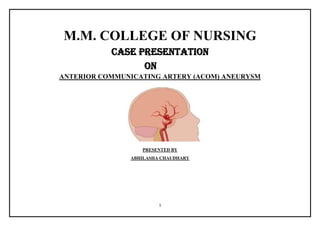
- 1. 1 M.M. COLLEGE OF NURSING CASE PRESENTATION ON ANTERIOR COMMUNICATING ARTERY (ACOM) ANEURYSM PRESENTED BY ABHILASHA CHAUDHARY
- 3. 3 HISTORY TAKING Identification of the patient Patient name- Mustkeen Age / gender- 45 years/ male Religion- Hindu Marital status- Married Occupation- labour (farming) Date of admission -07 march, 2017 Ward- NeuroICU Ip no-170307137 Address -Saharanpur Education- Illiterate Provisional diagnosis- Anterior Communicating artery (ACOM) aneurysm Final diagnosis-Pterional Craniotomy with right ACOM aneurysm clipping Chief complain Complain duration Headache since 2days
- 4. 4 History of present illness Patient was apparently well 5 days back. There was sudden intense headache on mid frontal area for half an hour . patient was taken to nearby hospital of saharnapur and medication was given and CT scan was done but they were not able to clearly diagnose and was referred to Mullana after 1 day of stay on 07 march. In emergency patient had chief complain of headache and Investigations were done along with CT scan and was diagnosed as ACOM aneurysm then patient was admitted to Neuro ICU at 3: 00 pm via emergency. Medical history Past medical history No any history of Hypertension, kidney disesease , heart disease or any other disease. Past surgical history No any surgical history Personal history and habits Hygiene and exercise- Hygiene satisfactorily maintained.Clothes clean. Active and Passive exercise done daily . Activities and hobbies Patient is active and there is no particular habit. Patient can ambulate with the help of support. Sleeping pattern Sleeping pattern is good . patient sleeps 6-7 hr at night and 1-2 hr at day. History of substance use No any history of alcoholism History of smoking(Bidi) since 30 year. Take 10- 12 bidi per day.
- 5. 5 Dietary pattern Dietary pattern is disturbed . patient is on soft diet. Patient take (daliya, porridge, curd, biscuits, milk, tea) Bowel and bladder habit Urine output is normal . patient is on catheterization. Bowel habit disturbed Stool not passed since 5 days. Family history Index Female Male Patient 45 yr 38 yr 19 yr No family history of related illness. Patient has adopted child because of infertility.
- 7. 7 PHYSICAL EXAMINATION General appearance and behaviour Body built- healthy Gait – normal Activity – normal but decreased Appearance – fatigue Vital Temperature-98.6 degree Fahrenheit Pulse -78beats per minute Respiratory rate-24 breaths/minute Blood pressure-120/70 mm of hg NEUROLOGICAL EXAMINATION GLASSGOW COMA SCALE E-4 V-5 M-6 GCS-15/15
- 8. 8 Assessment Findings of physical examination 1)MENTAL STATUS EXAMINATION Observe appearance and movement Posture Gait Motor movements Dress Hygiene Facial expression Speech Observe mood Feelings Expressions Observe thought,process and perceptions Clarity and content Perceptions Observe cognition Level of consciousness Memory Abstract reasoning – Relaxed with shoulder back and both feet stable and was in supine position. Coordinated and smooth with slight shuffling while walking. Smooth, coordinated movements Clothes fit and are appropriate for occasion and weather . Skin clean, nails clean and trimmed Good eye contact, smiles/frowns appropriately Clear with moderate pace Responds appropriately to topic discussed; expresses feelings appropriate to situations. Expresses good feelings about self, others, and life verbalizes positive coping mechanisms (I have a good family and will be back to work once I am fit ) Expresses full and free flowing thoughts during interview Follows directions accurately; perceptions realistic . Oriented to time, place and person(was able to say it’s day time, he is in hospital, and I am sister) Remote Memory was intact( was not able to say exact birthdate and marriage date as patient verbalize that” who remember those dates “ but was able to recall when he had adopted child, where he had worked. Recent memory intact (was able to recall what he had ate day before yesterday and 24 hour activities) Immediate memory intact- ( 1,3,5) able to recall Abstract reasoning intact “pet mae chuhae kudna”- patient replied “vuk lagna”
- 9. 9 Ability make sound judgement Ability to identify similarities – 2)ASSESSMENT OF CRANIAL NERVES Cranial nerve- I(olfactory) CN-II(Optic) CN-III, IV,VI(Oculomotor) Trochlear,Abducens) CN-V(trigeminal) CN-VII(Facial) Nach na jane agan tedha- “ no reply” Normal Judgement (why did you come to hospital?) Patient- “ I had severe headache that’s why to visit the doctor. What is the similarities between bird and aeroplane? Patient- “both fly in sky” What is difference between pen and copy? Patient- pen sae likhtae hae aur copy mae likhte hae” Patient was asked to smell spirit swab with each nostril Unable to identify correct odor. To assess vision Bilateral normal vision with no any diplopia, blurr vision Normal visual field Extraocular movements(Eye moves in a smooth ,coordinated motion in all directions i.e 6 cardinal fields) Pupillary response(pupil equal, round and reactive to light and accommodation) Ptosis, nystagmus absent Corneal reflex(Eyelid blink bilaterally) Identifies light ,cold and sharp touch sensation to forehead , cheeks and chin) On assessing masseter and temporal muscles as client clenches teeth(muscles contract bilaterally) Identifies taste of food correctly Symmetrical facial movements.
- 10. 10 CNVIII(Acoustic) CNIX(glossopharyngeal) CN-X(Vagus) CN-XI(accessory) CN-XII(hypoglossal) 3)MOTOR ASSESSMENT 4)SENSORY NERVE ASSESSMENT 5)CEREBELLAR ASSESSMENT 6)REFLEX ASSESSMENT Not done as patient had bandage over head . Bilateral symmetrical rise of soft palate and uvula, normal gag reflex and identifies correct taste.) Not done (to avoid forceful activity) Symmetrical tongue with smooth outward movement) Relaxed and contracted muscles bilaterally. Normal muscle tone. Power 5/5 bilaterally(normal strength) in upper and lower limb. Primary sensation Identifies area of light ,dull and sharp touch. Cortical and discriminatory sensation Sterogenesis(identifies correct object i.e key) Graphesthesia (identifies correct stroke drawn on palm) Kinesthesia( Identifies correct direction body part is moved) Tap forefinger to thumb And touches each finger to thumb Romberg test,tandem walk,hop on each foot (not done as patient was unable to balance properly) Bicep reflex +2 bilaterally Brachioradialis reflex +2 bilaterally
- 11. 11 BRUDZINSKI SIGN KERNIG SIGN Tricep reflex +2 bilaterally Patellar reflex +2 bilaterally Achilles reflex +2 bilaterally Babinski reflex not normal (fanning out of toes) absent absent 2)RESPIRATORY SYSTEM Assessment Findings after physical examination Inspection Palpation Percussion Auscultation Normal colour(pinkish),no bulging and retraction. Swallow breathing, symmetrical chest movement,symmetrical chest expansion Respiratory rate- 24 breaths/min No pain and tenderness Resonance heard over bilaterally Vesicular sound heard over both lungs
- 12. 12 3)GASTROINTESTINAL SYSTEM Assessment Findings of physical examination Inspection Auscultation Percussion Palpation No any lesion and prominent abdomen vein. Sunken, centrally located umbilicus Contour- round Symmetrical movement No distension present Soft click sound over upper right quadrant 4 per minute) Absent of clicking sound over other quadrant.Decreased bowel sound. Generalized tympany overall 4 quadrant Non tender, no any masses, smooth Liver and spleen not palpable 4)INTEGUMENTARY SYSTEM Assessment Findings of physical examination SKIN NAIL Colour- fair Moisture- Dry Temperature- 98.6 degree Fahrenheit Texture – Rough and dry Turgor – decreased Edema – present over upper limb in right hand and both legs(minimal) Clubbing Colour-yellow and blackish
- 13. 13 HAIR AND SCALP Shape of the skull- normal ,round Scalp – suture over frontal area, Positive findings CN I (olfactory)-was not able to differentiate smell. Decreased bowel sound Suture over frontal area of head Edema present in upper limb(right hand) and minimal present in lower limb.
- 15. 15 ANATOMY CEREBROVASCULAR CIRCULATION The brain receives approximately 750 mL/min of blood, or 15% to 20% of the total resting cardiac output. The brain is supplied by two pairs of arteries: the two internal carotid arteries and the two vertebral arteries. Cerebral circulation is also divided into the anterior and the posterior circulation. The anterior circulationrefers to the common carotids and their distal branches including the internal carotid arteries, the middle cerebral arteries, and the anterior cerebral arteries. The posterior circulation refers to the vertebral arteries, the basilar artery, and the posterior arteries. Circle of Willis The circle of Willis, which is located at the base of the skull, is divided into anterior (carotid portion) and posterior. (vertebrobasilar portion) circulation . The composition of each portion includes these elements: • Middle cerebral arteries, the anterior cerebral arteries, and the anterior communicating artery, which connects the two anterior cerebral arteries • Two posterior cerebral arteries; two posterior communicating arteries connect the middle cerebral arteries with the posterior cerebral arteries, thus uniting the internal carotid system with the vertebral-basilar system.
- 16. 16
- 17. 17 DISEASE PROCESSS CEREBRAL ANEURYSM A cerebral aneurysm is a saccular outpouching of a cerebral artery. Intracranial saccular aneurysms or berry aneurysms account for approximately 80% to 90% of all intracranial aneurysms. These small, berry-like projections occur at arterial bifurcations in the circle of Willis, with other shapes such as pedunculated, sessile, and multilobulated aneurysms occasionally seen. ETIOLOGY Book picture Patient picture Congenital developmental defect Absent Hypertension Absent Atherosclerosis Absent Head trauma Absent Bacterial and fungal infection Absent Cigarette smoking Present (since 30 years,10-12 bidi/day) Alcohol Absent Connective tissue disorder Absent CLASSIFICATION Book picture Patient picture ACCORDING TO SIZE - Small: to 10 mm Small (ACOM-6.7*6.7mm),MCA-4mm) Medium: 10 to 15 mm - Large: 15 to 25 mm - Giant: 25 to 50 mm - Super-giant:larger than 50 mm -
- 18. 18 ACCORDING TO SHAPE AND ETIOLOGY Berry aneurysm(berry or saccular shaped with a neck or stem) ACOM sacular aneurysm is present Fusiform aneurysm(an outpouching of an arterial wall, without a stem) MCA fusiform aneurysm is present) Traumatic aneurysm( aneurysm resulting from a traumatic head injury) - Mycotic (infectious) aneurysm(caused by septic emboli from infections, such as bacterial endocarditis; may lead to aneurysmal formation) - Charcot-Bouchard aneurysm(microscopic aneurysmal formation associated with hypertension; involves the basal ganglia and brainstem) - LOCATION Cerebral aneurysms usually occur at the bifurcations and branches of the large arteries at the base of the brain (circle of Willis). Book picture Patient picture 85% to 95% in carotid system A-com -30% Pos-com-25% MCA-20% Anterior communicating artery (ACOM) and middle cerebral artery(MCA) is is affected. 5-15%in posterior circulation in vertebrobasilar arteries Vertebral artery-10% Basilar artery-5% - 20-30% may have multiple aneurysm -
- 19. 19 SIGN AND SYMPTOMS OF UNRUPTURED ANEURYSM Book picture Patient picture Headache Present Facial pain Absent Neck stiffness and pain Absent Alteration in consciousness Absent Seizure Absent Autonomic disturbances(vomiting,sweating,fever,chills) Absent Visual symptoms (blurring of vision,diplopia) Absent Symptoms asociated with ACOM aneurysm Usually silent Visual field defect Akinetic mutism(neither movement nor speak) Amnestic syndrome Hypothalamic dysfunction Absent Absent Absent Absent Absent Absent Symptoms asociated with MCA aneurysm Aphasia Hemiparesis Hemisensory loss Anosognosia(deficit of self awareness) Visual field defect Absent Absent Absent Absent Absent INVESTIGATION Book picture Patient picture History taking and physical examination Done CT scan Done (F/S/O saccular aneurysm arising from anterior communicating artery with small fusiform aneurysm from left middle cerebral artery(MCA). MRI Not done
- 20. 20 Cerebral angiography saccular aneurysm arising from anterior communicating artery with small fusiform aneurysm from left middle cerebral artery(MCA). Lumbar puncture Not done Transcranial doppler Not done ECG Done (no report(report lost) BLOOD STUDIES Fibronegen and platelet Done White blood cell count(WBC) Done Hematocrit Not done Electrolytes Done Blood gases Done Glucose Done -126 mg/dl OTHER INVESTIGATIONS DONE IN THE PATIENT Clinical parameters Normal value Patient value Remarks LFT(LIVER FUNCTION TEST) ( (Date-07 march,2017 (Date- 08march,2017 Date (10 march,2017) Total bilirubin 0-1.20mg/dl 0.30 - - Normal Direct bilirubin 0-0.20 mg/dl 0.13 - - Normal SGOT(serum glutamic oxaloacetic transaminase) 2-31U/L 62 - - Increased SGPT(serum glutamic pyruvic transaminase) Up to 45 U/L 45 - - Normal Alkaline phosphatase 54-141 U/L 68 - - Normal Total protein 6.60-8.70g/dl 7 - - Normal Albumin 3.50-5.20g/dl 3.4 - - Hypoalbunemia RFT(RENAL FUNCTION TEST) Urea 13-43mg/dl 53 37 - Uremia Creatnine 0.70-1.40mg/dl 0.71 0.75 - Normal Uric acid 3.60-7.70mg/dl 4.4(normal 3.4(decreased) - Sodium 135- 145mmol/L 139 139 138 Normal
- 21. 21 Potassium 3.5-5.5 mmol/L 3.3 Hypokalemia 3.4 Hypokalemia) 4.3 Chloride 98-110 mmol/L 102 102 106 Normal BLEEDING TIME (BT) BT(bleeding time ) Up to 6 min 2.30 2.30 - Normal BLOOD PROTHROMBIN TIME Prothrombin time 11-16sec 18 15.0 - Prothrombin time control 11-16sec 13 13.0 - Normal INR 1.7 1.2 - Normal CLOTTING TIME(CT) 6.30 - DLC(DIFFERENTIAL LEUCOCYTE COUNT) Neutrophils 40-80% 66 72 - Normal Lymphocytes 20-40% 30 24 - Normal Eosinophils 1-6% 04 04 - Normal HAEMOGLOBIN 13-17g/dl 13.6 10.5 - Normal PLATELET COUNT 1.5-4.5 lakh/cumm 209*1000 140*1000 - TLC(Total leucocyte count) 4000- 10000/cumm 8.9*1000 9.4*1000 - Normal HHH(Hbsag, HCV,HIV) - Negative - - Negative CROSS MATCHING - Blood group –O, Rh positive -- - ABG 08/03/2017 09/03/2017 Ph 7.35-7.45 - 7.455 7.47 Pco2 35-45 - 37.9 44.1 PO2 - 187.4 120.4 SO2% 95-100 - 99.7 98.9 Hct - 37 46 Hb - 12.5 15.4 BEecf - 2.9 9.3 BEb - 3.6 9.0
- 22. 22 SBC -- 27.7 32.8 HCO3 22-26 -- 27.0(met.alk) 33.1(met.alk) TCO2 - 28.1 34.4 A - 98.0 90.6 A-aDo2 - 1 1 a/A - 1.9 1.3 CT SCAN 07 march, 2017 There is c/o focal outpouching measuring approz 6.7*6.7 mm in size arising eccentrically from ACOM showing wide neck with e/o saccular aneurysm. It shows intense enhancement on post contrast scan. No c/o non enhancing thrombus seen.no e/o calcification is seen within its walls. A2 segment is seen to arise from the aneurysm and shows normal caliber and enhancement. There is fusiform dilatation of M1 segment of left MCA just before the bifurcation for a segment 4mm. 09 march , 2017 There is a focal hypodense area seen in the right posterior frontal region with associated partial effacement of ipsilateral ventricle s/o infarct. Aneurysm clips noted in right paraclinoid region Extra-axial air seen in b/l frontal reion. A small streak of intraaxial fluid collection seen in right fronto temporal region Craniotomy defect seen in rightfronto tempo parietak region. Retained secretion noted in nasopharynx. Impression – infarct right frontal region with other post op changes as described. 10 march, 2017 Small focal infarct(17*15mm) in right posteriofrontal region with other post op changes as described.
- 23. 23 MANAGEMENT Book picture Patient picture MEDICAL MANAGEMENT Calcium channel blocker(Nimodipine) Given tab.nimodipine 60mg*6hrly Anticonvulsant (phenytoin) Given tab eptoin 100 mg*TDS Stool softner Syp.evuct(lactulose 6 tsp with water HS Corticosteroids Not given Antacid Tab.pantop 40 mg OD Sedatives Not given Analgesic Given (diclofenac 75mg sos) Heparin Not given Replacement of minerals mg,potassium,calcium IV fluid NS/RL 500 ml SURGICAL MANAGEMENT Surgical clipping Rt.pterional craniotomy for ACOM aneurysm clipping (Endovascular treatment)Coiling Not done Ballon remodeling technique Not done Stent placement Not done
- 24. 24 Other medication used for the patient Name of drug Generic name Dose Route Frequency Class Action Supacef Cefuroxime 750mg IV TDS 2nd generation cephalosporin Binds to protein and causes cell wall death of bacteria. Amikacin Amikacin 750mg IV OD Aminoglycoside Inhibits protein synthesis of bacteria esp.gram negative bacteria Justin Diclofenac 75mg IV SOS NSAIDS To control pain(headache) Mannitol Mannitol 100ml IV BD Diuretic (osmotic agents) Decreases cerebral edema and improves CBF PCM Paracetamol Oral SOS Acetaminophen Reduces fever and antinflammatory action Nimodipine nimodipine 60mg Oral QID Calcium channel blocker Prevent vasospasm and cross blood brain barrier. Eptoin Phenytoin 100mg Oral QID Antiepileptic class Given as a prophylaxis to prevent seizure Pantop Pantoprazole 40mg Oral BD Proton pump inhibitor To prevent gastric irritation Supradyn multivitamin Oral BD Multivitamin To replace essential minerals Evict Lactulose 6tsp Oral HS Laxatives prevent constipation and straining at stool, which may leads to increas ICP Dextran dextran 500 ML IV OD Volume expander Highly colloidal starch structure lowers patelet and RBC adhesion. Neb.Duolin Salbutamol+ipratropium bromide 1:3ml Inhaltion TDS Bronchodilator
- 25. 25 Trade name Pharmacological name Action Dose and route Indication Side effects Nursing responsibility Inj. Mannitol MANNITOL Mannitol elevates blood plasma osmolality, resulting in enhanced flow of water from tissues, including the brain and cerebrospinal fluid, into interstitial fluid and plasma. As a result, cerebral edema, elevated intracranial pressure, and cerebrospinal fluid volume and pressure may be reduced. Mannitol may also be used for the promotion of diuresis before irreversible renal failure becomes established Adult Oliguria: 50-100 g as a 5-25% solution. Intracranial/Intraocular pressure: 0.25-2 g/kg as 15-25% solution administered for 30-60 minutes. Children Oliguria: 0.25-2 g/kg as a 15-20% solution for 2-6 hours Intracranial/Intraocular pressure: 1-2 g/kg as a 15-20% solution administered for 30-60 minutes Promotion of diuresis in acute renal failure . Reduction of intracranial pressure and brain mass. Reduction of high intraocular pressure Promotion of urinary excretion of toxic materials Pulmonary congestion, fluid and electrolyte imbalance, acidosis, electrolyte loss, dryness of mouth, thirst, marked diuresis, urinary retention, edema, headache, blurred vision, convulsions, nausea, vomiting Vital signs Intake and output Central venous pressure Signs and symptoms of dehydration (e.g. poor skin turgor, dry skin, fever, thirst) Signs of electrolyte imbalance/deficit (e.g. muscular weakness, paresthesia, numbness, confusion, tingling sensation of extremity and excessive thirst)(for increase ICP) Neurologic status and intracranial pressure readings
- 26. 26 Trade name Pharmacolo gical name Action Dose and route Indication Side effects Nursing responsibility Tab. Eptoin PHENYTOI N central nervous system agent; anticonvulsant; hydration Pregnancy Category: D Reduced voltage, frequency, and spread of electrical discharges within the motor cortex. Adult: PO 15– 18 mg/kg or 1 g loading dose, then 300 mg/d in 1–3 divided doses, may be gradually increased by 100 mg/wk until seizures are controlled IV 15–18 mg/kg or 1 g loading dose, then 100 mg t.i.d. Patient: PO/IV 15–20 mg/kg loading dose, then 5 mg/kg or 250 mg/m2 in 2–3 divided doses. To control tonic- clonic (grand mal) seizures, psychomotor and nonepileptic seizures (e.g., Reye's syndrome, after head trauma). Also used to prevent or treat seizures occurring during or after neurosurgery. Is not effective for absence seizures. CNS: Nystagmus, drowsiness, ataxia, dizziness, mental confusion, tremors, insomnia, headache, seizures. CV: Bradycardia, hypotension, cardiovascular collapse, ventricular fibrillation, conjunctivitis, diplopia, blurred vision. GI: Gingival hyperplasia, nausea, vomiting, constipation, epigastric pain, dysphagia, loss of taste, Continuously monitor vital signs and symptoms during IV infusion and for an hour afterward. Watch for respiratory depression. Constant observation and a cardiac monitor are necessary with older adults or patients with cardiac disease. Observe patient closely for neurologic adverse effects following IV administration. Have on hand oxygen, atropine, vasopressor, assisted ventilation, seizure precaution equipment (mouth gag, nonmetal airway, suction apparatus). Be aware that gingival hyperplasia appears most commonly in patient and adolescents and never occurs in patients without teeth. Make sure patients on prolonged therapy have adequate intake of vitamin D-containing foods and sufficient exposure to sunlight.
- 27. 27 Metabolic: Fever, hyperglycemia, glycosuria, weight gain, edema, transient increase in serum thyrotropic (TSH) level, osteomalacia. Skin: Alopecia, hirsutism (especially in young female); Urogenital: Acute renal failure, Peyronie's disease. Respiratory: Acute pneumonitis, pulmonary fibrosis. Monitor diabetics for loss of glycemic control. Patient & Family Education Be aware that drug may make urine pink or red to red-brown. Report symptoms of fatigue, dry skin, deepening voice when receiving long-term therapy because phenytoin can unmask a low thyroid reserve. Do not alter prescribed drug regimen. Stopping drug abruptly may precipitate seizures and status epilepticus. Trade name Pharmacol ogical name Action Dose and route Indication Side effects Nursing responsibility Inj. AMIKACI Aminoglycosid Adult: IV/IM Bacterial CNS: ataxia, Assess patient for signs and
- 28. 28 Amikacin N SULFATE eantibiotics. 5–7.5 mg/kg loading dose, then 7.5 mg/kg q12h Patient: IV/IM 5–7.5 mg/kg loading dose, then 5 mg/kg q8h or 7.5 mg/kg q12h Neonate: IV/IM 10 mg/kg loading dose, then 7.5 mg/kg q12– 24h Infections. Treatment of serious gram- negative bacillary infections and infections caused by staphylococci when penicillin or other less toxic drugs are contraindicated. vertigo. EENT: ototoxicity (vestibular and cochlear). GU: nephrotoxicity. F and E: hypomagnesemia. MS: muscle paralysis (high parenteral doses). Neuro: increase neuromuscular blockade. Resp: apnea. Misc: hypersensitivity reactions. symptoms of infection prior to and throughout therapy. Instruct patient to continue taking medication around the clock until finished completely, even if feeling better. Keep patient hydrated during therapy. Instruct patient to report signs of hypersensitivity.
- 30. 30 Nursing Diagnosis:- 1. Pain related to meningeal irritation secondary to bloodvessel swelling and surgery. 2. Impaired cerebral perfusion related to decrease blood supply secondary to aneurysm. 3. Risk for ineffective breathing pattern related to post intubation and prolong bedridden 4. Risk of infection related to surgery done. 5. Risk for fluid volume deficit less than body requirement related to disease condition and decrease fluid intakke 6. Anxiety related lack of knowledge regarding disease condition, the need for surgery. 7. Disturbed Body Image related to hair loss, and changes in the structure and function of the boy.
- 31. 31 ASSESSMENT DIAGNOSIS GOAL INTERVENTION RATIONAL EVALUATION Subjective data: - patient looks restless not able to give proper history and gives history of decreased sleep. Objective data:- Restlessness, irritability, changes in vital sign. BP-120/70mmhg Temp.-98.6. f Pulse:- 78beats/min. Resp.:- 22breaths/min. Presence of ACOM aneurysm and MCS aneurysm Impaired cerebral perfusion related to decrease blood supply secondary to aneurysm. Patient will maintain brain function and would not place any sign of sign of ICP. Neurological assessment done every 2 hours to 4 hourly. Assess vital signs every 2 hours to 4 hours, record the irregular breathing and heart rate and rhythm and extent of the pressure pulse. Elevate the head of the bed 30 degrees. Prepare oxygen and suction equipment in place during the period of disturbance of consciousness (LOC). Changes in cranial nerve function showed ICP. Assessment of vital signs are as often as possible will help detect early signs of ICP (such as tachycardia, fluctuating blood pressure, and respiration Cheyne-Stokes) or the signs of the development of ICP. Elevation head in the bed allow gravity to increase the flow of cerebral,will help to decrease ICP. Equipment and suction mucus is required when the patient has seizures or apnea. Patient maintained normal brain functioning .
- 32. 32 ASSESSMENT DIAGNOSIS GOAL INTERVENTION RATIONAL EVALUATION Objective data: - patient has suture on right frontal area Risk of infection related to surgery done. Patient will demonstrate the absence of infection associated with surgical procedure characterized by a temperature less than 100 º F (37.8 º C) and no signs of swelling of the wound incision. Assess the patient's body temperature , decreased LOC, loss of appetite, vomiting, increased white blood cells, and swelling or redness over the surgical site. Monitor patient's body temperature every 4 hours. Review the incision area every 4 hours, see the drainage of fluid from the wound and the swelling. Record the number and type of discharge from the incision wound. Give antibiotics (amikacin,supracef) as directed. This sign gives an indication of infection. Increase in body temperature is an early sign of infection. The position where the head in the right position helps prevent damage to the skin or around the surgical site. Swelling around surgical incision, pus in drainage- may be an early sign of infection in the site Antibiotic which prophylaxis is usually given during surgery and continued at 48 to 72 hours after surgery. Patient demonstrate the absence of infection associated with surgical procedure characterized by a temperature less than 100 º F (37.8 º C) and no signs of swelling of the wound incision. No fluid comes out through incision. .
- 33. 33 ASSESSMENT DIAGNOSIS GOAL INTERVENTION RATIONAL EVALUATION Subjective data:- Family members of patient and patient asking about prognosis , Objective data:- Patient looks anxious and worried Anxiety related to lack of knowledge refarding disease condition. Family members and patient will express less worriness about disease condition and surgery performed. Explaining about disease condition, the basic purpose of the surgery. Also explain the purpose of the various actions recommended diagnostic tests and procedures to be performed. Give peri-operative measures, including: · Fasting · Define an action of intra venous medicines · Transport the patient to the surgery room · Surgery timetable · Long expected surgery · Recovery room · Monitoring vital signs. Tell the family members to help your patient to prepare to stay hospitalization and surgery Give a strengthening of the explanation surgeon. 1. Provide an explanation will help decrease fear and anxiety and increase acceptance of the patient's condition. 2. The explanation of this activity will reassure family members/patient that they should be aware that the patient will live it and help to provide encouragement to participate in preparatory activities, if possible. 3. Family members need time to adjust to the information so they can form the questions and express fears and concerns. 4. Diagrams, videos, books, and discussions may be more appropriate in older patient. 5. Family members and patient often receive too much information in a short time. Repeat explanations to help understanding the patient's condition. The parent’s anxiety is reduced to some extent naw, as they are able to express their doubts about their patient’s condition.
- 34. 34 PROGNOSIS 10/03/2017 11/03/2017 12/03/2017 13/03/2017 14/03/2017 Admission day Post-op day GCS-(EVM) Vitals Temperature Pulse Respiration Blood pressure MAP Spo2 Input/output- RBS Care provided Foley’s catheter CVP line 4rth day 2nd day (E-4,M-6,V-5)-15/15 98.6degree Fahrenheit 78beats/min 24 breath/min 120/70 mm of hg 96%without Oxygen 3840/3700=140+ve balance 107mg/dl Oral care Chest physiotherapy Active exercise 4rth day Patent ,4rth day 5th day 3rd day (E-4,M-6,V-5)-15/15 98degree Fahrenheit 82beats/min 26breath/min 140/80 mm of hg 100%without Oxygen 3350/2800=550+ve balance 127mg/dl Oral care Chest physiotherapy Active exercise 5th day Patent ,5th day 6th day 4 rth day (E-4,M-6,V-5)-15/15 98.8degree Fahrenheit 80beats/min 28breath/min 130/70 mm of hg 98%without Oxygen 2900/2200=700+ve balance 106mg/dl Oral care Chest physiotherapy Active exercise Ambulation Foley’s removed Patent ,6th day 7th day 5th day (E-4,M-6,V-5)-15/15 97.6degree Fahrenheit 76beats/min 24 breath/min 120/70 mm of hg 98%without Oxygen 2750/2600=150+ve balance 110mg/dl Oral care Chest physiotherapy Active exercise Ambulation - Patent ,8th day 8th day 6th day (E-4,M-6,V-5)-15/15 98 degree Fahrenheit 82beats/min 20 breath/min 130/80 mm of hg 98%without Oxygen 2300/2000=300+ve balance 120mg/dl Oral care Chest physiotherapy Active exercise Ambulation - Patent ,9th day
- 35. 35 Drain Diet – Chief complain – Specific instruction- Red in colour(approx.50 ml) Semi solid diet Headache Drain out Removed Semi solid diet Headache,unable to pass stool Iv fluid NS/RL stopped - Normal diet Headache,unable to pass stool CST - Normal diet Headache,unable to pass stool CST - Normal diet Headache,unable to pass stool Added lactulose
- 36. 36 BIBLIOGRAPHY Suddarth’s& Brunner,” textbook of medical surgical nursing”, volume-2,13th edition. wolters-kluwer publishers;2003. page no.1250- 1275. Black JM,Hawks JH, editors.Medical surgical nursing.8th ed.StLouis,Missouri:Mosby,An imprint of Elsevier:2009.P. 1488-1510 Clinical neuroanatomy ,by snell, 7th edition Clinical neuroanatomy by stephen g. Waxman, 25th edtion Neuroanatomical basis of clinical neurology,byorhanarslan DoengesMe,MoorhouseMF,MurrAc,editors.Nursing care plan,9th ed.Philadelphia:FA Davis company;2014 P.75-85