X ray film reading for bone tumour.
•Download as PPTX, PDF•
49 likes•5,094 views

health&medicine

Recommended
More Related Content
What's hot
What's hot (20)
Similar to X ray film reading for bone tumour.
Similar to X ray film reading for bone tumour. (20)
More from Abdellah Nazeer
More from Abdellah Nazeer (20)
Recently uploaded
Recently uploaded (20)
X ray film reading for bone tumour.
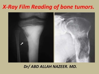
- 1. Dr/ ABD ALLAH NAZEER. MD. X-Ray Film Reading of bone tumors.
- 3. Radiographic appearance of osteochondroma. Lesions are most common in the metaphysis of long bones and may be pedunculated (A) or flat (B).
- 5. Hereditary multiple exostosis (HME).
- 7. Two cases with osteochondroma and malignant transformation
- 9. Enchondroma.
- 11. Enchondroma. Enchondroma with fracture.
- 13. A) Enchondroma of the proximal phalanx of the little finger; (B) Enchondroma at the lower metaphysis of the femur with cartilage calcification- rings and arcs; (C) low grade chondrosarcoma in mid shaft of femur
- 17. a X-ray of right proximal humeral cyst in a nine-year-old boy presenting with pathological fracture. b After two ABMI the cyst showed signs of healing. c Reactivation and enlargement of the cyst with pathological fracture. d After healing of the fracture the cyst became latent.
- 19. (Left) Typical UBC is shown in the humerus, the most common location for this lesion. It is several millimeters from the open growth plate . The metaphysis appears slightly widened , but its diameter does not exceed the width of the growth plate. (Right) Typical UBC is shown in the proximal femur, the 2nd most common location. The lesion extends to the open growth plate and, although it has thinned the cortex , the profile of the bone is not significantly widened.
- 21. Radiograph demonstrates an aneurysmal bone cyst in the hand.
- 23. Aneurysmal bone cyst (ABC) - tibia
- 25. OSTEOID OSTEOMA.
- 29. Imaging characteristics of spinal osteoid Osteoma (OO) and osteoblastoma (OB). Typical examples of a spinal OO (left and middle column) and a spinal OB (right column).
- 31. Osteoblastoma of left L3 and L4 transverse process.
- 37. Core Biopsy taken for Histopathology had features suggestive of giant cell tumor.
- 39. Benign epiphyseal chondroblastoma A) x-ray & CT picture of tibia with a lytic lesion restricted to the epiphysis; (B) X ray of a large chondroblastoma of femoral head (right), transgressing part of epiphyseal plate and causing destruction of sub epiphyseal bone and trochanter.
- 41. Chondrobalstoma.
- 45. Fibrous dysplasia involvement of the right femur.
- 59. Ewing Sarcoma.
- 61. Ewing Sarcoma.
- 63. Ewing Sarcoma.
- 65. Plain films show an expansile lesion. The MR shows cortical destruction and soft tissue extension. Diagnosis: Chondrosarcoma, proximal radius
- 67. Chondrosarcoma of the proximal phalanx of the ring finger
- 69. Peripheral chondrosarcoma in the femur.
- 73. Osteosarcoma with Codman’s triangle.
- 75. Osteosarcoma
- 78. Lymphoma. (A) Anteroposterior and oblique radiographs of the right humerus of a 20-year-old man show a long lesion exhibiting permeative and moth-eaten type of bone destruction. Periosteal reaction is secondary to the pathologic fracture. (B) Sagittal reformatted CT image demonstrates endosteal scalloping and early callus formation at the site of a pathologic fracture (arrows).
- 80. Metastasis
- 81. Thank You.