SlideShare a Scribd company logo
1 of 50
Radiological Imaging in Spinal Dysraphism.
Dr/ ABD ALLAH NAZEER. MD.
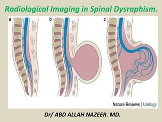
Spinal dysraphism is a broad term given to a group of anomalies
where there are malformations in the dorsum of the embryo.
Neural tube defects come under this group as well.
Pathology
There is often abnormal fusion of the midline embryonic neural,
vertebral and mesenchymal structures.
Sub types
Spinal dysraphism can be broadly divided to into two different
pathological entities:
open spinal dysraphism
meningocoele
myelomeningocoele
closed spinal dysraphism
dorsal dermal sinus
lipomyelomeningcoele
diastematomyelia
neurenteric cyst
thickened filum terminale
Prevalence
The estimated incidence of spinal dysraphism is about 1–3/1000 live births. The
prevalence of spinal dysraphism has been in decline the world over in the last few
decades due to the better nutrition for women, folic acid supplementation, improved
antenatal care and high-resolution ultrasound for prenatal screening and biochemical
markers.
Embryology
Embryogenesis in the first 2 months of gestation can be divided into 23 stages. Neural
plate is formed in stage 8 around the 18th day, followed by neural folds and their
fusion. The expansion of the neural tube and subsequent closure is completed by day
28. Open defects occur when the caudal neuropore fails to close. The secondary
neurulation sets the spinal cord formation. Defects at this stage result in occult
dysraphism connecting the epidermis and the mesenchymal tissues, leading to variety
of anomalies and tethered cord.
Failure of primary neurulation leads to open dysraphism posing the risks of CSF
leakage and exposure of neural placode. Extent and severity of neurological deficit
depends on the degree of malformation of the neural placode and also the level of the
defect. The higher the level, usually worse is the prognosis. A spectrum of neurological
abnormalities like hydrocephalus, Chiari malformation, syrinx, gyral malformations,
skeletal malformations and uro-vesical defects can be associated. In occult
dysraphism, the overlying skin is intact but the spinal cord is anchored to various
tissues starting from skin, subcutaneous tissue, adipose tissue or cartilage.
Etiology
Spina bifida has a multifactorial causation, involving both genetic and
environmental. Recent information stressed on the importance of maternal
nutrition and folic acid supplementation which has contributed to its
reduction. In India, certain regional factors and consanguinity seem to play
a role [hospital-based epidemiological study from NIMHANS (unpublished).
Symptomatology
Open dysraphism presents with a swelling over the back which is noticed at
birth. Symptoms are primarily referable to CSF leak or the exposed spinal
cord. Since the skin over the swelling is poorly developed, it usually gives way
during labor, resulting in CSF leak, contamination and meningitis. Defects
predominantly involve thoracolumbar, lumbosacral, lumbar, thoracic, sacral
and cervical in the order of frequency of occurrence. Incidence of high cervical
lesions is about 3.9%. Neurological deficits include motor, sensory and
sphincter dysfunction, depending upon the severity and level. In severe cases,
hypotonic areflexic limbs, sphincter atonia with rectal prolapse may be
associated. Chiari malformation presents with lower brainstem and lower
cranial nerve dysfunction. The presence of large head usually indicates
hydrocephalus. The associated skeletal abnormalities are kyphosis, scoliosis,
and deformities of the long bones and feet, hemivertebrae, defective ribs.
Classification of spinal dysraphism.
Radiography
With spinal dysraphism, radiographs may show structural
vertebral anomalies such as hemivertebra, butterfly
vertebra, or incomplete fusion of posterior elements; it
does not allow imaging of the spinal cord. Radiographs of
the vertebrae provide information for early evaluation of
infants born with myelomeningocele. Congenital spinal
deformities need to be tracked closely. Paralytic spinal
deformities require imaging based on clinical examination
findings; these deformities should be followed up
frequently during times of rapid growth. Plain radiographs
of patients with myelomeningocele demonstrate
incomplete fusion of posterior elements and increased
interpedicular distance.
Posteroanterior (PA) chest radiograph shows
defects of the laminae of the lower cervical spine.
Plain abdominal radiograph in the
same patient as in the previous image
shows spina bifida occulta of S1.
Myelograms in a 5-year-old patient show the dorsal region of the spine
and an anterior thoracic meningocele. Note the gross dorsal kyphosis.
Myelograms in a 4-year-old patient
show the lumbosacral region; a long,
tethered cord; and diastematomyelia.
Right, plain radiograph of the lumbar spine
shows diastematomyelia. Left, myelogram
in the same patient shows a filling defect at
the level of diastematomyelia.
Left, plain anteroposterior (AP) radiograph
of the lumbar spine shows spina bifida
occulta. Right, myelogram of the same
patient shows a thick tethered cord
Left, anteroposterior (AP) plain radiograph of the
lumbar spine shows a defect within the laminae of
S1 and S2. Right, myelograms in the same patient
show a markedly thickened, low tethered cord.
Plain anteroposterior (AP) lumbar spinal
radiograph in a 7-year-old patient shows
a defect within the laminae of L4-5 and
S1. Note the diastematomyelia.
Myelograms in the same patient as in the
previous image show a low, tethered cord.
Note also the diastematomyelia.
Left, plain radiograph of the lumbar spine
shows bony defects in the laminae of L2 to S1.
Right, myelogram shows a split cord.
Plain radiographs show
posterior scalloping.
Ultrasonongraphy:
Spinal sonography is usually not possible after the age of 6 months except
in cases of a persistent posterior spinal defect; in such cases, sonography
may be performed at any age In the neonate, the cord appears as a
tubular hypoechoic structure with hyperechoic walls. The central canal is
hyperechoic. The cord position is dependent. The subarachnoid space
around the cord is echo poor. In the neonate, the conus medullaris is
smooth and tapering and lies above the middle of L2; the range varies from
D10 to L2-L3. The cauda equina is seen as echogenic lines surrounding a
hyperechoic filum terminale with dependent positioning.
The normal filum terminale is 1.0-1.5 mm in diameter. The vertebral bodies
are seen as echogenic segmental structures lying anterior to the spinal
cord. In the normal infant, the cord lies one third to one half the distance
between the anterior and posterior walls of the spinal canal. There is
normal pulsatile movement of the cord. When the cord is viewed axially,
it appears round to oval and is surrounded by the fluid-filled subarachnoid
space. The cord is fixed within the spinal canal by the dentate ligaments,
which pass laterally from the cord. Below L2, the echogenic nerve roots are
identified with a vertical or oblique orientation
The spinal cord may be depicted throughout its length, allowing visualization of
the conus and free movement of the nerve roots. An absence of normal
transmitted pulsations and a lack of free movement of nerve roots on sonograms
suggest a tethered cord. In cases in which there is a low tethered cord, the conus is
low and the spinal cord is displaced dorsally. There is lack of normal cord
pulsatility, and the filum terminale is thickened to over 2 mm. The thickened filum
terminale may be fibrous or lipomatous. An abnormal cord may lie in a dorsal
position rather than being dependent. The clinical significance of a low cord
without tethering is unknown.
In a newborn term infant, the normal conus usually lies above the level of the mid
L2. In cases in which there is a skin-covered back mass, the contents of the mass
may be characterized. Axial spinal sonograms readily show the 2 hemicords and
the echogenic spur in cases of diastematomyelia. In cases of diastematomyelia,
sonography may show the spur. A dorsal dermal sinus may be depicted as an
echogenic tract deep to a hole in the skin.
It may be difficult to be confident in the sonographic findings if the tract
communicates with the spinal canal. However, a low-lying cord suggests tethering
by intraspinal extension of the sinus; sonograms may show abnormal echogenicity
at the depth of the tract, suggesting lipoma or dermoid. Alternatively, images
may show matted nerve roots, caused by arachnoiditis. No conus may be
identified if the cord terminates in a lipoma.
Antenatal ultrasonogram shows a lumbar meningocele.
Antenatal ultra sonogram shows a lemon sign and a banana sign.
Computed Tomography
CT myelography demonstrates splitting of the cord and, in some cases,
splitting of the meningeal sheath. In addition, other bony anomalies, such as
an intervertebral septum, and aberrant fibroneural bands may be depicted.
CT myelography allows better definition of cord expansion or deformity than
can be achieved by conventional myelography. In the case of intrinsic cord
tumors, repeat CT after 24 hours reveals intramedullary contrast
enhancement if associated syringomyelia is present. MRI is the imaging
procedure of choice.
Spinal lipomas, with their fatty tissue contents, are identifiable both on CT
scans and on MRIs. On CT scans, fatty tissue has a strongly hypoattenuating
appearance that may best be appreciated in comparison with CSF on soft
tissue windows and in comparison with air on bone windows. On MRIs, fatty
tissue is strongly hyperintense on images obtained with both short and long
repetition times. Newer techniques of fat suppression, such as short-tau
inversion recovery (STIR) imaging, may resolve any doubts. The 2 techniques
are complementary: On the one hand, CT better shows osseous abnormalities
associated with the lipomas; on the other, MRI is preferred because it allows
better depiction of detail and contrast resolution of soft tissues.
Axial CT scans through the lumbar spine with bone window setting in the same
patient as in the previous images show a bony bar due to diastematomyelia.
Axial CT scans through the upper lumbar spine show a split cord.
Axial CT scans through the lumbosacral junction shows absence of the posterior
spinal elements at L5-S1. Note sclerosis of the laminae and the wide spinal canal.
Spine Bifida
Dermal sinus.
T1- and T2-weighted sagittal MRIs of the cervical and dorsal
spine in the same patient as in the previous 3 images show
evidence of previous surgery for cervical meningocele. Note
the associated congenital fusion of C5 and C6.
Axial T2-weighted MRIs of the cervical spine in the same
patient as in the previous 4 images show a large spinal
canal, evidence of previous surgery, and a split cord.
Sagittal MRIs of the lumbar spine
show diastematomyelia. Note the
congenital fusion of L1 and L2.
Axial MRIs in the same patient as in the
previous image shows a hypo intense bar,
which is in an anteroposterior location because
of diastematomyelia that splits the cord.
T1- and T2-weighted sagittal MRIs of the lumbar spine show an
extradural spinal lipoma communicating with the subcutaneous fat.
T1- and T2-weighted
sagittal MRIs of the
lumbar spine show
an intradural sacral
lipoma. Note the
scalloping of the
posterior sacral
vertebral bodies
and the syrinx.
Terminal lipoma
Meningocele.
Tethered cord
Tethered cord.
Lipomyelomeningoceles.
Lipomyelomeningocele
MRI sagittal T2 weighted image showing myelocystocele with
hydromyelia in mid dorsal area with SCM Type1 at D7-8, D10, L2-3.
Isolated Terminal Myelocystocele:
Axial T1-weighted (A) and T2-weighted (B) MRI scans show a similar
depiction as sonography. Arrows point to the deformed and displaced
spinal cord. M = meningocele; S = syringocele; Vb = vertebral body.
Congenital spinal dysraphism with meningomyelocele and lipomas.
(A-C) and corresponding occult spinal dysraphism detected by sagittal, T1-weighted magnetic resonance imaging (MRI) studies of the
spinal cord (D-G). A, Sacral lipoma and deviated gluteal furrow (DGF); B, lumbar port-wine stain, lipoma, dermal sinus, and DGF; and C,
dorsal and lumbar unclassified hamartomas. D, Lipoma of the conus (arrow); E, dermal sinus (arrow); F, top of the lipoma of the filum
terminale (upper arrow) and fistula (lower arrow); G, multiple lipomas of the thoracic cord (upper arrow) and posterior conus (lower arrow).
T2-weighted sagittal (a) and coronal (b) magnetic resonance imaging of dorso-lumbar spine shows a
hemivertebrae from D10 to D12 level along with a bony spur at D11. Tethering of cord and
meningomyelocele is also visible. Continuation of left hemicord is appreciable in coronal view (b)
T2 weighted axial image shows a bony spur dividing the cord into two halves (a and b).
Herniation of right hemicord, meninges and cerebrospinal fluid is noted through the defect
in posterior element, forming a large hemi-meningomyelocele (c). Two hemicords are
visible below the level of herniation (d) which reunites at D12 level to form a single cord(e).
Sagittal MRI sequences (a) T1-weighted, (b) T2-weighted, (c) MR myelography, and (d) 3D-CISS
sequence; arrow points to the fluid containing hydromyelic cord herniating into the meningocele sac
Sagittal T2-weighted image with fat saturation (A), T1-weighted image (B), and contrast-enhanced T1-
weighted image (C) of L-spine MRI show hypoplastic S2 vertebra with agenesis of lower sacrum and coccyx.
The spinal cord terminates above L1 with club-shaped (chisel-shaped), blunted and angulated caudal end.
Separation of the anterior and posterior spinal roots of the cauda equina is also noted. Distended urinary
bladder with coarse trabeculation pattern is noted due to neurogenic bladder. Coronal T2-weighted
image (D) of L-spine MRI shows both iliac bones articulated with S1. MRI = magnetic resonance imaging.
MRI Sagittal T2w images of lumbo sacral spine shows: Hypoplastic S1 and S2 with failure of formation
of S3 and onwards. Cord ending at a higher level at D12-L1 with 'wedge' shaped termination. No cord
tethering. No terminal cord cyst or syrinx. Imaging diagnosis : Caudal Regression, Group I.
Axial T2-weighted MR images demonstrating spina
bifida occulta (left) with failure of fusion of the
posterior elements at the midline, and the lipoma-
placode interface (right, arrow).
(left) and T2-weighted (right) MR images
demonstrating a lipomyelomeningocele. Note
the lipomatous component extending in the
intradural and epidural spaces (arrows) as well
as the subcutaneous space.
Neuroenteric cyst MRI.
Persistent terminal ventricle.
Thank You.

More Related Content

What's hot

Posterior fossa malformations
Posterior fossa malformationsPosterior fossa malformations
Posterior fossa malformationsArchana Koshy
 
Imaging in Skull base
Imaging in Skull baseImaging in Skull base
Imaging in Skull baseRakesh Ca
 
Musculoskeletal radiology revision notes
Musculoskeletal radiology revision notesMusculoskeletal radiology revision notes
Musculoskeletal radiology revision notesTONY SCARIA
 
Presentation1.pptx, radiological imaging of skeletal dysplasia
Presentation1.pptx, radiological imaging of skeletal dysplasiaPresentation1.pptx, radiological imaging of skeletal dysplasia
Presentation1.pptx, radiological imaging of skeletal dysplasiaAbdellah Nazeer
 
54 DAVID SUTTON PICTURES NEURORADIOLOGY OF SPINE
54 DAVID SUTTON PICTURES NEURORADIOLOGY OF SPINE54 DAVID SUTTON PICTURES NEURORADIOLOGY OF SPINE
54 DAVID SUTTON PICTURES NEURORADIOLOGY OF SPINEDr. Muhammad Bin Zulfiqar
 
Presentation1, radiological imaging of developmental dysplasia of the hip joint.
Presentation1, radiological imaging of developmental dysplasia of the hip joint.Presentation1, radiological imaging of developmental dysplasia of the hip joint.
Presentation1, radiological imaging of developmental dysplasia of the hip joint.Abdellah Nazeer
 
Pineal region masses - radiology
Pineal region masses - radiologyPineal region masses - radiology
Pineal region masses - radiologyDr. Mohit Goel
 
Presentation1.pptx. radiological imaging of epilepsy.
Presentation1.pptx. radiological imaging of epilepsy.Presentation1.pptx. radiological imaging of epilepsy.
Presentation1.pptx. radiological imaging of epilepsy.Abdellah Nazeer
 
Radiology Spotters mixed collection ppt
Radiology Spotters mixed collection pptRadiology Spotters mixed collection ppt
Radiology Spotters mixed collection pptDr pradeep Kumar
 
radiology Spotters mixed bag
radiology Spotters mixed bagradiology Spotters mixed bag
radiology Spotters mixed bagAnish Choudhary
 
Larynx anatomy CT and MRI
Larynx anatomy CT and MRILarynx anatomy CT and MRI
Larynx anatomy CT and MRIDr. Mohit Goel
 
CRANIOVERTEBRAL JUNCTION ANATOMY, CRANIOMETRY, ANAMOLIES AND RADIOLOGY dr sum...
CRANIOVERTEBRAL JUNCTION ANATOMY, CRANIOMETRY, ANAMOLIES AND RADIOLOGY dr sum...CRANIOVERTEBRAL JUNCTION ANATOMY, CRANIOMETRY, ANAMOLIES AND RADIOLOGY dr sum...
CRANIOVERTEBRAL JUNCTION ANATOMY, CRANIOMETRY, ANAMOLIES AND RADIOLOGY dr sum...SUMIT KUMAR
 
Presentation1.pptx, supratentorial brain tumour
Presentation1.pptx, supratentorial brain tumourPresentation1.pptx, supratentorial brain tumour
Presentation1.pptx, supratentorial brain tumourAbdellah Nazeer
 
Presentation2, radiological imaging of phakomatosis.
Presentation2, radiological imaging of phakomatosis.Presentation2, radiological imaging of phakomatosis.
Presentation2, radiological imaging of phakomatosis.Abdellah Nazeer
 
Radiology Spots PPT- 2 by Dr Chandni Wadhwani
Radiology Spots PPT- 2 by Dr Chandni WadhwaniRadiology Spots PPT- 2 by Dr Chandni Wadhwani
Radiology Spots PPT- 2 by Dr Chandni WadhwaniChandni Wadhwani
 
Presentation1, radiological imaging of undescended testis.
Presentation1, radiological imaging of undescended testis.Presentation1, radiological imaging of undescended testis.
Presentation1, radiological imaging of undescended testis.Abdellah Nazeer
 
Presentation1, radiological film reading of knee joint.
Presentation1, radiological film reading of knee joint.Presentation1, radiological film reading of knee joint.
Presentation1, radiological film reading of knee joint.Abdellah Nazeer
 
Imaging in spinal dysraphism
Imaging in spinal dysraphismImaging in spinal dysraphism
Imaging in spinal dysraphismPooja Saji
 
Presentation1, radiological imaging of anal carcinoma.
Presentation1, radiological imaging of anal carcinoma.Presentation1, radiological imaging of anal carcinoma.
Presentation1, radiological imaging of anal carcinoma.Abdellah Nazeer
 

What's hot (20)

Posterior fossa malformations
Posterior fossa malformationsPosterior fossa malformations
Posterior fossa malformations
 
Imaging in Skull base
Imaging in Skull baseImaging in Skull base
Imaging in Skull base
 
Musculoskeletal radiology revision notes
Musculoskeletal radiology revision notesMusculoskeletal radiology revision notes
Musculoskeletal radiology revision notes
 
Presentation1.pptx, radiological imaging of skeletal dysplasia
Presentation1.pptx, radiological imaging of skeletal dysplasiaPresentation1.pptx, radiological imaging of skeletal dysplasia
Presentation1.pptx, radiological imaging of skeletal dysplasia
 
54 DAVID SUTTON PICTURES NEURORADIOLOGY OF SPINE
54 DAVID SUTTON PICTURES NEURORADIOLOGY OF SPINE54 DAVID SUTTON PICTURES NEURORADIOLOGY OF SPINE
54 DAVID SUTTON PICTURES NEURORADIOLOGY OF SPINE
 
Presentation1, radiological imaging of developmental dysplasia of the hip joint.
Presentation1, radiological imaging of developmental dysplasia of the hip joint.Presentation1, radiological imaging of developmental dysplasia of the hip joint.
Presentation1, radiological imaging of developmental dysplasia of the hip joint.
 
Pineal region masses - radiology
Pineal region masses - radiologyPineal region masses - radiology
Pineal region masses - radiology
 
Presentation1.pptx. radiological imaging of epilepsy.
Presentation1.pptx. radiological imaging of epilepsy.Presentation1.pptx. radiological imaging of epilepsy.
Presentation1.pptx. radiological imaging of epilepsy.
 
Radiology Spotters mixed collection ppt
Radiology Spotters mixed collection pptRadiology Spotters mixed collection ppt
Radiology Spotters mixed collection ppt
 
radiology Spotters mixed bag
radiology Spotters mixed bagradiology Spotters mixed bag
radiology Spotters mixed bag
 
Larynx anatomy CT and MRI
Larynx anatomy CT and MRILarynx anatomy CT and MRI
Larynx anatomy CT and MRI
 
CRANIOVERTEBRAL JUNCTION ANATOMY, CRANIOMETRY, ANAMOLIES AND RADIOLOGY dr sum...
CRANIOVERTEBRAL JUNCTION ANATOMY, CRANIOMETRY, ANAMOLIES AND RADIOLOGY dr sum...CRANIOVERTEBRAL JUNCTION ANATOMY, CRANIOMETRY, ANAMOLIES AND RADIOLOGY dr sum...
CRANIOVERTEBRAL JUNCTION ANATOMY, CRANIOMETRY, ANAMOLIES AND RADIOLOGY dr sum...
 
Presentation1.pptx, supratentorial brain tumour
Presentation1.pptx, supratentorial brain tumourPresentation1.pptx, supratentorial brain tumour
Presentation1.pptx, supratentorial brain tumour
 
Renal doppler
Renal dopplerRenal doppler
Renal doppler
 
Presentation2, radiological imaging of phakomatosis.
Presentation2, radiological imaging of phakomatosis.Presentation2, radiological imaging of phakomatosis.
Presentation2, radiological imaging of phakomatosis.
 
Radiology Spots PPT- 2 by Dr Chandni Wadhwani
Radiology Spots PPT- 2 by Dr Chandni WadhwaniRadiology Spots PPT- 2 by Dr Chandni Wadhwani
Radiology Spots PPT- 2 by Dr Chandni Wadhwani
 
Presentation1, radiological imaging of undescended testis.
Presentation1, radiological imaging of undescended testis.Presentation1, radiological imaging of undescended testis.
Presentation1, radiological imaging of undescended testis.
 
Presentation1, radiological film reading of knee joint.
Presentation1, radiological film reading of knee joint.Presentation1, radiological film reading of knee joint.
Presentation1, radiological film reading of knee joint.
 
Imaging in spinal dysraphism
Imaging in spinal dysraphismImaging in spinal dysraphism
Imaging in spinal dysraphism
 
Presentation1, radiological imaging of anal carcinoma.
Presentation1, radiological imaging of anal carcinoma.Presentation1, radiological imaging of anal carcinoma.
Presentation1, radiological imaging of anal carcinoma.
 

Viewers also liked

Imaging of congenital anomalies of spine and spinal cord
Imaging of congenital  anomalies of spine and spinal cord Imaging of congenital  anomalies of spine and spinal cord
Imaging of congenital anomalies of spine and spinal cord charusmita chaudhary
 
Presentation1.pptx, radiological imaging of congenital anomalies of the spine...
Presentation1.pptx, radiological imaging of congenital anomalies of the spine...Presentation1.pptx, radiological imaging of congenital anomalies of the spine...
Presentation1.pptx, radiological imaging of congenital anomalies of the spine...Abdellah Nazeer
 
Congenital Anomalies Of Spine And Spinal Cord
Congenital Anomalies Of Spine And Spinal CordCongenital Anomalies Of Spine And Spinal Cord
Congenital Anomalies Of Spine And Spinal CordRoshan Valentine
 
Spinal dysraphism
Spinal dysraphismSpinal dysraphism
Spinal dysraphismViral Patel
 
Spinal dysraphism
Spinal dysraphismSpinal dysraphism
Spinal dysraphismairwave12
 
Radiological findings of congenital anomalies of the spine and spinal cord
Radiological findings of congenital anomalies of the spine and spinal cordRadiological findings of congenital anomalies of the spine and spinal cord
Radiological findings of congenital anomalies of the spine and spinal cordDr. Armaan Singh
 
Imaging of spinal dysraphism
Imaging of spinal dysraphismImaging of spinal dysraphism
Imaging of spinal dysraphismMuthu Magesh
 
23 congenital anomalies of the spine on magnetic
23 congenital anomalies of the spine on magnetic23 congenital anomalies of the spine on magnetic
23 congenital anomalies of the spine on magneticDr. Muhammad Bin Zulfiqar
 
Mesenteric ischemia presentation by Dr.NOSHI Capital Hospital Islamabad Paki...
Mesenteric ischemia  presentation by Dr.NOSHI Capital Hospital Islamabad Paki...Mesenteric ischemia  presentation by Dr.NOSHI Capital Hospital Islamabad Paki...
Mesenteric ischemia presentation by Dr.NOSHI Capital Hospital Islamabad Paki...drfarhanali2008
 
GEMC: Mesenteric Ischemia: Resident Training
GEMC: Mesenteric Ischemia: Resident Training GEMC: Mesenteric Ischemia: Resident Training
GEMC: Mesenteric Ischemia: Resident Training Open.Michigan
 
Advanced usg lounge gynaecology imaging series
Advanced usg lounge gynaecology imaging seriesAdvanced usg lounge gynaecology imaging series
Advanced usg lounge gynaecology imaging seriesRitesh Mahajan
 
Acute Mesenteric Ischaemia: An Abdominal Calamity
Acute Mesenteric Ischaemia: An Abdominal CalamityAcute Mesenteric Ischaemia: An Abdominal Calamity
Acute Mesenteric Ischaemia: An Abdominal CalamityKETAN VAGHOLKAR
 
Ultrasound of spinal cord in neonates Dr. Muhammad Bin Zulfiqar
Ultrasound of spinal cord in neonates Dr. Muhammad Bin ZulfiqarUltrasound of spinal cord in neonates Dr. Muhammad Bin Zulfiqar
Ultrasound of spinal cord in neonates Dr. Muhammad Bin ZulfiqarDr. Muhammad Bin Zulfiqar
 
Neonatal spine ultrasound...normal and abnormal findings
Neonatal spine ultrasound...normal and abnormal findingsNeonatal spine ultrasound...normal and abnormal findings
Neonatal spine ultrasound...normal and abnormal findingsAhmed Bahnassy
 
ACUTE MESENTERIC ISCHAEMIA
ACUTE MESENTERIC ISCHAEMIAACUTE MESENTERIC ISCHAEMIA
ACUTE MESENTERIC ISCHAEMIAArkaprovo Roy
 
Mesenteric ischemia
Mesenteric ischemiaMesenteric ischemia
Mesenteric ischemiakrisshk1989
 
Congenital anomalies and Normal skeletal variants- Cervical spine
Congenital anomalies and Normal skeletal variants-  Cervical spineCongenital anomalies and Normal skeletal variants-  Cervical spine
Congenital anomalies and Normal skeletal variants- Cervical spineSanal Kumar
 
Embyrogenesis of the CNS and its disorders
Embyrogenesis of the CNS and its disordersEmbyrogenesis of the CNS and its disorders
Embyrogenesis of the CNS and its disordersAmr Hassan
 

Viewers also liked (20)

Imaging of congenital anomalies of spine and spinal cord
Imaging of congenital  anomalies of spine and spinal cord Imaging of congenital  anomalies of spine and spinal cord
Imaging of congenital anomalies of spine and spinal cord
 
Presentation1.pptx, radiological imaging of congenital anomalies of the spine...
Presentation1.pptx, radiological imaging of congenital anomalies of the spine...Presentation1.pptx, radiological imaging of congenital anomalies of the spine...
Presentation1.pptx, radiological imaging of congenital anomalies of the spine...
 
Congenital Anomalies Of Spine And Spinal Cord
Congenital Anomalies Of Spine And Spinal CordCongenital Anomalies Of Spine And Spinal Cord
Congenital Anomalies Of Spine And Spinal Cord
 
Spinal dysraphism
Spinal dysraphismSpinal dysraphism
Spinal dysraphism
 
Spinal dysraphism
Spinal dysraphismSpinal dysraphism
Spinal dysraphism
 
Radiological findings of congenital anomalies of the spine and spinal cord
Radiological findings of congenital anomalies of the spine and spinal cordRadiological findings of congenital anomalies of the spine and spinal cord
Radiological findings of congenital anomalies of the spine and spinal cord
 
Imaging of spinal dysraphism
Imaging of spinal dysraphismImaging of spinal dysraphism
Imaging of spinal dysraphism
 
23 congenital anomalies of the spine on magnetic
23 congenital anomalies of the spine on magnetic23 congenital anomalies of the spine on magnetic
23 congenital anomalies of the spine on magnetic
 
Congenital Diseases of the Spine
Congenital Diseases of the SpineCongenital Diseases of the Spine
Congenital Diseases of the Spine
 
Mesenteric ischemia presentation by Dr.NOSHI Capital Hospital Islamabad Paki...
Mesenteric ischemia  presentation by Dr.NOSHI Capital Hospital Islamabad Paki...Mesenteric ischemia  presentation by Dr.NOSHI Capital Hospital Islamabad Paki...
Mesenteric ischemia presentation by Dr.NOSHI Capital Hospital Islamabad Paki...
 
GEMC: Mesenteric Ischemia: Resident Training
GEMC: Mesenteric Ischemia: Resident Training GEMC: Mesenteric Ischemia: Resident Training
GEMC: Mesenteric Ischemia: Resident Training
 
Advanced usg lounge gynaecology imaging series
Advanced usg lounge gynaecology imaging seriesAdvanced usg lounge gynaecology imaging series
Advanced usg lounge gynaecology imaging series
 
Acute Mesenteric Ischaemia: An Abdominal Calamity
Acute Mesenteric Ischaemia: An Abdominal CalamityAcute Mesenteric Ischaemia: An Abdominal Calamity
Acute Mesenteric Ischaemia: An Abdominal Calamity
 
Mesenteric ischemia
Mesenteric ischemia Mesenteric ischemia
Mesenteric ischemia
 
Ultrasound of spinal cord in neonates Dr. Muhammad Bin Zulfiqar
Ultrasound of spinal cord in neonates Dr. Muhammad Bin ZulfiqarUltrasound of spinal cord in neonates Dr. Muhammad Bin Zulfiqar
Ultrasound of spinal cord in neonates Dr. Muhammad Bin Zulfiqar
 
Neonatal spine ultrasound...normal and abnormal findings
Neonatal spine ultrasound...normal and abnormal findingsNeonatal spine ultrasound...normal and abnormal findings
Neonatal spine ultrasound...normal and abnormal findings
 
ACUTE MESENTERIC ISCHAEMIA
ACUTE MESENTERIC ISCHAEMIAACUTE MESENTERIC ISCHAEMIA
ACUTE MESENTERIC ISCHAEMIA
 
Mesenteric ischemia
Mesenteric ischemiaMesenteric ischemia
Mesenteric ischemia
 
Congenital anomalies and Normal skeletal variants- Cervical spine
Congenital anomalies and Normal skeletal variants-  Cervical spineCongenital anomalies and Normal skeletal variants-  Cervical spine
Congenital anomalies and Normal skeletal variants- Cervical spine
 
Embyrogenesis of the CNS and its disorders
Embyrogenesis of the CNS and its disordersEmbyrogenesis of the CNS and its disorders
Embyrogenesis of the CNS and its disorders
 

Similar to Presentation1.pptx, radiological imaging of spinal dysraphism.

The fetal musculoskeletal system
The fetal musculoskeletal systemThe fetal musculoskeletal system
The fetal musculoskeletal systemVrishit Saraswat
 
USG Spine.pptx
USG Spine.pptxUSG Spine.pptx
USG Spine.pptxappleforu
 
FETAL SKELETAL ANOMALIES GROUP 3.pptx
FETAL SKELETAL ANOMALIES GROUP 3.pptxFETAL SKELETAL ANOMALIES GROUP 3.pptx
FETAL SKELETAL ANOMALIES GROUP 3.pptxDeogratiusGivenOkodi
 
Spinal dysraphism adnan al banna
Spinal dysraphism adnan al bannaSpinal dysraphism adnan al banna
Spinal dysraphism adnan al bannaadnan al-banna
 
Cranio-vertrable junction anamolies
Cranio-vertrable junction anamoliesCranio-vertrable junction anamolies
Cranio-vertrable junction anamoliesAbhay Mange
 
Cv Junction Anomaly
Cv Junction AnomalyCv Junction Anomaly
Cv Junction Anomalyrajasekar
 
Ultrasound image gallery
Ultrasound image galleryUltrasound image gallery
Ultrasound image galleryRitesh Mahajan
 
1- Why isn't Jamie allowed unlimited use of aspirin for pain- 2- Why d.docx
1- Why isn't Jamie allowed unlimited use of aspirin for pain- 2- Why d.docx1- Why isn't Jamie allowed unlimited use of aspirin for pain- 2- Why d.docx
1- Why isn't Jamie allowed unlimited use of aspirin for pain- 2- Why d.docxKevinjrHWatsono
 
Vgt voorjaar2010 kind_deel2
Vgt voorjaar2010 kind_deel2Vgt voorjaar2010 kind_deel2
Vgt voorjaar2010 kind_deel2vgtrad
 
Sonographic evaluation of msk dysplasia Dr. Muhammad Bin Zulfiqar
Sonographic evaluation of msk dysplasia Dr. Muhammad Bin ZulfiqarSonographic evaluation of msk dysplasia Dr. Muhammad Bin Zulfiqar
Sonographic evaluation of msk dysplasia Dr. Muhammad Bin ZulfiqarDr. Muhammad Bin Zulfiqar
 
POTT’S SPINE-1676656384.pptx
POTT’S  SPINE-1676656384.pptxPOTT’S  SPINE-1676656384.pptx
POTT’S SPINE-1676656384.pptxMisStrom
 
Neurology 12th disorders of the spine and spinal cord
Neurology 12th disorders of the spine and spinal cordNeurology 12th disorders of the spine and spinal cord
Neurology 12th disorders of the spine and spinal cordRamiAboali
 
developmental dysplasia of the hip
 developmental dysplasia of the hip  developmental dysplasia of the hip
developmental dysplasia of the hip Amr Mansour Hassan
 
Congenital chest wall anomalies
Congenital chest wall anomaliesCongenital chest wall anomalies
Congenital chest wall anomaliesTarek Mohsen
 
SKELTAL DYSPLASIA.pptx
SKELTAL DYSPLASIA.pptxSKELTAL DYSPLASIA.pptx
SKELTAL DYSPLASIA.pptxssuserf420c11
 
Lesiones del cuello en la infancia
Lesiones del cuello en la infanciaLesiones del cuello en la infancia
Lesiones del cuello en la infanciaLizbet Marrero
 
Leg Calve Perthes disease
Leg Calve Perthes disease Leg Calve Perthes disease
Leg Calve Perthes disease darshanck89
 

Similar to Presentation1.pptx, radiological imaging of spinal dysraphism. (20)

The fetal musculoskeletal system
The fetal musculoskeletal systemThe fetal musculoskeletal system
The fetal musculoskeletal system
 
USG Spine.pptx
USG Spine.pptxUSG Spine.pptx
USG Spine.pptx
 
FETAL SKELETAL ANOMALIES GROUP 3.pptx
FETAL SKELETAL ANOMALIES GROUP 3.pptxFETAL SKELETAL ANOMALIES GROUP 3.pptx
FETAL SKELETAL ANOMALIES GROUP 3.pptx
 
Spinal dysraphism adnan al banna
Spinal dysraphism adnan al bannaSpinal dysraphism adnan al banna
Spinal dysraphism adnan al banna
 
Cranio-vertrable junction anamolies
Cranio-vertrable junction anamoliesCranio-vertrable junction anamolies
Cranio-vertrable junction anamolies
 
Cv Junction Anomaly
Cv Junction AnomalyCv Junction Anomaly
Cv Junction Anomaly
 
Ultrasound image gallery
Ultrasound image galleryUltrasound image gallery
Ultrasound image gallery
 
1- Why isn't Jamie allowed unlimited use of aspirin for pain- 2- Why d.docx
1- Why isn't Jamie allowed unlimited use of aspirin for pain- 2- Why d.docx1- Why isn't Jamie allowed unlimited use of aspirin for pain- 2- Why d.docx
1- Why isn't Jamie allowed unlimited use of aspirin for pain- 2- Why d.docx
 
Vgt voorjaar2010 kind_deel2
Vgt voorjaar2010 kind_deel2Vgt voorjaar2010 kind_deel2
Vgt voorjaar2010 kind_deel2
 
Sonographic evaluation of msk dysplasia Dr. Muhammad Bin Zulfiqar
Sonographic evaluation of msk dysplasia Dr. Muhammad Bin ZulfiqarSonographic evaluation of msk dysplasia Dr. Muhammad Bin Zulfiqar
Sonographic evaluation of msk dysplasia Dr. Muhammad Bin Zulfiqar
 
Thumb hypoplasia(4).pptx
Thumb hypoplasia(4).pptxThumb hypoplasia(4).pptx
Thumb hypoplasia(4).pptx
 
POTT’S SPINE-1676656384.pptx
POTT’S  SPINE-1676656384.pptxPOTT’S  SPINE-1676656384.pptx
POTT’S SPINE-1676656384.pptx
 
Neurology 12th disorders of the spine and spinal cord
Neurology 12th disorders of the spine and spinal cordNeurology 12th disorders of the spine and spinal cord
Neurology 12th disorders of the spine and spinal cord
 
developmental dysplasia of the hip
 developmental dysplasia of the hip  developmental dysplasia of the hip
developmental dysplasia of the hip
 
Congenital chest wall anomalies
Congenital chest wall anomaliesCongenital chest wall anomalies
Congenital chest wall anomalies
 
SKELTAL DYSPLASIA.pptx
SKELTAL DYSPLASIA.pptxSKELTAL DYSPLASIA.pptx
SKELTAL DYSPLASIA.pptx
 
MR maging In Orthopaedics
MR maging In OrthopaedicsMR maging In Orthopaedics
MR maging In Orthopaedics
 
Nitin perthes
Nitin perthesNitin perthes
Nitin perthes
 
Lesiones del cuello en la infancia
Lesiones del cuello en la infanciaLesiones del cuello en la infancia
Lesiones del cuello en la infancia
 
Leg Calve Perthes disease
Leg Calve Perthes disease Leg Calve Perthes disease
Leg Calve Perthes disease
 

More from Abdellah Nazeer

Muculoskeletal Pediatic Imaging..pptx
Muculoskeletal Pediatic Imaging..pptxMuculoskeletal Pediatic Imaging..pptx
Muculoskeletal Pediatic Imaging..pptxAbdellah Nazeer
 
Presentation1, Ultrasound of the bowel loops and the lymph nodes..pptx
Presentation1, Ultrasound of the bowel loops and the lymph nodes..pptxPresentation1, Ultrasound of the bowel loops and the lymph nodes..pptx
Presentation1, Ultrasound of the bowel loops and the lymph nodes..pptxAbdellah Nazeer
 
Presentation1 Short cases MD..pptx
Presentation1 Short cases MD..pptxPresentation1 Short cases MD..pptx
Presentation1 Short cases MD..pptxAbdellah Nazeer
 
Presentation1, MD MCQ Cases..pptx
Presentation1, MD MCQ Cases..pptxPresentation1, MD MCQ Cases..pptx
Presentation1, MD MCQ Cases..pptxAbdellah Nazeer
 
Presentation1, Short Cases Quiz..pptx
Presentation1, Short Cases Quiz..pptxPresentation1, Short Cases Quiz..pptx
Presentation1, Short Cases Quiz..pptxAbdellah Nazeer
 
Presentation1, radiological imaging of lateral hindfoot impingement.
Presentation1, radiological imaging of lateral hindfoot impingement.Presentation1, radiological imaging of lateral hindfoot impingement.
Presentation1, radiological imaging of lateral hindfoot impingement.Abdellah Nazeer
 
Presentation2, radiological anatomy of the liver and spleen.
Presentation2, radiological anatomy of the liver and spleen.Presentation2, radiological anatomy of the liver and spleen.
Presentation2, radiological anatomy of the liver and spleen.Abdellah Nazeer
 
Presentation1, artifacts and pitfalls of the wrist and elbow joints.
Presentation1, artifacts and pitfalls of the wrist and elbow joints.Presentation1, artifacts and pitfalls of the wrist and elbow joints.
Presentation1, artifacts and pitfalls of the wrist and elbow joints.Abdellah Nazeer
 
Presentation1, artifact and pitfalls of the knee, hip and ankle joints.
Presentation1, artifact and pitfalls of the knee, hip and ankle joints.Presentation1, artifact and pitfalls of the knee, hip and ankle joints.
Presentation1, artifact and pitfalls of the knee, hip and ankle joints.Abdellah Nazeer
 
Presentation1, radiological imaging of artifact and pitfalls in shoulder join...
Presentation1, radiological imaging of artifact and pitfalls in shoulder join...Presentation1, radiological imaging of artifact and pitfalls in shoulder join...
Presentation1, radiological imaging of artifact and pitfalls in shoulder join...Abdellah Nazeer
 
Presentation1, radiological imaging of internal abdominal hernia.
Presentation1, radiological imaging of internal abdominal hernia.Presentation1, radiological imaging of internal abdominal hernia.
Presentation1, radiological imaging of internal abdominal hernia.Abdellah Nazeer
 
Presentation11, radiological imaging of ovarian torsion.
Presentation11, radiological imaging of ovarian torsion.Presentation11, radiological imaging of ovarian torsion.
Presentation11, radiological imaging of ovarian torsion.Abdellah Nazeer
 
Presentation1, musculoskeletal anatomy.
Presentation1, musculoskeletal anatomy.Presentation1, musculoskeletal anatomy.
Presentation1, musculoskeletal anatomy.Abdellah Nazeer
 
Presentation1, new mri techniques in the diagnosis and monitoring of multiple...
Presentation1, new mri techniques in the diagnosis and monitoring of multiple...Presentation1, new mri techniques in the diagnosis and monitoring of multiple...
Presentation1, new mri techniques in the diagnosis and monitoring of multiple...Abdellah Nazeer
 
Presentation1, radiological application of diffusion weighted mri in neck mas...
Presentation1, radiological application of diffusion weighted mri in neck mas...Presentation1, radiological application of diffusion weighted mri in neck mas...
Presentation1, radiological application of diffusion weighted mri in neck mas...Abdellah Nazeer
 
Presentation1, radiological application of diffusion weighted images in breas...
Presentation1, radiological application of diffusion weighted images in breas...Presentation1, radiological application of diffusion weighted images in breas...
Presentation1, radiological application of diffusion weighted images in breas...Abdellah Nazeer
 
Presentation1, radiological application of diffusion weighted images in abdom...
Presentation1, radiological application of diffusion weighted images in abdom...Presentation1, radiological application of diffusion weighted images in abdom...
Presentation1, radiological application of diffusion weighted images in abdom...Abdellah Nazeer
 
Presentation1, radiological application of diffusion weighted imges in neuror...
Presentation1, radiological application of diffusion weighted imges in neuror...Presentation1, radiological application of diffusion weighted imges in neuror...
Presentation1, radiological application of diffusion weighted imges in neuror...Abdellah Nazeer
 
Presentation1, mr physics.
Presentation1, mr physics.Presentation1, mr physics.
Presentation1, mr physics.Abdellah Nazeer
 
Presentation1. ct physics.
Presentation1. ct physics.Presentation1. ct physics.
Presentation1. ct physics.Abdellah Nazeer
 

More from Abdellah Nazeer (20)

Muculoskeletal Pediatic Imaging..pptx
Muculoskeletal Pediatic Imaging..pptxMuculoskeletal Pediatic Imaging..pptx
Muculoskeletal Pediatic Imaging..pptx
 
Presentation1, Ultrasound of the bowel loops and the lymph nodes..pptx
Presentation1, Ultrasound of the bowel loops and the lymph nodes..pptxPresentation1, Ultrasound of the bowel loops and the lymph nodes..pptx
Presentation1, Ultrasound of the bowel loops and the lymph nodes..pptx
 
Presentation1 Short cases MD..pptx
Presentation1 Short cases MD..pptxPresentation1 Short cases MD..pptx
Presentation1 Short cases MD..pptx
 
Presentation1, MD MCQ Cases..pptx
Presentation1, MD MCQ Cases..pptxPresentation1, MD MCQ Cases..pptx
Presentation1, MD MCQ Cases..pptx
 
Presentation1, Short Cases Quiz..pptx
Presentation1, Short Cases Quiz..pptxPresentation1, Short Cases Quiz..pptx
Presentation1, Short Cases Quiz..pptx
 
Presentation1, radiological imaging of lateral hindfoot impingement.
Presentation1, radiological imaging of lateral hindfoot impingement.Presentation1, radiological imaging of lateral hindfoot impingement.
Presentation1, radiological imaging of lateral hindfoot impingement.
 
Presentation2, radiological anatomy of the liver and spleen.
Presentation2, radiological anatomy of the liver and spleen.Presentation2, radiological anatomy of the liver and spleen.
Presentation2, radiological anatomy of the liver and spleen.
 
Presentation1, artifacts and pitfalls of the wrist and elbow joints.
Presentation1, artifacts and pitfalls of the wrist and elbow joints.Presentation1, artifacts and pitfalls of the wrist and elbow joints.
Presentation1, artifacts and pitfalls of the wrist and elbow joints.
 
Presentation1, artifact and pitfalls of the knee, hip and ankle joints.
Presentation1, artifact and pitfalls of the knee, hip and ankle joints.Presentation1, artifact and pitfalls of the knee, hip and ankle joints.
Presentation1, artifact and pitfalls of the knee, hip and ankle joints.
 
Presentation1, radiological imaging of artifact and pitfalls in shoulder join...
Presentation1, radiological imaging of artifact and pitfalls in shoulder join...Presentation1, radiological imaging of artifact and pitfalls in shoulder join...
Presentation1, radiological imaging of artifact and pitfalls in shoulder join...
 
Presentation1, radiological imaging of internal abdominal hernia.
Presentation1, radiological imaging of internal abdominal hernia.Presentation1, radiological imaging of internal abdominal hernia.
Presentation1, radiological imaging of internal abdominal hernia.
 
Presentation11, radiological imaging of ovarian torsion.
Presentation11, radiological imaging of ovarian torsion.Presentation11, radiological imaging of ovarian torsion.
Presentation11, radiological imaging of ovarian torsion.
 
Presentation1, musculoskeletal anatomy.
Presentation1, musculoskeletal anatomy.Presentation1, musculoskeletal anatomy.
Presentation1, musculoskeletal anatomy.
 
Presentation1, new mri techniques in the diagnosis and monitoring of multiple...
Presentation1, new mri techniques in the diagnosis and monitoring of multiple...Presentation1, new mri techniques in the diagnosis and monitoring of multiple...
Presentation1, new mri techniques in the diagnosis and monitoring of multiple...
 
Presentation1, radiological application of diffusion weighted mri in neck mas...
Presentation1, radiological application of diffusion weighted mri in neck mas...Presentation1, radiological application of diffusion weighted mri in neck mas...
Presentation1, radiological application of diffusion weighted mri in neck mas...
 
Presentation1, radiological application of diffusion weighted images in breas...
Presentation1, radiological application of diffusion weighted images in breas...Presentation1, radiological application of diffusion weighted images in breas...
Presentation1, radiological application of diffusion weighted images in breas...
 
Presentation1, radiological application of diffusion weighted images in abdom...
Presentation1, radiological application of diffusion weighted images in abdom...Presentation1, radiological application of diffusion weighted images in abdom...
Presentation1, radiological application of diffusion weighted images in abdom...
 
Presentation1, radiological application of diffusion weighted imges in neuror...
Presentation1, radiological application of diffusion weighted imges in neuror...Presentation1, radiological application of diffusion weighted imges in neuror...
Presentation1, radiological application of diffusion weighted imges in neuror...
 
Presentation1, mr physics.
Presentation1, mr physics.Presentation1, mr physics.
Presentation1, mr physics.
 
Presentation1. ct physics.
Presentation1. ct physics.Presentation1. ct physics.
Presentation1. ct physics.
 

Recently uploaded

Russian Call Girls Chickpet - 7001305949 Booking and charges genuine rate for...
Russian Call Girls Chickpet - 7001305949 Booking and charges genuine rate for...Russian Call Girls Chickpet - 7001305949 Booking and charges genuine rate for...
Russian Call Girls Chickpet - 7001305949 Booking and charges genuine rate for...narwatsonia7
 
Call Girls Jp Nagar Just Call 7001305949 Top Class Call Girl Service Available
Call Girls Jp Nagar Just Call 7001305949 Top Class Call Girl Service AvailableCall Girls Jp Nagar Just Call 7001305949 Top Class Call Girl Service Available
Call Girls Jp Nagar Just Call 7001305949 Top Class Call Girl Service Availablenarwatsonia7
 
Call Girls Jayanagar Just Call 7001305949 Top Class Call Girl Service Available
Call Girls Jayanagar Just Call 7001305949 Top Class Call Girl Service AvailableCall Girls Jayanagar Just Call 7001305949 Top Class Call Girl Service Available
Call Girls Jayanagar Just Call 7001305949 Top Class Call Girl Service Availablenarwatsonia7
 
Low Rate Call Girls Mumbai Suman 9910780858 Independent Escort Service Mumbai
Low Rate Call Girls Mumbai Suman 9910780858 Independent Escort Service MumbaiLow Rate Call Girls Mumbai Suman 9910780858 Independent Escort Service Mumbai
Low Rate Call Girls Mumbai Suman 9910780858 Independent Escort Service Mumbaisonalikaur4
 
Asthma Review - GINA guidelines summary 2024
Asthma Review - GINA guidelines summary 2024Asthma Review - GINA guidelines summary 2024
Asthma Review - GINA guidelines summary 2024Gabriel Guevara MD
 
Call Girl Service Bidadi - For 7001305949 Cheap & Best with original Photos
Call Girl Service Bidadi - For 7001305949 Cheap & Best with original PhotosCall Girl Service Bidadi - For 7001305949 Cheap & Best with original Photos
Call Girl Service Bidadi - For 7001305949 Cheap & Best with original Photosnarwatsonia7
 
Call Girls Service Noida Maya 9711199012 Independent Escort Service Noida
Call Girls Service Noida Maya 9711199012 Independent Escort Service NoidaCall Girls Service Noida Maya 9711199012 Independent Escort Service Noida
Call Girls Service Noida Maya 9711199012 Independent Escort Service NoidaPooja Gupta
 
Russian Call Girls in Pune Riya 9907093804 Short 1500 Night 6000 Best call gi...
Russian Call Girls in Pune Riya 9907093804 Short 1500 Night 6000 Best call gi...Russian Call Girls in Pune Riya 9907093804 Short 1500 Night 6000 Best call gi...
Russian Call Girls in Pune Riya 9907093804 Short 1500 Night 6000 Best call gi...Miss joya
 
Book Call Girls in Yelahanka - For 7001305949 Cheap & Best with original Photos
Book Call Girls in Yelahanka - For 7001305949 Cheap & Best with original PhotosBook Call Girls in Yelahanka - For 7001305949 Cheap & Best with original Photos
Book Call Girls in Yelahanka - For 7001305949 Cheap & Best with original Photosnarwatsonia7
 
Call Girls Hebbal Just Call 7001305949 Top Class Call Girl Service Available
Call Girls Hebbal Just Call 7001305949 Top Class Call Girl Service AvailableCall Girls Hebbal Just Call 7001305949 Top Class Call Girl Service Available
Call Girls Hebbal Just Call 7001305949 Top Class Call Girl Service Availablenarwatsonia7
 
Call Girl Koramangala | 7001305949 At Low Cost Cash Payment Booking
Call Girl Koramangala | 7001305949 At Low Cost Cash Payment BookingCall Girl Koramangala | 7001305949 At Low Cost Cash Payment Booking
Call Girl Koramangala | 7001305949 At Low Cost Cash Payment Bookingnarwatsonia7
 
Low Rate Call Girls Pune Esha 9907093804 Short 1500 Night 6000 Best call girl...
Low Rate Call Girls Pune Esha 9907093804 Short 1500 Night 6000 Best call girl...Low Rate Call Girls Pune Esha 9907093804 Short 1500 Night 6000 Best call girl...
Low Rate Call Girls Pune Esha 9907093804 Short 1500 Night 6000 Best call girl...Miss joya
 
College Call Girls Pune Mira 9907093804 Short 1500 Night 6000 Best call girls...
College Call Girls Pune Mira 9907093804 Short 1500 Night 6000 Best call girls...College Call Girls Pune Mira 9907093804 Short 1500 Night 6000 Best call girls...
College Call Girls Pune Mira 9907093804 Short 1500 Night 6000 Best call girls...Miss joya
 
Call Girls ITPL Just Call 7001305949 Top Class Call Girl Service Available
Call Girls ITPL Just Call 7001305949 Top Class Call Girl Service AvailableCall Girls ITPL Just Call 7001305949 Top Class Call Girl Service Available
Call Girls ITPL Just Call 7001305949 Top Class Call Girl Service Availablenarwatsonia7
 
High Profile Call Girls Jaipur Vani 8445551418 Independent Escort Service Jaipur
High Profile Call Girls Jaipur Vani 8445551418 Independent Escort Service JaipurHigh Profile Call Girls Jaipur Vani 8445551418 Independent Escort Service Jaipur
High Profile Call Girls Jaipur Vani 8445551418 Independent Escort Service Jaipurparulsinha
 
Call Girls In Andheri East Call 9920874524 Book Hot And Sexy Girls
Call Girls In Andheri East Call 9920874524 Book Hot And Sexy GirlsCall Girls In Andheri East Call 9920874524 Book Hot And Sexy Girls
Call Girls In Andheri East Call 9920874524 Book Hot And Sexy Girlsnehamumbai
 
97111 47426 Call Girls In Delhi MUNIRKAA
97111 47426 Call Girls In Delhi MUNIRKAA97111 47426 Call Girls In Delhi MUNIRKAA
97111 47426 Call Girls In Delhi MUNIRKAAjennyeacort
 
Call Girls Hosur Just Call 7001305949 Top Class Call Girl Service Available
Call Girls Hosur Just Call 7001305949 Top Class Call Girl Service AvailableCall Girls Hosur Just Call 7001305949 Top Class Call Girl Service Available
Call Girls Hosur Just Call 7001305949 Top Class Call Girl Service Availablenarwatsonia7
 
Bangalore Call Girls Marathahalli 📞 9907093804 High Profile Service 100% Safe
Bangalore Call Girls Marathahalli 📞 9907093804 High Profile Service 100% SafeBangalore Call Girls Marathahalli 📞 9907093804 High Profile Service 100% Safe
Bangalore Call Girls Marathahalli 📞 9907093804 High Profile Service 100% Safenarwatsonia7
 
College Call Girls Vyasarpadi Whatsapp 7001305949 Independent Escort Service
College Call Girls Vyasarpadi Whatsapp 7001305949 Independent Escort ServiceCollege Call Girls Vyasarpadi Whatsapp 7001305949 Independent Escort Service
College Call Girls Vyasarpadi Whatsapp 7001305949 Independent Escort ServiceNehru place Escorts
 

Recently uploaded (20)

Russian Call Girls Chickpet - 7001305949 Booking and charges genuine rate for...
Russian Call Girls Chickpet - 7001305949 Booking and charges genuine rate for...Russian Call Girls Chickpet - 7001305949 Booking and charges genuine rate for...
Russian Call Girls Chickpet - 7001305949 Booking and charges genuine rate for...
 
Call Girls Jp Nagar Just Call 7001305949 Top Class Call Girl Service Available
Call Girls Jp Nagar Just Call 7001305949 Top Class Call Girl Service AvailableCall Girls Jp Nagar Just Call 7001305949 Top Class Call Girl Service Available
Call Girls Jp Nagar Just Call 7001305949 Top Class Call Girl Service Available
 
Call Girls Jayanagar Just Call 7001305949 Top Class Call Girl Service Available
Call Girls Jayanagar Just Call 7001305949 Top Class Call Girl Service AvailableCall Girls Jayanagar Just Call 7001305949 Top Class Call Girl Service Available
Call Girls Jayanagar Just Call 7001305949 Top Class Call Girl Service Available
 
Low Rate Call Girls Mumbai Suman 9910780858 Independent Escort Service Mumbai
Low Rate Call Girls Mumbai Suman 9910780858 Independent Escort Service MumbaiLow Rate Call Girls Mumbai Suman 9910780858 Independent Escort Service Mumbai
Low Rate Call Girls Mumbai Suman 9910780858 Independent Escort Service Mumbai
 
Asthma Review - GINA guidelines summary 2024
Asthma Review - GINA guidelines summary 2024Asthma Review - GINA guidelines summary 2024
Asthma Review - GINA guidelines summary 2024
 
Call Girl Service Bidadi - For 7001305949 Cheap & Best with original Photos
Call Girl Service Bidadi - For 7001305949 Cheap & Best with original PhotosCall Girl Service Bidadi - For 7001305949 Cheap & Best with original Photos
Call Girl Service Bidadi - For 7001305949 Cheap & Best with original Photos
 
Call Girls Service Noida Maya 9711199012 Independent Escort Service Noida
Call Girls Service Noida Maya 9711199012 Independent Escort Service NoidaCall Girls Service Noida Maya 9711199012 Independent Escort Service Noida
Call Girls Service Noida Maya 9711199012 Independent Escort Service Noida
 
Russian Call Girls in Pune Riya 9907093804 Short 1500 Night 6000 Best call gi...
Russian Call Girls in Pune Riya 9907093804 Short 1500 Night 6000 Best call gi...Russian Call Girls in Pune Riya 9907093804 Short 1500 Night 6000 Best call gi...
Russian Call Girls in Pune Riya 9907093804 Short 1500 Night 6000 Best call gi...
 
Book Call Girls in Yelahanka - For 7001305949 Cheap & Best with original Photos
Book Call Girls in Yelahanka - For 7001305949 Cheap & Best with original PhotosBook Call Girls in Yelahanka - For 7001305949 Cheap & Best with original Photos
Book Call Girls in Yelahanka - For 7001305949 Cheap & Best with original Photos
 
Call Girls Hebbal Just Call 7001305949 Top Class Call Girl Service Available
Call Girls Hebbal Just Call 7001305949 Top Class Call Girl Service AvailableCall Girls Hebbal Just Call 7001305949 Top Class Call Girl Service Available
Call Girls Hebbal Just Call 7001305949 Top Class Call Girl Service Available
 
Call Girl Koramangala | 7001305949 At Low Cost Cash Payment Booking
Call Girl Koramangala | 7001305949 At Low Cost Cash Payment BookingCall Girl Koramangala | 7001305949 At Low Cost Cash Payment Booking
Call Girl Koramangala | 7001305949 At Low Cost Cash Payment Booking
 
Low Rate Call Girls Pune Esha 9907093804 Short 1500 Night 6000 Best call girl...
Low Rate Call Girls Pune Esha 9907093804 Short 1500 Night 6000 Best call girl...Low Rate Call Girls Pune Esha 9907093804 Short 1500 Night 6000 Best call girl...
Low Rate Call Girls Pune Esha 9907093804 Short 1500 Night 6000 Best call girl...
 
College Call Girls Pune Mira 9907093804 Short 1500 Night 6000 Best call girls...
College Call Girls Pune Mira 9907093804 Short 1500 Night 6000 Best call girls...College Call Girls Pune Mira 9907093804 Short 1500 Night 6000 Best call girls...
College Call Girls Pune Mira 9907093804 Short 1500 Night 6000 Best call girls...
 
Call Girls ITPL Just Call 7001305949 Top Class Call Girl Service Available
Call Girls ITPL Just Call 7001305949 Top Class Call Girl Service AvailableCall Girls ITPL Just Call 7001305949 Top Class Call Girl Service Available
Call Girls ITPL Just Call 7001305949 Top Class Call Girl Service Available
 
High Profile Call Girls Jaipur Vani 8445551418 Independent Escort Service Jaipur
High Profile Call Girls Jaipur Vani 8445551418 Independent Escort Service JaipurHigh Profile Call Girls Jaipur Vani 8445551418 Independent Escort Service Jaipur
High Profile Call Girls Jaipur Vani 8445551418 Independent Escort Service Jaipur
 
Call Girls In Andheri East Call 9920874524 Book Hot And Sexy Girls
Call Girls In Andheri East Call 9920874524 Book Hot And Sexy GirlsCall Girls In Andheri East Call 9920874524 Book Hot And Sexy Girls
Call Girls In Andheri East Call 9920874524 Book Hot And Sexy Girls
 
97111 47426 Call Girls In Delhi MUNIRKAA
97111 47426 Call Girls In Delhi MUNIRKAA97111 47426 Call Girls In Delhi MUNIRKAA
97111 47426 Call Girls In Delhi MUNIRKAA
 
Call Girls Hosur Just Call 7001305949 Top Class Call Girl Service Available
Call Girls Hosur Just Call 7001305949 Top Class Call Girl Service AvailableCall Girls Hosur Just Call 7001305949 Top Class Call Girl Service Available
Call Girls Hosur Just Call 7001305949 Top Class Call Girl Service Available
 
Bangalore Call Girls Marathahalli 📞 9907093804 High Profile Service 100% Safe
Bangalore Call Girls Marathahalli 📞 9907093804 High Profile Service 100% SafeBangalore Call Girls Marathahalli 📞 9907093804 High Profile Service 100% Safe
Bangalore Call Girls Marathahalli 📞 9907093804 High Profile Service 100% Safe
 
College Call Girls Vyasarpadi Whatsapp 7001305949 Independent Escort Service
College Call Girls Vyasarpadi Whatsapp 7001305949 Independent Escort ServiceCollege Call Girls Vyasarpadi Whatsapp 7001305949 Independent Escort Service
College Call Girls Vyasarpadi Whatsapp 7001305949 Independent Escort Service
 

Presentation1.pptx, radiological imaging of spinal dysraphism.

  • 1. Radiological Imaging in Spinal Dysraphism. Dr/ ABD ALLAH NAZEER. MD.
  • 2. Spinal dysraphism is a broad term given to a group of anomalies where there are malformations in the dorsum of the embryo. Neural tube defects come under this group as well. Pathology There is often abnormal fusion of the midline embryonic neural, vertebral and mesenchymal structures. Sub types Spinal dysraphism can be broadly divided to into two different pathological entities: open spinal dysraphism meningocoele myelomeningocoele closed spinal dysraphism dorsal dermal sinus lipomyelomeningcoele diastematomyelia neurenteric cyst thickened filum terminale
  • 3. Prevalence The estimated incidence of spinal dysraphism is about 1–3/1000 live births. The prevalence of spinal dysraphism has been in decline the world over in the last few decades due to the better nutrition for women, folic acid supplementation, improved antenatal care and high-resolution ultrasound for prenatal screening and biochemical markers. Embryology Embryogenesis in the first 2 months of gestation can be divided into 23 stages. Neural plate is formed in stage 8 around the 18th day, followed by neural folds and their fusion. The expansion of the neural tube and subsequent closure is completed by day 28. Open defects occur when the caudal neuropore fails to close. The secondary neurulation sets the spinal cord formation. Defects at this stage result in occult dysraphism connecting the epidermis and the mesenchymal tissues, leading to variety of anomalies and tethered cord. Failure of primary neurulation leads to open dysraphism posing the risks of CSF leakage and exposure of neural placode. Extent and severity of neurological deficit depends on the degree of malformation of the neural placode and also the level of the defect. The higher the level, usually worse is the prognosis. A spectrum of neurological abnormalities like hydrocephalus, Chiari malformation, syrinx, gyral malformations, skeletal malformations and uro-vesical defects can be associated. In occult dysraphism, the overlying skin is intact but the spinal cord is anchored to various tissues starting from skin, subcutaneous tissue, adipose tissue or cartilage.
  • 4. Etiology Spina bifida has a multifactorial causation, involving both genetic and environmental. Recent information stressed on the importance of maternal nutrition and folic acid supplementation which has contributed to its reduction. In India, certain regional factors and consanguinity seem to play a role [hospital-based epidemiological study from NIMHANS (unpublished). Symptomatology Open dysraphism presents with a swelling over the back which is noticed at birth. Symptoms are primarily referable to CSF leak or the exposed spinal cord. Since the skin over the swelling is poorly developed, it usually gives way during labor, resulting in CSF leak, contamination and meningitis. Defects predominantly involve thoracolumbar, lumbosacral, lumbar, thoracic, sacral and cervical in the order of frequency of occurrence. Incidence of high cervical lesions is about 3.9%. Neurological deficits include motor, sensory and sphincter dysfunction, depending upon the severity and level. In severe cases, hypotonic areflexic limbs, sphincter atonia with rectal prolapse may be associated. Chiari malformation presents with lower brainstem and lower cranial nerve dysfunction. The presence of large head usually indicates hydrocephalus. The associated skeletal abnormalities are kyphosis, scoliosis, and deformities of the long bones and feet, hemivertebrae, defective ribs.
  • 6.
  • 7. Radiography With spinal dysraphism, radiographs may show structural vertebral anomalies such as hemivertebra, butterfly vertebra, or incomplete fusion of posterior elements; it does not allow imaging of the spinal cord. Radiographs of the vertebrae provide information for early evaluation of infants born with myelomeningocele. Congenital spinal deformities need to be tracked closely. Paralytic spinal deformities require imaging based on clinical examination findings; these deformities should be followed up frequently during times of rapid growth. Plain radiographs of patients with myelomeningocele demonstrate incomplete fusion of posterior elements and increased interpedicular distance.
  • 8. Posteroanterior (PA) chest radiograph shows defects of the laminae of the lower cervical spine. Plain abdominal radiograph in the same patient as in the previous image shows spina bifida occulta of S1.
  • 9. Myelograms in a 5-year-old patient show the dorsal region of the spine and an anterior thoracic meningocele. Note the gross dorsal kyphosis.
  • 10. Myelograms in a 4-year-old patient show the lumbosacral region; a long, tethered cord; and diastematomyelia. Right, plain radiograph of the lumbar spine shows diastematomyelia. Left, myelogram in the same patient shows a filling defect at the level of diastematomyelia.
  • 11. Left, plain anteroposterior (AP) radiograph of the lumbar spine shows spina bifida occulta. Right, myelogram of the same patient shows a thick tethered cord Left, anteroposterior (AP) plain radiograph of the lumbar spine shows a defect within the laminae of S1 and S2. Right, myelograms in the same patient show a markedly thickened, low tethered cord.
  • 12. Plain anteroposterior (AP) lumbar spinal radiograph in a 7-year-old patient shows a defect within the laminae of L4-5 and S1. Note the diastematomyelia. Myelograms in the same patient as in the previous image show a low, tethered cord. Note also the diastematomyelia.
  • 13. Left, plain radiograph of the lumbar spine shows bony defects in the laminae of L2 to S1. Right, myelogram shows a split cord. Plain radiographs show posterior scalloping.
  • 14. Ultrasonongraphy: Spinal sonography is usually not possible after the age of 6 months except in cases of a persistent posterior spinal defect; in such cases, sonography may be performed at any age In the neonate, the cord appears as a tubular hypoechoic structure with hyperechoic walls. The central canal is hyperechoic. The cord position is dependent. The subarachnoid space around the cord is echo poor. In the neonate, the conus medullaris is smooth and tapering and lies above the middle of L2; the range varies from D10 to L2-L3. The cauda equina is seen as echogenic lines surrounding a hyperechoic filum terminale with dependent positioning. The normal filum terminale is 1.0-1.5 mm in diameter. The vertebral bodies are seen as echogenic segmental structures lying anterior to the spinal cord. In the normal infant, the cord lies one third to one half the distance between the anterior and posterior walls of the spinal canal. There is normal pulsatile movement of the cord. When the cord is viewed axially, it appears round to oval and is surrounded by the fluid-filled subarachnoid space. The cord is fixed within the spinal canal by the dentate ligaments, which pass laterally from the cord. Below L2, the echogenic nerve roots are identified with a vertical or oblique orientation
  • 15. The spinal cord may be depicted throughout its length, allowing visualization of the conus and free movement of the nerve roots. An absence of normal transmitted pulsations and a lack of free movement of nerve roots on sonograms suggest a tethered cord. In cases in which there is a low tethered cord, the conus is low and the spinal cord is displaced dorsally. There is lack of normal cord pulsatility, and the filum terminale is thickened to over 2 mm. The thickened filum terminale may be fibrous or lipomatous. An abnormal cord may lie in a dorsal position rather than being dependent. The clinical significance of a low cord without tethering is unknown. In a newborn term infant, the normal conus usually lies above the level of the mid L2. In cases in which there is a skin-covered back mass, the contents of the mass may be characterized. Axial spinal sonograms readily show the 2 hemicords and the echogenic spur in cases of diastematomyelia. In cases of diastematomyelia, sonography may show the spur. A dorsal dermal sinus may be depicted as an echogenic tract deep to a hole in the skin. It may be difficult to be confident in the sonographic findings if the tract communicates with the spinal canal. However, a low-lying cord suggests tethering by intraspinal extension of the sinus; sonograms may show abnormal echogenicity at the depth of the tract, suggesting lipoma or dermoid. Alternatively, images may show matted nerve roots, caused by arachnoiditis. No conus may be identified if the cord terminates in a lipoma.
  • 16. Antenatal ultrasonogram shows a lumbar meningocele.
  • 17. Antenatal ultra sonogram shows a lemon sign and a banana sign.
  • 18. Computed Tomography CT myelography demonstrates splitting of the cord and, in some cases, splitting of the meningeal sheath. In addition, other bony anomalies, such as an intervertebral septum, and aberrant fibroneural bands may be depicted. CT myelography allows better definition of cord expansion or deformity than can be achieved by conventional myelography. In the case of intrinsic cord tumors, repeat CT after 24 hours reveals intramedullary contrast enhancement if associated syringomyelia is present. MRI is the imaging procedure of choice. Spinal lipomas, with their fatty tissue contents, are identifiable both on CT scans and on MRIs. On CT scans, fatty tissue has a strongly hypoattenuating appearance that may best be appreciated in comparison with CSF on soft tissue windows and in comparison with air on bone windows. On MRIs, fatty tissue is strongly hyperintense on images obtained with both short and long repetition times. Newer techniques of fat suppression, such as short-tau inversion recovery (STIR) imaging, may resolve any doubts. The 2 techniques are complementary: On the one hand, CT better shows osseous abnormalities associated with the lipomas; on the other, MRI is preferred because it allows better depiction of detail and contrast resolution of soft tissues.
  • 19. Axial CT scans through the lumbar spine with bone window setting in the same patient as in the previous images show a bony bar due to diastematomyelia.
  • 20. Axial CT scans through the upper lumbar spine show a split cord.
  • 21. Axial CT scans through the lumbosacral junction shows absence of the posterior spinal elements at L5-S1. Note sclerosis of the laminae and the wide spinal canal.
  • 24. T1- and T2-weighted sagittal MRIs of the cervical and dorsal spine in the same patient as in the previous 3 images show evidence of previous surgery for cervical meningocele. Note the associated congenital fusion of C5 and C6. Axial T2-weighted MRIs of the cervical spine in the same patient as in the previous 4 images show a large spinal canal, evidence of previous surgery, and a split cord.
  • 25.
  • 26. Sagittal MRIs of the lumbar spine show diastematomyelia. Note the congenital fusion of L1 and L2. Axial MRIs in the same patient as in the previous image shows a hypo intense bar, which is in an anteroposterior location because of diastematomyelia that splits the cord.
  • 27. T1- and T2-weighted sagittal MRIs of the lumbar spine show an extradural spinal lipoma communicating with the subcutaneous fat.
  • 28. T1- and T2-weighted sagittal MRIs of the lumbar spine show an intradural sacral lipoma. Note the scalloping of the posterior sacral vertebral bodies and the syrinx.
  • 35. MRI sagittal T2 weighted image showing myelocystocele with hydromyelia in mid dorsal area with SCM Type1 at D7-8, D10, L2-3.
  • 37. Axial T1-weighted (A) and T2-weighted (B) MRI scans show a similar depiction as sonography. Arrows point to the deformed and displaced spinal cord. M = meningocele; S = syringocele; Vb = vertebral body.
  • 38. Congenital spinal dysraphism with meningomyelocele and lipomas.
  • 39. (A-C) and corresponding occult spinal dysraphism detected by sagittal, T1-weighted magnetic resonance imaging (MRI) studies of the spinal cord (D-G). A, Sacral lipoma and deviated gluteal furrow (DGF); B, lumbar port-wine stain, lipoma, dermal sinus, and DGF; and C, dorsal and lumbar unclassified hamartomas. D, Lipoma of the conus (arrow); E, dermal sinus (arrow); F, top of the lipoma of the filum terminale (upper arrow) and fistula (lower arrow); G, multiple lipomas of the thoracic cord (upper arrow) and posterior conus (lower arrow).
  • 40. T2-weighted sagittal (a) and coronal (b) magnetic resonance imaging of dorso-lumbar spine shows a hemivertebrae from D10 to D12 level along with a bony spur at D11. Tethering of cord and meningomyelocele is also visible. Continuation of left hemicord is appreciable in coronal view (b)
  • 41. T2 weighted axial image shows a bony spur dividing the cord into two halves (a and b). Herniation of right hemicord, meninges and cerebrospinal fluid is noted through the defect in posterior element, forming a large hemi-meningomyelocele (c). Two hemicords are visible below the level of herniation (d) which reunites at D12 level to form a single cord(e).
  • 42. Sagittal MRI sequences (a) T1-weighted, (b) T2-weighted, (c) MR myelography, and (d) 3D-CISS sequence; arrow points to the fluid containing hydromyelic cord herniating into the meningocele sac
  • 43. Sagittal T2-weighted image with fat saturation (A), T1-weighted image (B), and contrast-enhanced T1- weighted image (C) of L-spine MRI show hypoplastic S2 vertebra with agenesis of lower sacrum and coccyx. The spinal cord terminates above L1 with club-shaped (chisel-shaped), blunted and angulated caudal end. Separation of the anterior and posterior spinal roots of the cauda equina is also noted. Distended urinary bladder with coarse trabeculation pattern is noted due to neurogenic bladder. Coronal T2-weighted image (D) of L-spine MRI shows both iliac bones articulated with S1. MRI = magnetic resonance imaging.
  • 44. MRI Sagittal T2w images of lumbo sacral spine shows: Hypoplastic S1 and S2 with failure of formation of S3 and onwards. Cord ending at a higher level at D12-L1 with 'wedge' shaped termination. No cord tethering. No terminal cord cyst or syrinx. Imaging diagnosis : Caudal Regression, Group I.
  • 45. Axial T2-weighted MR images demonstrating spina bifida occulta (left) with failure of fusion of the posterior elements at the midline, and the lipoma- placode interface (right, arrow). (left) and T2-weighted (right) MR images demonstrating a lipomyelomeningocele. Note the lipomatous component extending in the intradural and epidural spaces (arrows) as well as the subcutaneous space.
  • 46.
  • 49.