Presentation1, basic principle of ultrasound.
•Download as PPTX, PDF•
111 likes•47,676 views

health&Medicine

Recommended
More Related Content
What's hot
What's hot (20)
Similar to Presentation1, basic principle of ultrasound.
Similar to Presentation1, basic principle of ultrasound. (20)
More from Abdellah Nazeer
More from Abdellah Nazeer (20)
Recently uploaded
Recently uploaded (20)
Presentation1, basic principle of ultrasound.
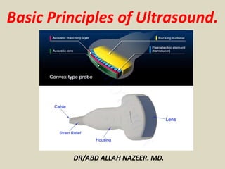
- 1. DR/ABD ALLAH NAZEER. MD. Basic Principles of Ultrasound.
- 3. Sound is a mechanical, longitudinal wave that travels in a straight line and measured by cycle per second by Hertz(Hz) unit. Audible Sound 20-20,000 Hz. Sound requires a medium through which to travel. Ultrasound is a mechanical, longitudinal pressure wave with a frequency exceeding the upper limit of human hearing, which is 20,000 Hz or 20 kHz. Medical Ultrasound is > 2MHz,(2MHz to 16MHz).
- 4. Basic Ultrasound Physics Amplitude oscillations/sec = frequency - expressed in Hertz (Hz). Wave length &frequency
- 5. ULTRASOUND – How is it produced? Produced by passing an electrical current through a piezoelectrical (material that expands and contracts with current) crystal.
- 9. Ultrasound Production. Transducer produces ultrasound pulses (transmit 1% of the time) These elements convert electrical energy into a mechanical ultrasound wave. Reflected echoes return to the scan head which converts the ultrasound wave into an electrical signal
- 10. Frequency vs. Resolution The frequency also affects the QUALITY of the ultrasound image The HIGHER the frequency, the BETTER the resolution The LOWER the frequency, the LESS the resolution A 12 MHz transducer has very good resolution, but cannot penetrate very deep into the body A 3 MHz transducer can penetrate deep into the body, but the resolution is not as good as the 12 MHz Low Frequency 3MHz. High frequency 12 MHz.
- 11. Image Formation. Electrical signal produces ‘dots’ on the screen. Brightness of the dots is proportional to the strength of the returning echoes Location of the dots is determined by travel time. The velocity in tissue is assumed constant at 1540m/sec Distance = Velocity Time
- 12. Interactions of Ultrasound with Tissue. •Acoustic impedance (AI) is dependent on the density of the material in which sound is propagated - the greater the impedance the denser the material. •Reflections comes from the interface of different AI’s • greater of the AI = more signal reflected • works both ways (send and receive directions)
- 13. Acoustic impedance(Z). Is the resistance to the propagation of sound and this depend on density and velocity of sound and measured by MegaRayls (Z). Air = 0.0004 Z.(Low acoustic impedance). Bone = 7.8 Z.(High acoustic impedance). Adipose = 1.34 Z. Liver = 1.65 Z.
- 14. Interactions of Ultrasound with Tissue Reflection. Refraction. Transmission. Attenuation.
- 15. Reflection The ultrasound reflects off tissue and returns to the transducer, the amount of reflection depends on differences in acoustic impedance. The ultrasound image is formed from reflected echoes.
- 16. Refraction
- 17. Transmission Some of the ultrasound waves continue deeper into the body. These waves will reflect from deeper tissue structures.
- 18. Attenuation Defined - the deeper the wave travels in the body, the weaker it becomes -3 processes: reflection, absorption, refraction Air (lung)> bone > muscle > soft tissue >blood > water. Speed of sound in Air= 330 m/s, Bone=4030 m/s, Tissues =1040m/s.
- 25. Ultrasound frequencies in diagnostic radiology range from 2 MHz to approximately 15 MHz. It is important to remember that higher frequencies of ultrasound have shorter wavelengths and are absorbed/attenuated more easily. Therefore, higher frequencies are not as penetrating. This explains why high frequencies are used for the superficial body structures and low frequencies are used for those that are deeper. The following frequencies are a guide to frequencies typically used for ultrasound examination: 2.5 MHz: deep abdomen, obstetric and gynecological imaging 3.5 MHz: general abdomen, obstetric and gynecological imaging 5.0 MHz: vascular, breast, pelvic imaging 7.5 MHz: breast, thyroid 10.0 MHz: breast, thyroid, superficial veins, superficial masses, musculoskeletal imaging. 15.0 MHz: superficial structures, musculoskeletal imaging.
- 34. Artifacts.
- 46. Terminology. Echogenicity. Hypo-Hyper-Iso-An Echoic The circle is Hyperechoic to the surrounding tissue. The circle is Hypoechoic to the surrounding tissue.
- 47. The circle is Isoechoic to the surrounding tissue. The circle is An Echoic to the surrounding tissue.
- 50. Thank You.