
Empfohlen
Weitere ähnliche Inhalte
Ähnlich wie Cardiovascular Physiology
Ähnlich wie Cardiovascular Physiology (20)
Mehr von ZahidSubhani3
Mehr von ZahidSubhani3 (18)
Kürzlich hochgeladen
Kürzlich hochgeladen (20)
Cardiovascular Physiology
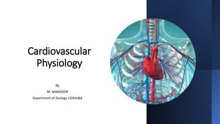
- 1. Cardiovascular Physiology By M. MANSOOR Department of Zoology UOKAJ&K
- 2. Location of Heart The heart is located in the thoracic cavity Posterior to the sternum Superior to the diaphragm SE BA Between the lungs The tip of the heart is called the ‘apex’
- 3. Anatomy of Heart • The heart has: • 3 layers • pericardium • endocardium • Myocardium • 4 chambers • 2 atrium • 2 ventricles • 4 valves • Mitral • Aortic • Tricuspid • Pulmonary
- 4. Function of heart • The heart pumps oxygen and nutrient rich blood to the organs, tissues and cells of the body, and eliminates waste products • Blood is carried from the heart to the organs through arteries, arterioles and capillaries • Blood returns to the heart through venules and veins
- 5. Layers of the Heart-Pericardium • The heart is surrounded by a fibro serous sac called the pericardium • The function of the pericardium is: • To limit cardiac distension and restrict excessive movement • To protect and lubricate • The pericardium is composed of: • Visceral pericardium • Parietal pericardium
- 6. Layers of the Heart Endocardium Innermost/deepest layer of the heart Lines the heart chambers and the valves Smooth thin lining to reduce friction of blood flow through the chambers Cardiac conduction system located in this layer
- 7. Layers of the Heart • Myocardium: • Middle, thickest layer of the heart • Contains the muscle fibers which are responsible for pumping • Contraction of this layer allows blood to be pumped through to the blood vessels
- 8. Chambers of the heart • The heart is divided into four chambers: • A: Right Atrium • V: Right Ventricle • A: Left Atrium • V: Left Ventricle
- 9. Upper Chambers • The upper chambers are: • The atria - Right - Left
- 10. Upper Chambers • The right atrium: • Receives deoxygenated blood from the body through the: • superior vena cava (head and upper body) • inferior vena cava (legs and lower torso) • The left atrium: • Receives oxygenated blood from the lungs through the: • pulmonary vein
- 11. Lower Chambers • The lower chambers are: • The ventricles – Right – Left
- 12. Lower Chambers • The right ventricle: • Receives de-oxygenated blood as the right atrium contracts • The left ventricle: • Receives oxygenated blood as the left atrium contracts
- 13. Valves of the heart • The valves are located within the chambers of the heart. • The function of the valves: • Controls the direction of blood flow • Allows one way flow of blood • through chambers • from the heart to the body
- 14. Valves of the heart • The four valves are known as: the tricuspid valve the pulmonic or pulmonary valve the mitral valve the aortic valve
- 15. Valves of the heart • The tricuspid valve: • Is an atrioventricular valve, situated between the atria and the ventricle • Controls the opening between the right atrium and the right ventricle • The mitral valve: • Is an atrioventricular valve, situated between the atria and the ventricle • Controls the blood between the left atrium and the left ventricle
- 16. Valves of the heart • The pulmonic or pulmonary valve: • Is a semi lunar valve which controls the blood leaving the heart • Situated between the right ventricle and the pulmonary artery • Controls the flow of blood from the right ventricle • Prevents blood flow back to the right ventricle, as it relaxes
- 17. Valves of the heart • The aortic valve: • Is a semi lunar valve which controls the blood leaving the heart • Controls blood flow between the left atrium and the aorta
- 18. Blood Vessels • Closed circulatory system • Arteries • Arterioles • Capillaries • Venules • Veins
- 19. The Vessels • Functions: • Distribution of blood • Exchange of materials with tissues • Return of blood to the heart • Structure: • Most have the same basic structure: • 3 layers surrounding a hollow lumen
- 20. General structures of Blood vessels • Arteries and veins are composed of three layers: 1. tunica interna 2. tunica media 3. tunica externa • Capillaries are composed of endothelium.
- 21. The Anatomy Of Blood Vessels • Layers 1. Tunica interna (intima): • Endothelial layer that lines the lumen of all vessels. • In vessels larger than 1 mm, a subendothelial connective tissue basement membrane is present 2. Tunica media: • Smooth muscle and elastic fiber layer, regulated by sympathetic nervous system • Controls vasoconstriction/vasodilation of vessels 3. Tunica externa (adventitia): • Collagen fibers that protect and reinforce vessels • Larger vessels contain vasa vasorum
- 24. Types of Vessels • Arteries: • Carry blood away from the heart • The elastic tissue in the artery wall allows the vessel to ‘give’ as blood surges through. • So, the artery wall first stretches as a result of the high blood pressure, before an elastic recoil of the wall pushes the blood on its way. • This swelling can be felt as a pulse where arteries travel near the skin surface
- 26. Types of Vessels • Veins: • Carry blood back to the heart • Thin muscular walls • Little elastic tissue in the wall • Relatively large lumen • Blood under low pressure • Blood flow is slow • No pulse • Valves prevent backflow of blood
- 27. Types of Vessels • Capillaries: • Link up arteries and veins in the tissues • No muscle • Wall made up of one cell thick endothelium • Small lumen – just large enough for a red blood cell to squeeze through • Pressure falls as blood passes along capillary • Blood flowing is slowing down • No pulse • No valves
- 28. Types of Vessels • Arterioles: • Diameter of 0.3 mm or less • smallest arteries; lead to capillary beds. • close to capillaries - single layer of muscle spiralling around the endothelial lining • Venules • Are formed when capillary beds unite • Allow fluids and WBCs to pass from the bloodstream to tissues
- 29. ELECTRICAL ACTIVITY OF THE HEART • The automatic nature of the heartbeat is referred to as automaticity. • In heart, there are three regions that can spontaneously generate action potentials and thereby function as pacemakers. • In the normal heart, only one of these, the sinoatrial node (SA node) is a small, flattened, ellipsoid strip of specialized cardiac muscle, functions as the pacemaker. • The SA node is located in the right atrium near the opening of the superior vena cava and serves as the primary (normal) pacemaker of the heart. • The two potential, or secondary, pacemaker regions—the AV node and Purkinje fibers (parts of the conduction network)—are normally suppressed by action potentials originating in the SA node.
- 31. Auto rhythmicity of the Sinus Fibers • The “resting membrane potential” of the sinus nodal fiber between discharges has a negativity of about -55 to -60 millivolts • The cause of this lesser negativity is that the cell membranes of the sinus fibers are naturally leaky to sodium and calcium ions • Cardiac muscle has three types of membrane ion channels that play important roles in causing the voltage changes of the action potential. • They are 1. fast sodium channels 2. slow sodium-calcium channels 3. potassium channels
- 32. Auto rhythmicity of the Sinus Fibers • Opening of the fast sodium channels for a few 10,000ths of a second is responsible for the rapid upstroke spike of the action potential observed in ventricular muscle. • Then the “plateau” of the ventricular action potential is caused primarily by slower opening of the slow sodium-calcium channels, which lasts for about 0.3 second. • Finally, opening of potassium channels allows diffusion of large amounts of positive potassium ions in the outward direction through the fiber membrane and returns the membrane potential to its resting level.
- 34. Self-Excitation of Sinus Nodal Fibers • Because of the moderate number of already open sodium channels, positive sodium ions from outside the fibers normally tend to leak to the inside. • Therefore, between heartbeats, influx of positively charged sodium ions causes a slow rise in the resting membrane potential in the positive direction. • When the potential reaches a threshold voltage of about -40 millivolts, the sodium-calcium channels become “activated,” thus causing the action potential. • Therefore, basically, the inherent leakiness of the sinus nodal fibers to sodium and calcium ions causes their self-excitation.
- 36. Introduction • The body is a good conductor of electricity because tissue fluids have a high concentration of ions that move (creating a current) in response to potential differences. • Potential differences generated by the heart are conducted to the body surface, where they can be recorded by surface electrodes placed on the skin. • The recording thus obtained is called an electrocardiogram (ECG or EKG); the recording device is called an electrocardiograph.
- 37. THE ELECTROCARDIOGRAM (ECG) • Each cardiac cycle produces three distinct ECG waves, designated P, QRS, and T. • The P wave is caused by electrical potentials generated when the atria depolarize before atrial contraction begins. • The QRS complex is caused by potentials generated when the ventricles depolarize before contraction, that is, as the depolarization wave spreads through the ventricles. Therefore, both the P wave and the components of the QRS complex are depolarization waves • The T wave is caused by potentials generated as the ventricles recover from the state of depolarization. This process normally occurs in ventricular muscle 0.25 to 0.35 second after depolarization, and the T wave is known as a repolarization wave
- 39. THE ELECTROCARDIOGRAM (ECG) • There are two types of ECG recording electrodes, or “leads.” • The bipolar limb leads record the voltage between electrodes placed on the wrists and legs • These bipolar leads include lead I (right arm to left arm), lead II (right arm to left leg), and lead III (left arm to left leg). The right leg is used as a ground lead • In the unipolar leads, voltage is recorded between a single “exploratory electrode” placed on the body and an electrode that is built into the electrocardiograph and maintained at zero potential (ground)
- 41. THE ELECTROCARDIOGRAM (ECG) • The unipolar limb leads are placed on the right arm, left arm, and left leg, and are abbreviated AVR, AVL, and AVF, respectively. • The unipolar chest leads are labeled 1 through 6, starting from the midline position. • Thus, a total of 12 standard ECG leads “view” the changing pattern of the heart’s electrical activity from different perspectives. • This is important because certain abnormalities are best seen with particular leads and may not be visible at all with other leads
- 46. Rate of Heartbeat as Determined from the Electrocardiogram • The rate of heartbeat can be determined easily from an electrocardiogram because the heart rate is the reciprocal of the time interval between two successive heartbeats. • If the interval between two beats as determined from the time calibration lines is 1 second, the heart rate is 60 beats per minute. • The normal interval between two successive QRS complexes in the adult person is about 0.83 second. • This is a heart rate of 60/0.83 times per minute, or 72 beats per minute.
- 47. HEMODYNAMICS
- 48. Hemodynamics • Hemodynamics describes the physical behavior of blood as a fluid. Hemodynamics examines the interrelationships between flow, pressure gradients, resistance, vessel cross-sectional area, and velocity. • Terms • F = flow = volume movement with respect to time • P = pressure = force exerted over a surface divided by its area • R = resistance = impediment to flow, expressed in resistance units • V = velocity = distance traveled with respect to time • A = area = cross-sectional area
- 49. Relationship between blood flow, pressure and resistance: • Blood flow through a blood vessel is determined by two factors: 1. pressure difference of the blood between the two ends of the vessel, also sometimes called “pressure gradient” along the vessel, which is the force that pushes the blood through the vessel 2. the impediment to blood flow through the vessel, which is called vascular resistance. • Suppose a blood vessel segment located anywhere in the circulatory system.
- 50. Cont. • P1 represents the pressure at the origin of the vessel; at the other end, the pressure is P2. • Resistance occurs as a result of friction between the flowing blood and the intravascular endothelium all along the inside of the vessel. • The flow through the vessel can be calculated by the following formula, which is called Ohm’s law: • F is blood flow, ΔP is the pressure difference (P1 - P2) between the two ends of the vessel, and R is the resistance. • This formula states, in effect, that the blood flow is directly proportional to the pressure difference but inversely proportional to the resistance. • This interaction is complex, since a change in any one component can impact the other two
- 51. CARDIAC OUTPUT, VENOUS RETURN, AND THEIR REGULATION • Cardiac output is the quantity of blood pumped into the aorta each minute by the heart. This is also the quantity of blood that flows through the circulation. • Venous return is the quantity of blood flowing from the veins into the right atrium each minute. • Normal Values for Cardiac Output at Rest and During Activity • The following factors, among others, directly affect cardiac output: 1. the basic level of body metabolism 2. whether the person is exercising 3. the person’s age 4. size of the body • For young, healthy men, resting cardiac output averages about 5.6 L/min. • For women, this value is about 4.9 L/min.
- 52. Cardiac Index • Cardiac output increases approximately in proportion to the surface area of the body. • Therefore, cardiac output is frequently stated in terms of the cardiac index, which is the cardiac output per square meter of body surface area. • The normal human being weighing 70 kilograms has a body surface area of about 1.7 square meters, which means that the normal average cardiac index for adults is about 3 L/min/m2 of body surface area
- 53. Control of Cardiac Output by Venous Return— Frank-Starling Mechanism of the Heart • When one states that cardiac output is controlled by venous return, this means that it is not the heart itself that is the primary controller of cardiac output. • Instead, it is the various factors of the peripheral circulation that affect flow of blood into the heart from the veins, called venous return, that are the primary controllers • Heart has a built-in mechanism that normally allows it to pump automatically whatever amount of blood that flows into the right atrium from the veins. • This mechanism, called the Frank-Starling law of the heart.
- 54. Frank-Starling Mechanism of the Heart • Basically, this law states that when increased quantities of blood flow into the heart, the increased blood stretches the walls of the heart chambers. • As a result of the stretch, the cardiac muscle contracts with increased force, and this empties the extra blood that has entered from the systemic circulation. • Therefore, the blood that flows into the heart is automatically pumped without delay into the aorta and flows again through the circulation
- 55. Factors That Can Cause Hyper Effective Heart • Only two types of factors usually can make the heart a better pump than normal. 1. Nervous stimulation 2. hypertrophy of the heart muscle 1. Effect of Nervous Excitation to Increase Heart Pumping. • A combination of sympathetic stimulation and parasympathetic inhibition does two things to increase the pumping effectiveness of the heart: 1. it greatly increases the heart rate—sometimes, in young people, from the normal level of 72 beats/min up to 180 to 200 beats/min 2. it increases the strength of heart contraction to twice its normal strength
- 56. Factors That Can Cause Hyper Effective Heart 2. Heart Hypertrophy • Long-term increased workload causes the heart muscle to increase in mass and contractile strength in the same way that heavy exercise causes skeletal muscles to hypertrophy. • For instance, it is common for the hearts of marathon runners to be increased in mass by 50 to 75 per cent. • This increases the plateau level of the cardiac output curve, sometimes 60 to 100 per cent, and therefore allows the heart to pump much greater than usual amounts of cardiac output
- 57. Factors That Cause a Hypo effective Heart • Any factor that decreases the heart’s ability to pump blood causes hypoeffectivity. • Some of the factors that can do this are the following: 1. Coronary artery blockage, causing a “heart attack” 2. Inhibition of nervous excitation of the heart 3. Pathological factors that cause abnormal heart rhythm or rate of heartbeat 4. Valvular heart disease 5. Increased arterial pressure against which the heart must pump, such as in • hypertension • Congenital heart disease • Myocarditis • Cardiac hypoxia
- 58. THANK YOU