
Recommended
Recommended
More Related Content
What's hot
What's hot (20)
Similar to Deep vein thrombosis Ultrasound
Similar to Deep vein thrombosis Ultrasound (20)
More from Syed Yousaf Gilani
More from Syed Yousaf Gilani (13)
Recently uploaded
Recently uploaded (20)
Deep vein thrombosis Ultrasound
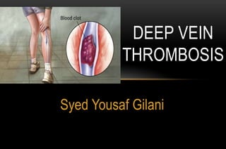
- 1. Syed Yousaf Gilani DEEP VEIN THROMBOSIS
- 2. WHAT IS VENOUS THROMBOEMBOLISM? • When a blood clot (thrombus) forms in a deep vein, this is called deep vein thrombosis (DVT) - If it happens it happens most frequently in the leg • Thromboembolism occurs when part of a clot breaks away and enters the bloodstream – When this happens the blood clot is called an embolus • Pulmonary embolism is the third most common cause of mortality by cardiovascular disease after coronary artery disease and stroke.
- 3. EPIDEMIOLOGY DVTs occur in about 1 per 1000 persons per year. 100,000 deaths may be directly or indirectly related to these diseases • In pregnant women, it has an incidence of 0.5 to 7 per 1,000 pregnancies, and is the second most common cause of maternal death in developed countries after bleeding Journal of Internal Medicine volume 232 Issue 2, Pages 155 - 160
- 4. PRESENTATION AND CLINICAL EXAMINATION • PAIN AND TENDERNESS • SWELLING (USUALLY IN ONE LIMB) • REDNESS • WARMTH • EDEMA • CYANOSIS • HOMANS SIGN ( dorsiflexion of foot while knee is extended)
- 5. ANATOMY OF LOWER EXTREMITY VEINS
- 6. VEINS OF LOWER LIMB Superficial veins Deep veins Perforating veins Superficial veins includes great and small sephanous veins and their tributaries. They drain into deep veins through perforating veins. The greater saphenous vein joins the femoral vein at afixed point in the groin 2.5 cm below and lateral to the pubic tubercle, and the lesser saphenous vein terminates at avariable site in the popliteal fossa. Blood passing up the superficial veins enters the deep veins at the saphenopopliteal and saphenofemoral junctions..
- 7. ULTRASONOGRAPHY color-flow Duplex scanningis the imaging test of choice for patients with suspected DVT inexpensive, noninvasive, widely available Ultrasound can also distinguish other causes of leg swelling, such as tumor, popliteal cyst, abscess, aneurysm, or hematoma.
- 8. COMMON FEMORAL VEIN- NORMAL AND DVT
- 9. FEMORAL VEIN superficial femoral vein
- 10. POPLITEAL VEIN Leg allowed to hang over the edge off the bed with the probe positioned in the popliteal fossa Ma OJ, Mateer JR. Blaivas M. Emergency Ultrasound, 2nd Edition
- 11. NORMAL VENOUS FLOW 1. Spontaneity: Spontaneous flow without augmentation 2. Phasicity: Flow changes with respiration 3. Compression: Transverse plane 4. Augmentation: Compression distal to site of examination patency below site of examination patency below site of examination 5. Valsalva: Deep breath strain while holding breath patency of abdominal & pelvic vein
- 12. PHASICITY FLOW CHANGES WITH RESPIRATION Rapid Slow Apnea
- 13. EXTERNAL COMPRESSION OF THE VEINS
- 15. PATIENT POSITION LEG BENT AT THE KNEE AND ROTATED OUTWARD BEST EXPOSURE OF THE FEMORAL VEINS AND THE POPLITEAL FOSSA
- 17. COMPRESSION TEST AT LEVEL OF ADDUCTOR CANAL Compression test inadequate at level of adductor canal. Rather, examiner additionally presses the vein against transducer from below the flat hand.
- 18. NORMAL POSTERIOR TIBIAL VEINS Diastole Systole Augmentation
- 19. TRIPLE POSTERIOR TIBIAL VEINS
- 20. THROMBUS IN THE CFV Relaxation Compression
- 21. FREE- FLOATING THROMBUS Free-floating thrombus in LFV extending into CFV Hamper UM et al. Radiol Clin N Am 2007 : 45: 525 – 547/
- 23. ACCURACY OF US FOR DIAGNOSIS OF LOWER EXTREMITIES DVT
- 24. US diagnostic criteria of DVT Acute and chronic thrombus Signs interpreted according to clinical history
- 25. CLOT FORMATION IN THE VEIN 25 CHRONIC CALCIFIC THROMBUS IN CALF VEIN
- 26. CFV THROMBUS 26
- 27. DIFFERENTIAL DIAGNOSIS OF DVT Useche JN et al. Radiographics 2008: 28 : 1785-1797. • 7 of 10 patients could have cause other than DVT. • Ancillary findings detected in only 10% of Doppler study. • 90% of incidental findings related to patients symptoms. • Anatomic approach is the most useful strategy for dd. Make every effort to establish a diagnosis when DVT is ruled out
- 28. DIFFERENTIAL DIAGNOSIS OF DVT ANATOMIC APPROACH • Groin From inguinal ligament to 10cm below. • Thigh From this line to Hunter canal. • Popliteal From hunter canal to 10cm below pop crease. • Lower leg 10cm from popliteal crease to ankle Useche JH et al. RadioGraphics 2008 : 28 : 1785 – 1797.
- 29. DIFFERENTIAL DIAGNOSIS OF DVT Region Differential Diagnosis 1. Inguinal Hernias: Femoral – Inguinal. Hiopsoas & Heopectineal bursitis. Adenopathy (inflammatory & neoplastic) Pseudo aneurysm – AVI – anticoagulation hematoma. 2. Thigh Sports-related lesions (conclusions, muscle tears, hematoma) Muscle herniation – myositis – abscess. 3. Popliteal Ruptured Baker’s cyst. Para meniscal cyst – pes anserinus bursitis. Popliteal artery thrombosis –aneurysm – adventitial cyst. 4. Lower Leg PA entrapment syndrome – thrombophlebitis Tennis Leg Cardiac and renal failure. Useche JH et al. RadioGraphics 2008 : 28 : 1785 – 1797.
- 30. NORMAL INGINAL ANATOMY RT INGUINAL REGION – PARALLEL TO & CRANIAL TO INGUINAL LIGAMENT Jamadar DA et al. AJR 2007: 188 : 1356-1364.
- 31. RT INGUINAL REGION – PARALLEL TO & CRANIAL TO INGUINAL LIGAMENT INDIRECT INGUINAL HERNIA Pre-Valsalva maneuver Post-Valsalva maneuver
- 32. ENLARGED LYMPH NODE Grey Scale Color Doppler
- 33. MUSCULAR ABSCESS Abscess Normal femoral vessels
- 35. BAKER’S CYST Anechoic fluid distends SM – GC bursa Characteristics neck between SM tendon & medial GC muscle & tendon,
- 36. POPLITEAL ARTERY ANEURYSM PARTIAL THROMBOSIS Transverse color Doppler US Sagittal color Doppler US
- 37. THANK YOU