SlideShare ist ein Scribd-Unternehmen logo
1 von 178
Objectives
1. To recognize the types of asexual and sexual reproduction.
2. To understand the meaning of asexual and sexual reproduction.
3. To understand the alternation of generation of plants.
4. To understand the life history of pteridophytes and angiospermae.
5. To comprehend the structure and function of male and female reproductive
organs.
6. To comprehend the formation and structure of sperm and ovum.
7. To comprehend female menstrual cycle and the influence between hormone and
menstrual cycle.
8. To understand the fertilization and its process.
9. To understand the human embryonic development
10. To understand the process of labour.
Main points
• Double fertilization
• Formation of sperm and ovule
• Menstruation cycles and hormones
• Fertilization and the development of zygote
Difficult points
• Importance of sexual reproduction
• Alternative of generation in plants
• Sexual reproduction in seed plants
• Menstruation cycles and hormones
Objectives
1. To understand the importance of reproduction.
2. To recognize the types of asexual reproduction.
3. To understand the meaning of asexual reproduction.
Chapter 17 Reproduction
Ong Yee Sing
2018
Notice the sleeping movement of the plants
Rewind… remember Junior High and Chapter 16
• How does a plant form more cells and grow?
• How does a plant produce seeds?
• Why does the sunflower plant needs to produce seed?
Reproduction
• Reproduction is the process of
organisms producing offspring后代
• Continuation of species
• Avoid extinction
• Basic characteristic of living organisms.
Types of reproduction
• Asexual reproduction无性繁殖
• No fusion of reproductive cells /
gametes配子.
• produce a new individual directly
from the parent body.
• Sexual reproduction有性繁殖
• fusion of two gametes to form a
zygote合子.
• a new individual is form through
the development of zygote.
Gametes配子
• from Greek gamos ‘marriage’
• a mature haploid male or female
germ cell which is able to unite
with another of the opposite sex
in sexual reproduction to form a
zygote.
Zygotes合子
• from Greek zugoun ‘to yoke’.
• a diploid cell resulting from the
fusion of two haploid gametes.
yoke
Quiz
• Which of the following statements about asexual reproduction is NOT
true?
A. It does not improve genetic variation.
B. It produces clones of the original cell.
C. It does not require a mate for reproduction.
D. It is a seldom used form of reproduction.
17.1.1 Asexual
reproduction
Asexual reproduction
• Binary fission
• Budding
• Sporulation
• Fragmentation
• Vegetative propagation
Binary fission分裂生殖
• The parent cell divides to form
genetically identical offspring with the
same size and shape.
• Simple fission二分裂 produces two
offspring.
• bacteria and amoeba
• Multiple fission复分裂 produces many
offspring.
• Plasmodium疟原虫
裂殖体
Budding出芽生殖
• A new individual is derived from an
outgrowth (bud).
• When the bud matures, the new
individual separates from the body of
the parent to form a new individual.
• Sometimes the offspring do not
separate from the parent before
budding again.
• Examples: yeast and hydra水螅.
Sporulation孢子生殖
• Spores are stable, dormant cells
that are produced from vegetative
cells.
• When the conditions are
favourable, the spores germinate
to form new individuals.
• Example: most of the fungi and
algae.
• Some spores have flagella鞭毛that
can swim - zoospores单胞藻
Fragmentation断裂生殖
• Some parts of the organism break off and subsequently differentiate
and develop into new individuals.
• Example: spirogyra水绵, cnidarians腔肠动物 (e.g. hydra水螅) and annelids
环节动物.
spirogyra
Vegetative propagation
营养生殖
• New individuals develop from a part of
nutritive organs (root, stem and leaves)
of plants.
• Example, tubers of potatoes, bulbs of
onion
Characters of asexual reproduction
• Characters of the parents are maintained
• New generations are genetically identical to the parents
• Poorly adapt to new environment
• Lack of diversity and genetic variation in the offspring population
• Fast growing
• Do not required complicated maturation process from zygote
• Able to colonise an area quickly when the environment is favourable
Quiz
• Which of these statements is true of asexual reproduction?
A. It produces offspring genetically identical to each other and requires
one parent.
B. It produces offspring genetically identical to each other and requires
two parents.
C. It produces offspring genetically different from each other and
requires one parent.
D. It produces offspring genetically different from each other and
requires two parents.
Quiz
• When an organism undergoes a period of growth then splits in two
separate organisms, it is called _____________
A. Budding
B. Fission
C. Fragmentation
D. Parthenogenesis
Quiz
• Which of the following statements about gametes are true?
I. In sexual reproduction, each parent provides genetic material for the
formation of a gamete cell.
II. Gametes are produced by mitosis.
III. If a sexually reproducing organism is diploid, the gametes are haploid.
A. I, II and III
B. I and III
C. II and III
D. I and II
• https://www.youtube.com/watch?v=i9zj9V8OWRk
Objectives
1. To recognize the types of sexual reproduction.
2. To understand the meaning of sexual reproduction.
3. To compare and contrast the characters of asexual and sexual
reproduction.
Sexual reproduction
• Isogamy
• Anisogamy
• Oogamy
Sexual reproduction有性生殖
• Two reproductive cells (gametes配子) (N
+ N) to form a zygote合子 (2N).
• Zygotes develop into new individual.
• Sexual reproduction can be divided
into isogamy, anisogamy and oogamy
according to their morphological
appearance.
Characters of sexual reproduction
• More genetic variation in offsprings
• Heredity characters of both parents
• Crossovers
• Better adaptive to changing environment
lsogamy同配生殖
• Production and fusion of two
gametes that are similar in size
and structure.
• Generally flagellated and mobile
• Examples: lower animals and
plants, such as certain protozoa
原生动物, algae (Chlamydomonas单
胞藻) and fungi.
Anisogamy异配生殖
• The two gametes that fused to form
zygotes are dissimilar in size.
• Large gametes = female gametes = ova /
egg cells 雌配子
• Small gametes = male gamates = sperm雄
配子
• Example: Pandorina空球藻.
Oogamy卵式生殖
• Egg cell – female gametes
• Sessile固定,不能运动
• Large
• Provide nutrient for the zygotes受精卵,合子
• Sperm – male gametes
• Mobile
• Small
• Search for the ova
Parthenogenesis孤雌生殖/单性生殖
• Greek παρθένος, parthenos,
'virgin' + γένεσις, genesis,
'creation‘
• A natural form of asexual
reproduction in which growth
and development of embryos
occur without fertilization.
• In animals, parthenogenesis is
the development of an
embryo from an unfertilized
egg cell.
• In plants parthenogenesis is a
component process of
apomixis无融合生殖.
Haploid parthenogenesis单倍孤雌生殖
Diploid
parthenogenesis
双倍孤雌生殖
• In the spring / early
summer
• Lots of food
• Produce 2N egg cells
• Offspring born viviparity卵胎
生
• In the late summer / autum
• Food is scarce
• Produce offspring that have
two sexes that will mate and
produce fertilized eggs
• Dormant in the winter
aphid 蚜虫
17.2 Alternation of generation in
plants
Alternation of generation in plants
• All plants undergo a life
cycle that takes them
through both haploid and
diploid generations.
• This fluctuation between
these diploid and haploid
stages that occurs in plants
is called the alternation of
generations世代交替.
Sporophyte and gametophyte
• Sporophyte孢子体
• grow from a zygote
• diploid
• produces spores through
meiotic division.
• Gametophyte配子体
• grow from a spore
• haploid
• produces gametes through
mitosis
Quiz: fill in the blanks
Generalized Life Cycle Pattern For Animals & Plants
4
3
2
1
5
Human Life Cycle
• In the human life cycle (and the life
cycles of most multicellular
animals), the only cells that are
haploid are the sperm and egg.
• From the zygote to the diploid
mother cells inside the sex organs,
all the cells are diploid with two
sets of chromosomes.
• Humans are dioecious species with
separate male and female
individuals in the diploid
population.
Evolution of plants
general evolutionary trend
• gradual increase in the
diploid sporophyte phase
• decrease in the haploid
gametophyte phase
Moss
Moss Life Cycle
• The dominant (conspicuous) part of
the life cycle is the haploid, leafy N
gametophyte.
• The gametophytes are photosynthetic.
• Most moss gametophytes are
dioecious, with separate male and
female individuals in the population.
• Female sex organs = archegonia.
• Male sex organ = antheridium
• Fertilization involves a motile,
biflagellate sperm that swims through
water to reach the egg on female
plants.
Moss Life Cycle
• The non-photosynthetic diploid
sporophyte consists of a sporangium-
bearing stalk that grows directly out of
the gametophyte.
• Spore mother cells within the
sporangium undergo meiosis,
producing numerous haploid spores
that fall to the ground like tiny
particles of dust.
• Since the sporophyte is without
chlorophyll, it is completely dependent
on the gametophyte for its water,
minerals and carbohydrate nutrition.
• Consequently, the sporophyte of the
moss is heterotrophic and parasitic on
the gametophyte.
Quiz
Fern sporophyte
Fern gametophyte (prothallus)
Fern life cycle
• The dominant (conspicuous)
part of the life cycle is the
diploid, leaf-bearing
sporophyte.
• The sporophytes of ferns are
photosynthetic and autotrophic.
• Spore mother cells within the
sporangium undergo meiosis,
producing numerous haploid
spores.
• The sporangia split open at
maturity, releasing millions of
spores that fall to the ground
like tiny particles of dust.
Fern life cycle
• Each spore germinates and
grows into a heart-shaped
gametohyte (prothallus).
• The gametophytes of ferns are
photosynthetic and autotrophic.
• male sex organs (antheridia)
• female sex organs (archegonia)
• Ferns are typically monoecious
(=bisexual) with both male and
female sex organs on the same
gametophytes.
• Fertilization involves a
multiciliate sperm that swims
through water to reach the egg.
• Young sporophyte developed
from the old grametophyte.
Quiz
Fertilization
Mitosis (2n)
Leaf of young
sporophyte
growing from
gametophyte
indusium
sorus
Meiosis
Mitosis (n)
Spore
Gametophyte
Sporophyte
Embryo
Sporangium
Angiosperm
• Also known as
angiospermophyta or the
flowering plant.
• Greek angos –ανγοσ
“box” + sperma –σπέρμα
“seed” + phyto –φυτό
Characters of life cycle of angiosperms
• Gametophyte reduce
• Gametophyte reply on
sporophyte to survive
• Does not need H20 to reproduce
– pollen tube
• Double fertilization
• Embryo nourished with
endosperm
Life cycle of
angiosperm
• Sporophyte = the plant
• Gametophyte
• Megagametophyte = ovule
• Microgametophyte = pollen grain
• Forms in flower
Structure of a flower
雌蕊
花药
花丝 雌蕊
柱头
花柱
子房
胚珠
花瓣
花萼
花托
花梗
Quiz: label the flower. 1
2
3
4
5
6
7
8
9
Formation of microspore
• Stamen = anther + filament
• Each anther has four pollen sacs with many 2N
pollen mother cells.
• 2N pollen mother cells meiosis to form four N
pollen grains = microspores.
Growth of microspore
• The nucleus of each
microspore undergo mitosis.
• A N generative cell生殖细胞
without cell wall is formed
within the N vegetative cell营
养细胞
• Generative cell will mitosis into
two N sperm cells.
• Vegetative cell provides
nutrients and will form the
tube cell.
Mature pollen grain
• N mature pollen grain = N male gametophyte
• 1 vegetative cell
• 1 generative cell OR 2 sperm cells
• A pollen grain contain germ pore on the
surface.
• Pollen tube grows from these pores.
A. Cross-section through an anther of Lilie (Lilium)sp.) with on the left and
the right side two loculi each. In the loculi sporemothercells (SMCs) can be
seen from which the four spores develop through meiosis I and II.
Inbetween the loculi of each pair a thin layer of cells (arrow) is visible
along which the loculus can burst open atmaturity and release the pollen
grains. In the middle the cross-sectioned filament (Fi) to which the anther
is attached is indicated. In the upper part the vascular bundle (v) of the
loculus can be distinguished.
B. Loculus. The lumen contains developing pollen. On the inner wall (w) of
the loculus a layer constitued of block-shaped single cells is present, the
tapetum (t). The tapetum feeds the developing spore and -later- pollen.
C. Tetrad stage during pollen development. After the two meiotic divisions
the four daughter cells are still interconnected and form a tetrad. They are
still surrounded by the wall (arrow) of the original cell, the microspore
mothercell (MMC).
D. Mitotic division in the spore leading to the formation of a
microgametophyte or pollen. Only the metaphase is shown here. The
chromosomes lay in the equatorial plane of the cell.
E. Nearly ripe pollen grain: visible are a vegetative cell with nucleus (VN),
which later will form the pollen tube, and a generative cell with its own
nucleus (GN), which later will divide into two sperm cells.
F. Ripe pollen grain in which the texture of the outer cell wall, the exine,
can be recognized. The grainy dark purple structure in the middle of the
pollen grain is the vegetative nucleus.
G. Diagram in 3 parts: Ripe pollen grain consisting of the vegetative cell
(VC) and therein the smaller generative cell (GC). After landing on the
stigma (St) the pollen grain germinates and forms a pollen tube. In the
pollen tube the generative cell divides into two sperm cells (SC). The pollen
tube grows to the embryo sac (ES) and delivers the two sperm cells that
are involved in double fertilization.
Structure of a pistil
• Pistil = stigma + style + ovary
• Inside the ovary, there may be one
or numerous 2N ovule
• Ovule = megasporangia
雌蕊 = 柱头 + 花柱 + 子房
Structure of an ovary
• Funicle: to support, projection
and conduction
• Nucellus: where development
of female gametophyte occurs
• Integument: to protect nucellus
and embryo sac
(megagametophyte)
• Micropyle: for a passage for
pollen tube to enter the ovule
• Chalaza: transfer nutrients to
nucellus
Formation of a megaspore
• The 2N megasporocyte
(megaspore mother cell)大
孢子母细胞 undergoes
meiosis
• Four N megaspores大孢子
are form
• Three megaspore that are
closer to the micropyle
degenerate
• Only one megaspore珠孔
remaining functional
micropyle
Formation of an megagametophyte
• After 3 mitosis
• 7 N cells with 8 nucleus are form
• = embryo sac胚囊
• = megagametophyte
Megagametophyte = embryo sac
• At the microphylar end
• 1 egg cell卵细胞
• 2 synergids助细胞
• Middle
• 1 central cell中央细胞 with
2 polar nuclei极核
• May fuse to form a 2N
cell
• At the chalazal end
• 3 antipodals cells反足细胞
Function of cells in the embryo sac
• The central cell, after fertilization,
develops into the endosperm胚乳,
which produce nutrients to the zygote.
• The synergids are thought to help the
pollen nucleus reach the egg cell for
fertilization.
• The antipodal cells nourishes the
embryo sac
Pollination
• Pollination is the act of
transferring pollen grains from
the male anther of a flower to
the female stigma.
Growth of pollen tube
• A pollen grain germinate in response
to a sugary fluid and lipids secreted by
the mature stigma.
• The vegetative cell then produces the
pollen tube, a tubular protrusion from
the pollen grain, which carries the
sperm cells within its cytoplasm.
• The germinated pollen tube must then
drill its way through the nutrient-rich
style and curl to the bottom of the
ovary to reach the ovule.
• Once the pollen tube reaches an
ovule, it bursts to deliver the two
sperm cells.
Double fertilization
• One of the sperm fertilizes
the egg cell which develops
into an 2N embryo胚, which
will become the future plant.
• The other sperm cell fuses
with both polar nuclei of the
central cell to form the 3N
endosperm胚乳, which serves
as the embryo's food supply.
• The ovary will
develop into a fruit.
• The ovules will
develop into seeds.
Woodland
Strawberry
Fragaria vesca
17.3.1 Male reproductive system
• Human male reproductive
system includes testes,
epididymis, vas deferens
(sperm duct), seminal vesicles,
prostate gland, bulbourethral
glands and penis etc.
Testes
• Human male reproductive system consists of two testes which both
are enclosed by scrotum.
• The functions of the testes are to produce both sperm and
androgens, primarily testosterone.
Tubules of testes
• Within the testes are very fine coiled
tubes called seminiferous tubules.
• Primary cell types within the
seminiferous tubules includes germ
cells, sertoli and peritubular myoid
cells.
• Cells between the seminiferous
tubules are called interstitial cells),
primarily Leydig cells.
• All the tubules in a testis are joined to
a single tubes called epididymis.
• Epididymis is the site for temporary
storing of sperms and for the sperm to
develop until mature.
Lumen
Primary cell types within
the seminiferous tubules
• The germ cells give rise to sperm
through spermatogenesis.
• The sertoli cells are the true
epithelium of the seminiferous
epithelium, and is critical for the
support of germ cell development into
spermatozoa. Sertoli cells also secrete
an inhibitory hormone called inhibin.
• Peritubular myoid cells are smooth
muscle cells surrounding the
seminiferous tubules.
Primary cell types
between the
seminiferous tubules
• The Leydig cells, which are
generally refers as interstitial
cells, secrete testosterone for
sexual development and
puberty, secondary sexual
characteristics, supporting
spermatogenesis and erectile
function.
Semen
• Semen is a fluid that is a
composition of sperms and other
secretions.
Gland/Site Features
Testis/Epididymis Sperm / spermatozoa
Seminal Vesicle Contains large amounts of fructose, which
is used by the sperm mitochondria to
generate ATP to allow movement through
the female reproductive tract.
Prostate Produce an alkaline, milky fluid that is
critical to first coagulate and then
decoagulate the semen following
ejaculation.
Bulbourethral
Glands
or Cowper’s glands
Produce a thick, salty mucus that
lubricates the end of the urethra and the
vagina, and helps to clean urine residues
from the penile urethra.
Ejaculation
• Ejaculation is the
discharge of semen
from the male
reproductory tract.
• When the male orgasm,
small arteries of erectile
tissue (spongy tissue)
dilate to increase the
blood supply, causes
penis to become hard
and erect.
Ejaculation
• Rhythmic contractions of
muscles will produce waves
of pressure within the
urethra.
• This enables sperm enter
urethra and mixes with the
fluid that secreted from
seminal vesicles, prostate
gland and bulbourethral
gland to form semen. The
resultant semen is forced out
of the penis by powerful
contractions of the urethra.
Summary
17.3.1.1 Formation of sperms
Spermatogenesis
• The process of the
formation of sperm in
the testes is called
spermatogenesis.
• Spermatogenesis
occurs in the
seminiferous tubules.
• One production cycle,
from spermatogonia
through formed
sperm, takes
approximately 64
days.
Spermatogenesis
• Mitosis of a spermatogonium
(stem cell) involves a single cell
division that results in two
identical, diploid daughter cells
called primary spermatocyte.
• Meiosis has two rounds of cell
division: primary spermatocyte
to secondary spermatocyte,
and then secondary
spermatocyte to spermatid.
• A total of four spermatids are
produced from each
spermatogonium.
Spermatogenesis
• Although haploid, early
spermatids look very similar to
cells in the earlier stages of
spermatogenesis, with a round
shape, central nucleus, and
large amount of cytoplasm.
• These early spermatids reduced
the cytoplasm to form sperm.
• Eventually, the sperm are
released into the lumen and are
moved along a series of ducts
in the testis toward a structure
called the epididymis for the
next step of sperm maturation.
Summary
17.3.1.2 Structure of a sperm
Three regions of a sperm
• Sperm have a distinctive head, mid-piece, and tail region.
• The head of the sperm contains the extremely compact haploid nucleus
with very little cytoplasm.
• An acrosome covers most of the head of the sperm cell as a “cap” that is
filled with lysosomal enzymes important for preparing sperm to
participate in fertilization.
• Tightly packed mitochondria fill the mid-piece of the sperm. ATP produced
by these mitochondria will power the flagellum.
• The tail is a flagellum , which extends from the neck and the mid-piece
through the tail of the sperm, enabling it to move the entire sperm cell.
17.3.1.3 Hormonal control of
male reproductive system
Hormonal Control of Male
Reproduction
• The hypothalamus monitors and causes the
release of hormones from the pituitary gland
using a negative feedback mechanism.
• When the reproductive hormone is required,
the hypothalamus sends a gonadotropin-
releasing hormone (GnRH) to the anterior
pituitary.
• This causes the release of gonadotrophic
hormones, i.e. follicle stimulating hormone
(FSH) and luteinizing hormone (LH) from the
anterior pituitary into the blood.
Negative feedback loop of spermatogenesis
• FSH enters the testes and stimulates the
Sertoli cells to begin facilitating
spermatogenesis.
• The Sertoli cells produce the
hormone inhibin, which is released into
the blood when the sperm count is too
high.
• This inhibits the release of GnRH and
FSH, which will cause spermatogenesis to
slow down.
• If the sperm count reaches 20 million/ml,
the Sertoli cells cease the release of
inhibin, and the sperm count increases.
Negative feedback loop of testosterone
• LH also enters the testes and stimulates
the interstitial cells of Leydig besides the
seminiferous tubule to make and release
testosterone into the testes and the blood.
• Testosterone, the hormone responsible for
the secondary sexual characteristics that
develop in the male during adolescence,
also stimulates spermatogenesis.
• A negative feedback system occurs in the
male with rising levels of testosterone
acting on the hypothalamus and anterior
pituitary to inhibit the release of GnRH,
FSH, and LH.
Micrograph showing a cluster of
Leydig cells (center of image). H&E
stain.
Histological section through testicular parenchyma of a boar. 1
Lumen of convoluted part of the seminiferous tubules, 2
spermatids, 3 spermatocytes, 4 spermatogonia, 5 Sertoli cell, 6
myofibroblasts, 7 Leydig cells, 8 capillaries
Quiz
• Which hormone causes Leydig cells to make testosterone?
A. FSH
B. LH
C. inhibin
D. estrogen
Quiz
• Which hormone causes FSH and LH to be released?
A. testosterone
B. estrogen
C. GnRH
D. progesterone
Summary
17.3.2 female reproductive organ
• https://www.youtube.com/watch?v=QyPAM0c4ZYA
The female reproductive system is located
primarily inside the pelvic cavity.
Vagina
• Serves as the entrance to the
reproductive tract.
• Serves as the exit from the uterus
during menses and childbirth.
• The vagina is home to a normal
population of microorganisms that
help to protect against infection by
pathogenic bacteria, yeast, or other
organisms that can enter the vagina.
• The vaginal bacteria secretes lactic
acid, and thus protects the vagina by
maintaining an acidic pH (below 4.5).
Ovaries
• The ovaries are the pair of female
gonads.
• The ovaries are supported by
ovarian ligament that contains the
ovarian blood and lymph vessels.
• The cortex of ovaries is composed
the germinal epithelium, that will
eventually forms oocyte.
• Beneath the cortex lies the inner
ovarian medulla, the site of blood
vessels, lymph vessels, and the
nerves of the ovary.
Oviduct / Fallopian tubes
• The oviducts serve as the conduit of
the oocyte from the ovary to the
uterus.
• Each of the two uterine tubes is close
to, but not directly connected to, the
ovary.
• The distal end (closer to the ovaries)
flares out with slender, finger-like
projections called fimbriae.
• The inner layer of the oviduct
contains ciliated cells that beat in the
direction of the uterus, producing a
current that move the oocyte.
The Uterus and Cervix
• The uterus is the muscular organ that nourishes
and supports the growing embryo.
• The cervix is the narrow inferior portion of the
uterus that projects into the vagina.
• The cervix produces mucus secretions that become
thin and stringy under the influence of high systemic
plasma estrogen concentrations, and these
secretions can facilitate sperm movement through
the reproductive tract.
• The innermost layer of the uterus is called
the endometrium.
• Structurally, the endometrium consists of two layers:
the stratum basalis and the stratum functionalis (the
basal and functional layers). The stratum basalis layer
does not shed during menses.
• In contrast, the thicker stratum functionalis grows
and thickens in response to increased levels of
estrogen and progesterone. This inner functional
layer provides the proper site of implantation for the
fertilized egg, and—should fertilization not occur—it
is only the stratum functionalis layer of the
endometrium that sheds during menstruation.
17.3.2.1 Oogenesis
Before birth
• The ovarian stem cells,
or oogonia (sg.
oogonium), are formed
during fetal
development, and
divide via mitosis, from
the germinal
epithelium.
• Oogonia form primary
oocytes in the fetal
ovary prior to birth.
• These primary oocytes
are then arrested in
prophase I of meiosis I.
After puberty
• Meiosis resumes.
• The cytoplasm is divided
unequally, and one
daughter cell is much
larger than the other.
• This larger cell is the
secondary oocyte.
• The secondary oocyte
arrests at metaphase II.
• The smaller cell, called
the first polar body,
eventually disintegrates.
After puberty
• Meiosis of a secondary
oocyte is completed only if a
sperm succeeds in
penetrating its barriers.
• Meiosis II then resumes,
producing one haploid
ovum that, at the instant of
fertilization by a (haploid)
sperm, becomes the first
diploid cell of the new
offspring (a zygote).
• Thus, the ovum can be
thought of as a brief,
transitional, haploid stage
between the diploid oocyte
and diploid zygote.
Follicle development
• Ovarian follicles are oocytes and their
supporting cells.
• Primordial follicles are resting follicle with a
single flat layer of support follicular cells,
that surround the oocyte, and they can stay
in this resting state for years—some until
right before menopause.
• After puberty, a few primordial follicles will
join a pool of immature growing follicles
called primary follicles, as the follicular cells
increase in size and proliferate.
• Secondary follicles increase in diameter,
adding a new outer layer of connective
tissue, blood vessels, and starts to produce
estrogens.
• Tertiary follicle has a thick fluid, called
follicular fluid, collected in a large pool.
Ovulation
• Several follicles reach the
tertiary stage at the same time,
and most of these will undergo
atresia (die).
• The one that does not die will
continue to grow and develop
until ovulation, when it will
expel its secondary oocyte
surrounded by several layers of
follicular cells from the ovary.
In this electron micrograph of a secondary
follicle, the oocyte, theca cells (thecae
folliculi), and developing antrum are
clearly visible. EM × 1100. (Micrograph
provided by the Regents of University of
Michigan Medical School © 2012)
Ovulation
• Several follicles reach the
tertiary stage at the same time,
and most of these will undergo
atresia (die).
• The one that does not die will
continue to grow and develop
until ovulation, when it will
expel its secondary oocyte
surrounded by several layers of
follicular cells from the ovary.
Afterwards
• The collapsed follicle is transformed
into a new endocrine structure called
the corpus luteum.
• Corpus luteum produces large
amounts of the sex steroid hormone
progesterone, a hormone that is
critical for the establishment and
maintenance of pregnancy and to
prevent development of new
dominant follicles develop at this
time.
• If pregnancy does not occur within 10
to 12 days, the corpus luteum will
stop secreting progesterone and
degrade into the corpus albicans, a
nonfunctional “whitish body” that will
disintegrate in the ovary over a period
of several months.
The ovarian cycle
• The ovarian cycle refers to the series of
changes in the ovary during which the
follicle matures, the ovum is shed, and
the corpus luteum develops.
• The follicular phase describes the
development of the follicle in response
to follicle stimulation hormone ( FSH ).
• As luteinizing hormone ( LH ) and FSH
levels increase they stimulate ovulation,
or the release of a mature oocyte into
the fallopian tubes.
• In the luteal phase, the corpus luteum
forms on the ovary and secretes many
hormones, most significantly
progesterone, which makes the
endometrium of the uterus ready for
implantation of an embryo.
• If implantation does not occur, the
corpus luteum will be degraded,
resulting in menstruation.
• If implantation occurs the corpus
luteum is maintained.
17.3.2.2 structure of an ovum
Ovum
• Ovum is a rounded and non-motile structure.
• The cytoplasm of the egg is called ooplasm.
• It contains a very little amount of yolk in man.
• In animals where huge amount of yolk is
present, the cytoplasm of egg consists of
lipoproteins, pigment granules, water, along
with other cytoplasmic organelles.
• The peripheral layer of ooplasm is known as
cortex and it contains many microvilli and
cortical granules.
• The cortical granules are most associated with
polyspermy prevention after the event of
fertilization.
• Microvilli are tubular outrushing of
plasmalemma for transportation of substances
into and out of egg cytoplasm.
Ovum
• In man the ovum is covered over
by a thin vitelline membrane
(made up of protein fibre) which is
further covered over by another
primary membrane known as zona
pellucida.
• During discharge of ovum from the
graafian follicle, several layer’ of
follicular cells adhere to the outer
surface of zona pellucida and are
arranged radially forming corona
radiata.
17.3.3 Menstrual cycle
Menstrual cycle
• Menstrual cycle is the series of changes
in which the uterine lining is shed,
rebuilds, and prepares for implantation.
• The cycle length is determined by
counting the days between the onset of
bleeding in two subsequent cycles.
• The average length of a woman’s
menstrual cycle is 28 days, but the
length of the menstrual cycle varies
among women, and even in the same
woman from one cycle to the next,
typically from 21 to 32 days.
Phases of the menstrual cycle
• The three distinct phases of the
menstrual cycle.
• The menses phase of the
menstrual cycle is the phase
during which the lining is shed.
• The endometrium begins to
proliferate again in
the proliferative phase.
• The secretory phase of the
menstrual cycle refers the
maintenance of the thickness of
the endometrium as the
endometrial lining prepares for
implantation.
Menses phase
• The menses phase occurs during
the early days of the follicular
phase of the ovarian cycle, when
progesterone, FSH, and LH levels
are low.
• Progesterone concentrations
decline as a result of the
degradation of the corpus luteum,
marking the end of the luteal
phase.
• This decline in progesterone
triggers the shedding of the
endometrium.
Proliferative phase
• The tertiary follicles begin to produce
increased amounts of estrogen that
stimulate the endometrial lining to
rebuild.
• The high estrogen concentrations will
lead to a decrease in FSH as a result of
negative feedback, resulting in atresia of
all but one of the developing tertiary
follicles.
• The switch to positive feedback—which
occurs with the elevated estrogen
production from the dominant follicle—
then stimulates the LH surge that will
trigger ovulation.
• In a typical 28-day menstrual cycle,
ovulation occurs on day 14.
• Ovulation marks the end of the
proliferative phase as well as the end of
the follicular phase.
Secretory Phase
• In the ovary, the collapsed follicle
forms the progesterone-producing
corpus luteum, marking the beginning
of the luteal phase of the ovarian
cycle.
• In the uterus, progesterone from the
corpus luteum begins the secretory
phase of the menstrual cycle, in which
the endometrial lining prepares for
implantation.
• Over the next 10 to 12 days, the
endometrial glands secrete a fluid rich
in glycogen that can nourish
developing zygote. At the same time,
the spiral arteries develop to provide
blood to the thickened endometrium.
Secretory Phase
• If no pregnancy occurs within
approximately 10 to 12 days, the
corpus luteum will degrade into the
corpus albicans.
• Levels of both estrogen and
progesterone will fall, and the
endometrium will grow thinner.
• Prostaglandins will be secreted that
cause constriction of the spiral
arteries, reducing oxygen supply.
• The endometrial tissue will die,
resulting in menses—or the first day of
the next cycle.
Summary
• The follicular phase begins with an
increase in follicle -stimulation
hormone (FSH), which causes
increases in luteinizing hormone (LH)
and gonadotropin-releasing hormone
(GnRH). Concurrent increases in
estrogen levels cause increases in
progesterone, stimulating
proliferation of the endometrium.
• A spike in LH and FSH (“LH surge”)
causes ovulation, following a
suppression of GnRH.
• Estrogen levels continue to rise
following ovulation and the corpus
luteum forms, which secretes
progesterone in significant levels and
causes decreases in LH and FSH
levels.
• Without implantation, estrogen and
progesterone levels will fall and the
corpus luteum will degrade.
17.3.3.2 Hormonal control in the
female reproductive system
Follicular phase
• The hypothalamus produces GnRH, a
hormone that signals the anterior
pituitary gland to produce the
gonadotropins FSH and LH.
• FSH stimulates the follicles to grow,
and the five or six tertiary follicles
expand in diameter.
• The release of LH also stimulates the
follicles to produce the sex steroid
hormone estradiol, a type of
estrogen.
Follicular phase
• The larger a follicle is, the more estrogen it
will produce in response to LH stimulation.
• As a result of these large follicles producing
large amounts of estrogen, systemic plasma
estrogen concentrations increase.
• Following a classic negative feedback loop,
the high concentrations of estrogen will
stimulate the hypothalamus and pituitary to
reduce the production of GnRH, LH, and
FSH.
• Because the large tertiary follicles require
FSH to grow and survive at this point, this
decline in FSH caused by negative feedback
leads most of them to die (atresia).
• Typically only one follicle, now called the
dominant follicle, will survive this reduction
in FSH, and this follicle will be the one that
releases an oocyte.
Ovulation
• The remaining follicle secrets a
high concentrations of estrogen
• This trigger the anterior pituitary
to responds by secreting large
amounts of LH and FSH into the
bloodstream.
• It is this large burst of LH (called
the LH surge) that leads to
ovulation of the dominant follicle.
Luteal phase
• The surge of LH also stimulates the
luteinization of collapsed follicle into
a new endocrine structure called
the corpus luteum.
• The corpus luteum begin to produce
large amounts of the sex steroid
hormone progesterone.
• Progesterone triggers negative
feedback at the hypothalamus and
pituitary, which keeps GnRH, LH, and
FSH secretions low, so no new
dominant follicles develop at this
time.
17.3.4 Development of human
embryo
Gestation
• From Latin gestare ‘carry, carry in
the womb’
• Gestation is the period of time
required for full development of
a fetus in utero.
• Prenatal development can be
subdivided into distinct
gestational periods:
• Pre-Embryonic / Germinal stage –
first two weeks
• Embryonic stage – weeks 3 -8
• Fetus stage – week 9 until birth
Pre-implantation Embryonic Development
• Following fertilization, the zygote and its
associated membranes (= conceptus),
continue to be projected toward the
uterus by peristalsis and beating cilia.
• During its journey to the uterus, the
zygote undergoes five or six rapid mitotic
cell divisions in the process called
cleavage.
• Although each cleavage results in more
cells, it does not increase the total
volume of the cells.
• Each daughter cell produced by cleavage
is called a blastomere (blastos = “germ,” in
the sense of a seed or sprout).
Pre-implantation Embryonic Development
• Approximately 3 days after
fertilization, a 16-cell conceptus
reaches the uterus.
• The cells that had been loosely
grouped are now compacted and
look more like a solid mass.
• The name given to this structure is
the morula (morula = “little
mulberry”).
Pre-implantation Embryonic Development
• Once inside the uterus, the conceptus
continues to divide, creating a ball of
approximately 100 cells, and
consuming nutritive endometrial
secretions called uterine milk while the
uterine lining thickens.
• The ball of now tightly bound cells
starts to secrete fluid and organize
themselves around a fluid-filled cavity,
the blastocoel.
• At this developmental stage, the
conceptus is referred to as
a blastocyst.
Pre-implantation Embryonic Development
• Within this structure, a group of cells forms
into an inner cell mass, which is fated to
become the embryo.
• The cells that form the outer shell are
called trophoblasts (trophe = “to feed” or “to
nourish”).
• As the blastocyst forms, the trophoblast excretes
enzymes that begin to degrade the zona pellucida
in a process called “hatching,”
• The conceptus breaks free of the zona pellucida in
preparation for implantation.
• The trophoblasts will develop into the extra-
embryonic structures.
Day 3 to 6
Implantation
• At the end of the first week, the
blastocyst comes in contact with
the uterine wall and adheres to
it, embedding itself in the
uterine lining via the
trophoblast cells.
• The trophoblast is invasive,
secreting proteolytic enzymes
that allow the blastocyst to
penetrate into the
endometrium.
Disorders from incorrect implantation
• In one to two percent
of cases, the embryo
implants either
outside the uterus
(an ectopic
pregnancy) or in a
region of uterus that
can create
complications for the
pregnancy.
Disorders from incorrect implantation
• If the embryo implants in
the inferior portion of the
uterus, the placenta can
potentially grow over the
opening of the cervix, a
condition call placenta
previa.
Embryonic Membranes
• During the second week
of development, with the
embryo implanted in the
uterus, cells within the
blastocyst start to
organize into layers.
• Some grow to form the
extra-embryonic
membranes needed to
support and protect the
growing embryo: the
amnion, the yolk sac, the
allantois, and the chorion.
Amnion
• At the beginning of the
second week, the cells of the
inner cell mass form into a
two-layered disc of
embryonic cells, and a
space—the amniotic cavity—
opens up between it and the
trophoblast.
• Cells from the upper layer of
the disc (the epiblast) extend
around the amniotic cavity,
creating a membranous sac
that forms into the amnion by
the end of the second week.
• The amnion fills with
amniotic fluid and eventually
grows to surround the
embryo.
Amniotic fluid
• Floating within the
amniotic fluid, the
embryo—and later, the
fetus—is protected
from trauma and rapid
temperature changes.
• During delivery, the
amniotic fluid also
lubricates the vagina.
Yolk sac
• On the ventral side of the embryonic
disc, opposite the amnion, cells in the
lower layer of the embryonic disk
(the hypoblast) extend into the
blastocyst cavity and form a yolk sac.
• The yolk sac supplies some nutrients
absorbed from the trophoblast and
also provides primitive blood
circulation to the developing embryo
for the second and third week of
development.
• When the placenta takes over
nourishing the embryo at
approximately week 4, the yolk sac
has been greatly reduced in size and
its main function is to serve as the
source of blood cells and germ cells
(cells that will give rise to gametes).
Allantois
• During week 3, a finger-like
outpocketing of the yolk sac
develops into the allantois,
a primitive excretory duct of
the embryo that will
become part of the urinary
bladder.
• Together, the stalks of the
yolk sac and allantois
establish the outer structure
of the umbilical cord.
Chorion
• The last of the extra-
embryonic membranes
is the chorion, which is
the one membrane
that surrounds all
others.
• The chorionic
membrane forms
finger-like structures
called chorionic
villi that burrow into
the endometrium like
tree roots, making up
the fetal portion of the
placenta.
Placenta
• The placenta is an organ that connects
the developing fetus to the uterine wall
to allow nutrient uptake, thermo-
regulation, waste elimination, and gas
exchange via the mother's blood supply;
to fight against internal infection; and to
produce hormones which support
pregnancy.
• The placenta attaches to the wall of the
uterus, and the fetus's umbilical cord
develops from the placenta.
Exchange of substances
• Some substances such as oxygen,
carbon dioxide, and any other lipid-
soluble substances move across
the placenta by simple diffusion.
• Other substances such as water-
soluble glucose move across by
facilitated diffusion.
• The fetus has a high demand for
amino acids and iron, and those
substances are moved across the
placenta by active transport.
Placenta blood barrier
• Maternal and fetal blood does not
commingle because blood cells
cannot move across the placenta.
• This separation prevents the
mother’s cytotoxic T cells from
reaching and subsequently
destroying the fetus, which bears
“non-self” antigens.
• Further, it ensures the fetal red
blood cells do not enter the
mother’s circulation and trigger
antibody development (if they
carry “non-self” antigens)—at
least until the final stages of
pregnancy or birth.
Functions of the Placenta
Nutrition and digestion Respiration Endocrine function
•Mediates diffusion of
maternal glucose, amino
acids, fatty acids, vitamins,
and minerals
•Stores nutrients during
early pregnancy to
accommodate increased
fetal demand later in
pregnancy
•Excretes and filters fetal
nitrogenous wastes into
maternal blood
•Mediates maternal-to-fetal
oxygen transport and fetal-
to-maternal carbon dioxide
transport
•Secretes several
hormones, including hCG,
estrogens, and
progesterone, to maintain
the pregnancy and
stimulate maternal and
fetal development
•Mediates the transmission
of maternal hormones into
fetal blood and vice versa
Umbilical cord
• The umbilical cord is a conduit
between the developing embryo or
fetus and the placenta.
• The umbilical cord develops from
and contains remnants of the yolk
sac and allantois.
• The umbilical cord is physiologically
normally contains two arteries (the
umbilical arteries) and one vein (the
umbilical vein.
• The umbilical vein supplies the
fetus with oxygenated, nutrient-
rich blood from the placenta.
• The fetal heart pumps
deoxygenated, nutrient-depleted
blood through the umbilical
arteries back to the placenta.
Germs layers
• As the third week of
development begins, the
two-layered disc of cells
becomes a three-layered
disc.
• The first layer is
the endoderm, a sheet of
cells that displaces the
hypoblast and lies adjacent
to the yolk sac.
• The second layer of cells fills
in as the middle layer,
or mesoderm.
• The ectoderm is the layer
adjacent to the amniotic
cavity.
Germs layers
• Each of these germ
layers will develop into
specific structures in
the embryo.
Germs layers
• The ectoderm gives rise to
cell lineages that
differentiate to become
the central and peripheral
nervous systems, sensory
organs, epidermis, hair,
and nails.
• Mesodermal cells
ultimately become the
skeleton, muscles,
connective tissue, heart,
blood vessels, and kidneys.
• The endoderm goes on to
form the epithelial lining of
the gastrointestinal tract,
liver, and pancreas, as well
as the lungs.
Growth of embryo
• A 4-week embryo is about 6 mm long and 0.01 gram in mass and the
embryo heart beat is starts.
• During 5th to 6th weeks, hands and legs grow out from the limb bud.
• During the 8th weeks, embryo’s head, face and four limbs etc. will
appear and look like a man, it is now known as a fetus.
•
Growth of fetus
• When the fetus develops until 12th weeks, all the internal organs and
systems are formed.
• It focuses on the growth and the differentiation of main organs and
tissues for the future development.
• The head of the fetus is normally tilted downwards after 7 months of
pregnancy.
• During the end of 9th months, the fetus is mature.
Birth
• Birth is the act or process of bearing
or bringing forth offspring.
• The normal process of childbirth
takes several hours and has three
stages.
The first stage
• The first stage starts with a series of involuntary
contractions of the muscular walls of the uterus
and gradual dilation of the cervix.
• The active phase of the first stage starts when
the cervix is dilated more than about 4 cm in
diameter and is when the contractions become
stronger and regular.
• The head (or the buttocks in a breech birth) of
the baby is pushed against the cervix, which
gradually dilates until is fully dilated at 10 cm
diameter.
• At some time, the amniotic sac bursts and the
amniotic fluid escapes (also known as rupture
of membranes or breaking the water).
The second stage
• In stage two, starting when the cervix
is fully dilated, strong contractions of
the uterus and active pushing by the
mother expels the baby out through
the vagina, which during this stage of
labour is called a birth canal as this
passage contains a baby, and the
baby is born with umbilical cord
attached.
• After birth, doctors will cut the
umbilical cord and apply a layer of
medicine onto the wound.
The third stage
• In stage three, which begins after the
birth of the baby, further
contractions expel the placenta,
amniotic sac, and the remaining
portion of the umbilical cord usually
within a few minutes.
• About 10 minutes later, placenta and
ectoderm of the fetus will be
expelled from the mother body. The
whole process of birth is completed.

Weitere ähnliche Inhalte

Was ist angesagt?

独中高中生物Chapter 8 transport in plants
独中高中生物Chapter 8 transport in plants独中高中生物Chapter 8 transport in plants
独中高中生物Chapter 8 transport in plantsYee Sing Ong
 
LIFE CYCLE AND REPRODUCTIVE STRUCTURES
LIFE CYCLE AND REPRODUCTIVE STRUCTURESLIFE CYCLE AND REPRODUCTIVE STRUCTURES
LIFE CYCLE AND REPRODUCTIVE STRUCTURESFasama H. Kollie
 
Structure of chromosomes final
Structure of chromosomes finalStructure of chromosomes final
Structure of chromosomes finalkadu7ajinkya
 
独中高中生物Chapter 10 defence of the body
独中高中生物Chapter 10 defence of the body独中高中生物Chapter 10 defence of the body
独中高中生物Chapter 10 defence of the bodyYee Sing Ong
 
Seedless plants BIO II Slides
Seedless plants BIO II SlidesSeedless plants BIO II Slides
Seedless plants BIO II SlidesLumen Learning
 
Plants2 plant parts roots & stems, specialized roots, tropisms, includes lab...
Plants2 plant parts roots & stems, specialized roots, tropisms,  includes lab...Plants2 plant parts roots & stems, specialized roots, tropisms,  includes lab...
Plants2 plant parts roots & stems, specialized roots, tropisms, includes lab...Robin Seamon
 
Organelles
OrganellesOrganelles
OrganellesOhMiss
 
Primary Plant Body (Root, Stem and Leaves)
Primary Plant Body (Root, Stem and Leaves)Primary Plant Body (Root, Stem and Leaves)
Primary Plant Body (Root, Stem and Leaves)Fasama H. Kollie
 
Chapter 23 Lecture- Roots, Stems, Leaves
Chapter 23 Lecture- Roots, Stems, LeavesChapter 23 Lecture- Roots, Stems, Leaves
Chapter 23 Lecture- Roots, Stems, LeavesMary Beth Smith
 
Introduction to Cell Division Notes
Introduction to Cell Division NotesIntroduction to Cell Division Notes
Introduction to Cell Division Notesericchapman81
 
2, origin, structure and function of eucaryotes cells 5 11-2012
2, origin, structure and function of eucaryotes cells 5 11-20122, origin, structure and function of eucaryotes cells 5 11-2012
2, origin, structure and function of eucaryotes cells 5 11-2012ganganaik
 

Was ist angesagt? (20)

独中高中生物Chapter 8 transport in plants
独中高中生物Chapter 8 transport in plants独中高中生物Chapter 8 transport in plants
独中高中生物Chapter 8 transport in plants
 
LIFE CYCLE AND REPRODUCTIVE STRUCTURES
LIFE CYCLE AND REPRODUCTIVE STRUCTURESLIFE CYCLE AND REPRODUCTIVE STRUCTURES
LIFE CYCLE AND REPRODUCTIVE STRUCTURES
 
Structure of chromosomes final
Structure of chromosomes finalStructure of chromosomes final
Structure of chromosomes final
 
独中高中生物Chapter 10 defence of the body
独中高中生物Chapter 10 defence of the body独中高中生物Chapter 10 defence of the body
独中高中生物Chapter 10 defence of the body
 
Thallophytes
ThallophytesThallophytes
Thallophytes
 
Seedless plants BIO II Slides
Seedless plants BIO II SlidesSeedless plants BIO II Slides
Seedless plants BIO II Slides
 
Plants2 plant parts roots & stems, specialized roots, tropisms, includes lab...
Plants2 plant parts roots & stems, specialized roots, tropisms,  includes lab...Plants2 plant parts roots & stems, specialized roots, tropisms,  includes lab...
Plants2 plant parts roots & stems, specialized roots, tropisms, includes lab...
 
The cell theory
The cell theoryThe cell theory
The cell theory
 
Organelles
OrganellesOrganelles
Organelles
 
Primary Plant Body (Root, Stem and Leaves)
Primary Plant Body (Root, Stem and Leaves)Primary Plant Body (Root, Stem and Leaves)
Primary Plant Body (Root, Stem and Leaves)
 
Chapter 23 Lecture- Roots, Stems, Leaves
Chapter 23 Lecture- Roots, Stems, LeavesChapter 23 Lecture- Roots, Stems, Leaves
Chapter 23 Lecture- Roots, Stems, Leaves
 
Introduction to Cell Division Notes
Introduction to Cell Division NotesIntroduction to Cell Division Notes
Introduction to Cell Division Notes
 
Animal Phyla
Animal PhylaAnimal Phyla
Animal Phyla
 
Plant tissues and organs
Plant tissues and organsPlant tissues and organs
Plant tissues and organs
 
Lec 1 intro to botany
Lec 1  intro to botanyLec 1  intro to botany
Lec 1 intro to botany
 
Plant movement
Plant movementPlant movement
Plant movement
 
Basic cytology
Basic cytologyBasic cytology
Basic cytology
 
2, origin, structure and function of eucaryotes cells 5 11-2012
2, origin, structure and function of eucaryotes cells 5 11-20122, origin, structure and function of eucaryotes cells 5 11-2012
2, origin, structure and function of eucaryotes cells 5 11-2012
 
Plant org lab tz
Plant org lab tzPlant org lab tz
Plant org lab tz
 
Planttissues by pooja
Planttissues   by poojaPlanttissues   by pooja
Planttissues by pooja
 

Ähnlich wie 独中生物 Chapter 17 reproduction

Reproduction in organisms.
Reproduction in organisms.Reproduction in organisms.
Reproduction in organisms.NinaAgnihotriChd
 
Reproduction in organisms
Reproduction in organismsReproduction in organisms
Reproduction in organismspooja singh
 
Reproduction in organism NCERT Question and Answer by mohanbio2013
Reproduction in organism NCERT Question and Answer by mohanbio2013Reproduction in organism NCERT Question and Answer by mohanbio2013
Reproduction in organism NCERT Question and Answer by mohanbio2013mohan bio
 
Reproduction in organism 2014 mohanbio
Reproduction in organism 2014 mohanbioReproduction in organism 2014 mohanbio
Reproduction in organism 2014 mohanbiomohan bio
 
Asexual reproduction, reproduction in organisms, neet biology
Asexual reproduction, reproduction in organisms, neet biologyAsexual reproduction, reproduction in organisms, neet biology
Asexual reproduction, reproduction in organisms, neet biologyAtoZBiology
 
BIOLOGY FORM 5 CHAPTER 4 - 4.1 Asexual &Sexual Reproduction
BIOLOGY FORM 5 CHAPTER 4 - 4.1  Asexual &Sexual ReproductionBIOLOGY FORM 5 CHAPTER 4 - 4.1  Asexual &Sexual Reproduction
BIOLOGY FORM 5 CHAPTER 4 - 4.1 Asexual &Sexual ReproductionNirmala Josephine
 
methods_of_reproduction_ppt-2 (1).ppt
methods_of_reproduction_ppt-2 (1).pptmethods_of_reproduction_ppt-2 (1).ppt
methods_of_reproduction_ppt-2 (1).pptRonjimmuelDeborja
 
Section 1 Notes-Word Before
Section 1 Notes-Word BeforeSection 1 Notes-Word Before
Section 1 Notes-Word BeforeRyan Baird
 
Reproduction in palnt and organisams
Reproduction in palnt and organisamsReproduction in palnt and organisams
Reproduction in palnt and organisamsJASJEET SINGH KHALSA
 
asexual_and_sexual_reproduction.ppt
asexual_and_sexual_reproduction.pptasexual_and_sexual_reproduction.ppt
asexual_and_sexual_reproduction.pptAmritaSingh984162
 
How Do Organisms Reproduce ? - Class 10 CBSE science (BIo)
How Do Organisms Reproduce ? - Class 10 CBSE  science (BIo)How Do Organisms Reproduce ? - Class 10 CBSE  science (BIo)
How Do Organisms Reproduce ? - Class 10 CBSE science (BIo)Amit Choube
 
Asexual vs Sexual Reproduction.pptx
Asexual vs Sexual Reproduction.pptxAsexual vs Sexual Reproduction.pptx
Asexual vs Sexual Reproduction.pptxntakirutimana valens
 
Types of reproduction & cuttings
Types of reproduction & cuttingsTypes of reproduction & cuttings
Types of reproduction & cuttingsskyujones
 

Ähnlich wie 独中生物 Chapter 17 reproduction (20)

Reproduction in organisms.
Reproduction in organisms.Reproduction in organisms.
Reproduction in organisms.
 
Reproduction in organisms
Reproduction in organismsReproduction in organisms
Reproduction in organisms
 
Reproduction in organism NCERT Question and Answer by mohanbio2013
Reproduction in organism NCERT Question and Answer by mohanbio2013Reproduction in organism NCERT Question and Answer by mohanbio2013
Reproduction in organism NCERT Question and Answer by mohanbio2013
 
Reproduction-PUC_II
Reproduction-PUC_IIReproduction-PUC_II
Reproduction-PUC_II
 
REPRODUCTION.pptx
REPRODUCTION.pptxREPRODUCTION.pptx
REPRODUCTION.pptx
 
Reproduction in organism 2014 mohanbio
Reproduction in organism 2014 mohanbioReproduction in organism 2014 mohanbio
Reproduction in organism 2014 mohanbio
 
Asexual reproduction, reproduction in organisms, neet biology
Asexual reproduction, reproduction in organisms, neet biologyAsexual reproduction, reproduction in organisms, neet biology
Asexual reproduction, reproduction in organisms, neet biology
 
Plant and Animal Reproduction
Plant and Animal ReproductionPlant and Animal Reproduction
Plant and Animal Reproduction
 
Reproduction
ReproductionReproduction
Reproduction
 
BIOLOGY FORM 5 CHAPTER 4 - 4.1 Asexual &Sexual Reproduction
BIOLOGY FORM 5 CHAPTER 4 - 4.1  Asexual &Sexual ReproductionBIOLOGY FORM 5 CHAPTER 4 - 4.1  Asexual &Sexual Reproduction
BIOLOGY FORM 5 CHAPTER 4 - 4.1 Asexual &Sexual Reproduction
 
methods_of_reproduction_ppt-2 (1).ppt
methods_of_reproduction_ppt-2 (1).pptmethods_of_reproduction_ppt-2 (1).ppt
methods_of_reproduction_ppt-2 (1).ppt
 
Section 1 Notes-Word Before
Section 1 Notes-Word BeforeSection 1 Notes-Word Before
Section 1 Notes-Word Before
 
Reproduction in palnt and organisams
Reproduction in palnt and organisamsReproduction in palnt and organisams
Reproduction in palnt and organisams
 
asexual_and_sexual_reproduction.ppt
asexual_and_sexual_reproduction.pptasexual_and_sexual_reproduction.ppt
asexual_and_sexual_reproduction.ppt
 
How Do Organisms Reproduce ? - Class 10 CBSE science (BIo)
How Do Organisms Reproduce ? - Class 10 CBSE  science (BIo)How Do Organisms Reproduce ? - Class 10 CBSE  science (BIo)
How Do Organisms Reproduce ? - Class 10 CBSE science (BIo)
 
Std10 Ch12-Life-cycle
Std10 Ch12-Life-cycleStd10 Ch12-Life-cycle
Std10 Ch12-Life-cycle
 
Life cycle
Life cycleLife cycle
Life cycle
 
Asexual vs Sexual Reproduction.pptx
Asexual vs Sexual Reproduction.pptxAsexual vs Sexual Reproduction.pptx
Asexual vs Sexual Reproduction.pptx
 
reproduction in organisms in cells division
reproduction in organisms in cells division reproduction in organisms in cells division
reproduction in organisms in cells division
 
Types of reproduction & cuttings
Types of reproduction & cuttingsTypes of reproduction & cuttings
Types of reproduction & cuttings
 

Mehr von Yee Sing Ong

统考生物 第二十一章 演化
统考生物  第二十一章 演化统考生物  第二十一章 演化
统考生物 第二十一章 演化Yee Sing Ong
 
Chapter 20 ecosystem chinese
Chapter 20 ecosystem chineseChapter 20 ecosystem chinese
Chapter 20 ecosystem chineseYee Sing Ong
 
统考生物 第十九章 遗传与变异
统考生物 第十九章 遗传与变异统考生物 第十九章 遗传与变异
统考生物 第十九章 遗传与变异Yee Sing Ong
 
统考生物 第十七章 生殖
统考生物 第十七章 生殖统考生物 第十七章 生殖
统考生物 第十七章 生殖Yee Sing Ong
 
统考生物 第二十二章 微生物
统考生物 第二十二章 微生物统考生物 第二十二章 微生物
统考生物 第二十二章 微生物Yee Sing Ong
 
统考生物 第十八章 生长与发育
统考生物 第十八章 生长与发育统考生物 第十八章 生长与发育
统考生物 第十八章 生长与发育Yee Sing Ong
 
统考生物 Chapter 19 Heredity and Variation
统考生物 Chapter 19 Heredity and Variation统考生物 Chapter 19 Heredity and Variation
统考生物 Chapter 19 Heredity and VariationYee Sing Ong
 
独中高中生物 Chapter 15 Part 2 Movement of Plants
独中高中生物 Chapter 15 Part 2 Movement of Plants独中高中生物 Chapter 15 Part 2 Movement of Plants
独中高中生物 Chapter 15 Part 2 Movement of PlantsYee Sing Ong
 
独中高中生物Chapter 13 Part 3 Peripheral nervous syste,
独中高中生物Chapter 13 Part 3 Peripheral nervous syste,独中高中生物Chapter 13 Part 3 Peripheral nervous syste,
独中高中生物Chapter 13 Part 3 Peripheral nervous syste,Yee Sing Ong
 
独中高中生物Chapter 12 Receptors and sensation
独中高中生物Chapter 12 Receptors and sensation独中高中生物Chapter 12 Receptors and sensation
独中高中生物Chapter 12 Receptors and sensationYee Sing Ong
 
独中高中生物Chapter 12 structure of different sense organs
独中高中生物Chapter 12 structure of different sense organs独中高中生物Chapter 12 structure of different sense organs
独中高中生物Chapter 12 structure of different sense organsYee Sing Ong
 
独中高中生物 Chapter 12 Part 3
独中高中生物 Chapter 12 Part 3独中高中生物 Chapter 12 Part 3
独中高中生物 Chapter 12 Part 3Yee Sing Ong
 
独中高中生物Chapter 12 Part 2 Hearing
独中高中生物Chapter 12 Part 2 Hearing独中高中生物Chapter 12 Part 2 Hearing
独中高中生物Chapter 12 Part 2 HearingYee Sing Ong
 
独中高中生物Chapter 11 Part 3 Temperature regulation
独中高中生物Chapter 11 Part 3 Temperature regulation独中高中生物Chapter 11 Part 3 Temperature regulation
独中高中生物Chapter 11 Part 3 Temperature regulationYee Sing Ong
 
独中高中生物Chapter 11 Part 2 Blood sugar homeostasis
独中高中生物Chapter 11 Part 2 Blood sugar homeostasis独中高中生物Chapter 11 Part 2 Blood sugar homeostasis
独中高中生物Chapter 11 Part 2 Blood sugar homeostasisYee Sing Ong
 
独中高中生物Chapter 9 transportation in animals
独中高中生物Chapter 9 transportation in animals独中高中生物Chapter 9 transportation in animals
独中高中生物Chapter 9 transportation in animalsYee Sing Ong
 
Chapter 9 endangered eosystem Form 4 Biology
Chapter 9 endangered eosystem Form 4 BiologyChapter 9 endangered eosystem Form 4 Biology
Chapter 9 endangered eosystem Form 4 BiologyYee Sing Ong
 
Chapter 8 dynamic ecosystem Form 4 Biology
Chapter 8 dynamic ecosystem Form 4 BiologyChapter 8 dynamic ecosystem Form 4 Biology
Chapter 8 dynamic ecosystem Form 4 BiologyYee Sing Ong
 
Chapter 7 gas exchange Senior 1 Biology 独中高一生物
Chapter 7 gas exchange Senior 1 Biology 独中高一生物Chapter 7 gas exchange Senior 1 Biology 独中高一生物
Chapter 7 gas exchange Senior 1 Biology 独中高一生物Yee Sing Ong
 
6.4.2 respiratory quotient UEC Biology Senior 1
6.4.2 respiratory quotient UEC Biology Senior 16.4.2 respiratory quotient UEC Biology Senior 1
6.4.2 respiratory quotient UEC Biology Senior 1Yee Sing Ong
 

Mehr von Yee Sing Ong (20)

统考生物 第二十一章 演化
统考生物  第二十一章 演化统考生物  第二十一章 演化
统考生物 第二十一章 演化
 
Chapter 20 ecosystem chinese
Chapter 20 ecosystem chineseChapter 20 ecosystem chinese
Chapter 20 ecosystem chinese
 
统考生物 第十九章 遗传与变异
统考生物 第十九章 遗传与变异统考生物 第十九章 遗传与变异
统考生物 第十九章 遗传与变异
 
统考生物 第十七章 生殖
统考生物 第十七章 生殖统考生物 第十七章 生殖
统考生物 第十七章 生殖
 
统考生物 第二十二章 微生物
统考生物 第二十二章 微生物统考生物 第二十二章 微生物
统考生物 第二十二章 微生物
 
统考生物 第十八章 生长与发育
统考生物 第十八章 生长与发育统考生物 第十八章 生长与发育
统考生物 第十八章 生长与发育
 
统考生物 Chapter 19 Heredity and Variation
统考生物 Chapter 19 Heredity and Variation统考生物 Chapter 19 Heredity and Variation
统考生物 Chapter 19 Heredity and Variation
 
独中高中生物 Chapter 15 Part 2 Movement of Plants
独中高中生物 Chapter 15 Part 2 Movement of Plants独中高中生物 Chapter 15 Part 2 Movement of Plants
独中高中生物 Chapter 15 Part 2 Movement of Plants
 
独中高中生物Chapter 13 Part 3 Peripheral nervous syste,
独中高中生物Chapter 13 Part 3 Peripheral nervous syste,独中高中生物Chapter 13 Part 3 Peripheral nervous syste,
独中高中生物Chapter 13 Part 3 Peripheral nervous syste,
 
独中高中生物Chapter 12 Receptors and sensation
独中高中生物Chapter 12 Receptors and sensation独中高中生物Chapter 12 Receptors and sensation
独中高中生物Chapter 12 Receptors and sensation
 
独中高中生物Chapter 12 structure of different sense organs
独中高中生物Chapter 12 structure of different sense organs独中高中生物Chapter 12 structure of different sense organs
独中高中生物Chapter 12 structure of different sense organs
 
独中高中生物 Chapter 12 Part 3
独中高中生物 Chapter 12 Part 3独中高中生物 Chapter 12 Part 3
独中高中生物 Chapter 12 Part 3
 
独中高中生物Chapter 12 Part 2 Hearing
独中高中生物Chapter 12 Part 2 Hearing独中高中生物Chapter 12 Part 2 Hearing
独中高中生物Chapter 12 Part 2 Hearing
 
独中高中生物Chapter 11 Part 3 Temperature regulation
独中高中生物Chapter 11 Part 3 Temperature regulation独中高中生物Chapter 11 Part 3 Temperature regulation
独中高中生物Chapter 11 Part 3 Temperature regulation
 
独中高中生物Chapter 11 Part 2 Blood sugar homeostasis
独中高中生物Chapter 11 Part 2 Blood sugar homeostasis独中高中生物Chapter 11 Part 2 Blood sugar homeostasis
独中高中生物Chapter 11 Part 2 Blood sugar homeostasis
 
独中高中生物Chapter 9 transportation in animals
独中高中生物Chapter 9 transportation in animals独中高中生物Chapter 9 transportation in animals
独中高中生物Chapter 9 transportation in animals
 
Chapter 9 endangered eosystem Form 4 Biology
Chapter 9 endangered eosystem Form 4 BiologyChapter 9 endangered eosystem Form 4 Biology
Chapter 9 endangered eosystem Form 4 Biology
 
Chapter 8 dynamic ecosystem Form 4 Biology
Chapter 8 dynamic ecosystem Form 4 BiologyChapter 8 dynamic ecosystem Form 4 Biology
Chapter 8 dynamic ecosystem Form 4 Biology
 
Chapter 7 gas exchange Senior 1 Biology 独中高一生物
Chapter 7 gas exchange Senior 1 Biology 独中高一生物Chapter 7 gas exchange Senior 1 Biology 独中高一生物
Chapter 7 gas exchange Senior 1 Biology 独中高一生物
 
6.4.2 respiratory quotient UEC Biology Senior 1
6.4.2 respiratory quotient UEC Biology Senior 16.4.2 respiratory quotient UEC Biology Senior 1
6.4.2 respiratory quotient UEC Biology Senior 1
 

Kürzlich hochgeladen

User Guide: Pulsar™ Weather Station (Columbia Weather Systems)
User Guide: Pulsar™ Weather Station (Columbia Weather Systems)User Guide: Pulsar™ Weather Station (Columbia Weather Systems)
User Guide: Pulsar™ Weather Station (Columbia Weather Systems)Columbia Weather Systems
 
Call Girls in Majnu Ka Tilla Delhi 🔝9711014705🔝 Genuine
Call Girls in Majnu Ka Tilla Delhi 🔝9711014705🔝 GenuineCall Girls in Majnu Ka Tilla Delhi 🔝9711014705🔝 Genuine
Call Girls in Majnu Ka Tilla Delhi 🔝9711014705🔝 Genuinethapagita
 
Pests of jatropha_Bionomics_identification_Dr.UPR.pdf
Pests of jatropha_Bionomics_identification_Dr.UPR.pdfPests of jatropha_Bionomics_identification_Dr.UPR.pdf
Pests of jatropha_Bionomics_identification_Dr.UPR.pdfPirithiRaju
 
Citronella presentation SlideShare mani upadhyay
Citronella presentation SlideShare mani upadhyayCitronella presentation SlideShare mani upadhyay
Citronella presentation SlideShare mani upadhyayupadhyaymani499
 
Environmental Biotechnology Topic:- Microbial Biosensor
Environmental Biotechnology Topic:- Microbial BiosensorEnvironmental Biotechnology Topic:- Microbial Biosensor
Environmental Biotechnology Topic:- Microbial Biosensorsonawaneprad
 
bonjourmadame.tumblr.com bhaskar's girls
bonjourmadame.tumblr.com bhaskar's girlsbonjourmadame.tumblr.com bhaskar's girls
bonjourmadame.tumblr.com bhaskar's girlshansessene
 
CHROMATOGRAPHY PALLAVI RAWAT.pptx
CHROMATOGRAPHY  PALLAVI RAWAT.pptxCHROMATOGRAPHY  PALLAVI RAWAT.pptx
CHROMATOGRAPHY PALLAVI RAWAT.pptxpallavirawat456
 
Microteaching on terms used in filtration .Pharmaceutical Engineering
Microteaching on terms used in filtration .Pharmaceutical EngineeringMicroteaching on terms used in filtration .Pharmaceutical Engineering
Microteaching on terms used in filtration .Pharmaceutical EngineeringPrajakta Shinde
 
《Queensland毕业文凭-昆士兰大学毕业证成绩单》
《Queensland毕业文凭-昆士兰大学毕业证成绩单》《Queensland毕业文凭-昆士兰大学毕业证成绩单》
《Queensland毕业文凭-昆士兰大学毕业证成绩单》rnrncn29
 
ECG Graph Monitoring with AD8232 ECG Sensor & Arduino.pptx
ECG Graph Monitoring with AD8232 ECG Sensor & Arduino.pptxECG Graph Monitoring with AD8232 ECG Sensor & Arduino.pptx
ECG Graph Monitoring with AD8232 ECG Sensor & Arduino.pptxmaryFF1
 
Four Spheres of the Earth Presentation.ppt
Four Spheres of the Earth Presentation.pptFour Spheres of the Earth Presentation.ppt
Four Spheres of the Earth Presentation.pptJoemSTuliba
 
trihybrid cross , test cross chi squares
trihybrid cross , test cross chi squarestrihybrid cross , test cross chi squares
trihybrid cross , test cross chi squaresusmanzain586
 
GLYCOSIDES Classification Of GLYCOSIDES Chemical Tests Glycosides
GLYCOSIDES Classification Of GLYCOSIDES  Chemical Tests GlycosidesGLYCOSIDES Classification Of GLYCOSIDES  Chemical Tests Glycosides
GLYCOSIDES Classification Of GLYCOSIDES Chemical Tests GlycosidesNandakishor Bhaurao Deshmukh
 
Manassas R - Parkside Middle School 🌎🏫
Manassas R - Parkside Middle School 🌎🏫Manassas R - Parkside Middle School 🌎🏫
Manassas R - Parkside Middle School 🌎🏫qfactory1
 
Ai in communication electronicss[1].pptx
Ai in communication electronicss[1].pptxAi in communication electronicss[1].pptx
Ai in communication electronicss[1].pptxsubscribeus100
 
OECD bibliometric indicators: Selected highlights, April 2024
OECD bibliometric indicators: Selected highlights, April 2024OECD bibliometric indicators: Selected highlights, April 2024
OECD bibliometric indicators: Selected highlights, April 2024innovationoecd
 
REVISTA DE BIOLOGIA E CIÊNCIAS DA TERRA ISSN 1519-5228 - Artigo_Bioterra_V24_...
REVISTA DE BIOLOGIA E CIÊNCIAS DA TERRA ISSN 1519-5228 - Artigo_Bioterra_V24_...REVISTA DE BIOLOGIA E CIÊNCIAS DA TERRA ISSN 1519-5228 - Artigo_Bioterra_V24_...
REVISTA DE BIOLOGIA E CIÊNCIAS DA TERRA ISSN 1519-5228 - Artigo_Bioterra_V24_...Universidade Federal de Sergipe - UFS
 
Organic farming with special reference to vermiculture
Organic farming with special reference to vermicultureOrganic farming with special reference to vermiculture
Organic farming with special reference to vermicultureTakeleZike1
 
Harmful and Useful Microorganisms Presentation
Harmful and Useful Microorganisms PresentationHarmful and Useful Microorganisms Presentation
Harmful and Useful Microorganisms Presentationtahreemzahra82
 

Kürzlich hochgeladen (20)

User Guide: Pulsar™ Weather Station (Columbia Weather Systems)
User Guide: Pulsar™ Weather Station (Columbia Weather Systems)User Guide: Pulsar™ Weather Station (Columbia Weather Systems)
User Guide: Pulsar™ Weather Station (Columbia Weather Systems)
 
Call Girls in Majnu Ka Tilla Delhi 🔝9711014705🔝 Genuine
Call Girls in Majnu Ka Tilla Delhi 🔝9711014705🔝 GenuineCall Girls in Majnu Ka Tilla Delhi 🔝9711014705🔝 Genuine
Call Girls in Majnu Ka Tilla Delhi 🔝9711014705🔝 Genuine
 
Pests of jatropha_Bionomics_identification_Dr.UPR.pdf
Pests of jatropha_Bionomics_identification_Dr.UPR.pdfPests of jatropha_Bionomics_identification_Dr.UPR.pdf
Pests of jatropha_Bionomics_identification_Dr.UPR.pdf
 
Citronella presentation SlideShare mani upadhyay
Citronella presentation SlideShare mani upadhyayCitronella presentation SlideShare mani upadhyay
Citronella presentation SlideShare mani upadhyay
 
Environmental Biotechnology Topic:- Microbial Biosensor
Environmental Biotechnology Topic:- Microbial BiosensorEnvironmental Biotechnology Topic:- Microbial Biosensor
Environmental Biotechnology Topic:- Microbial Biosensor
 
bonjourmadame.tumblr.com bhaskar's girls
bonjourmadame.tumblr.com bhaskar's girlsbonjourmadame.tumblr.com bhaskar's girls
bonjourmadame.tumblr.com bhaskar's girls
 
CHROMATOGRAPHY PALLAVI RAWAT.pptx
CHROMATOGRAPHY  PALLAVI RAWAT.pptxCHROMATOGRAPHY  PALLAVI RAWAT.pptx
CHROMATOGRAPHY PALLAVI RAWAT.pptx
 
Microteaching on terms used in filtration .Pharmaceutical Engineering
Microteaching on terms used in filtration .Pharmaceutical EngineeringMicroteaching on terms used in filtration .Pharmaceutical Engineering
Microteaching on terms used in filtration .Pharmaceutical Engineering
 
《Queensland毕业文凭-昆士兰大学毕业证成绩单》
《Queensland毕业文凭-昆士兰大学毕业证成绩单》《Queensland毕业文凭-昆士兰大学毕业证成绩单》
《Queensland毕业文凭-昆士兰大学毕业证成绩单》
 
ECG Graph Monitoring with AD8232 ECG Sensor & Arduino.pptx
ECG Graph Monitoring with AD8232 ECG Sensor & Arduino.pptxECG Graph Monitoring with AD8232 ECG Sensor & Arduino.pptx
ECG Graph Monitoring with AD8232 ECG Sensor & Arduino.pptx
 
Four Spheres of the Earth Presentation.ppt
Four Spheres of the Earth Presentation.pptFour Spheres of the Earth Presentation.ppt
Four Spheres of the Earth Presentation.ppt
 
trihybrid cross , test cross chi squares
trihybrid cross , test cross chi squarestrihybrid cross , test cross chi squares
trihybrid cross , test cross chi squares
 
GLYCOSIDES Classification Of GLYCOSIDES Chemical Tests Glycosides
GLYCOSIDES Classification Of GLYCOSIDES  Chemical Tests GlycosidesGLYCOSIDES Classification Of GLYCOSIDES  Chemical Tests Glycosides
GLYCOSIDES Classification Of GLYCOSIDES Chemical Tests Glycosides
 
Manassas R - Parkside Middle School 🌎🏫
Manassas R - Parkside Middle School 🌎🏫Manassas R - Parkside Middle School 🌎🏫
Manassas R - Parkside Middle School 🌎🏫
 
Ai in communication electronicss[1].pptx
Ai in communication electronicss[1].pptxAi in communication electronicss[1].pptx
Ai in communication electronicss[1].pptx
 
AZOTOBACTER AS BIOFERILIZER.PPTX
AZOTOBACTER AS BIOFERILIZER.PPTXAZOTOBACTER AS BIOFERILIZER.PPTX
AZOTOBACTER AS BIOFERILIZER.PPTX
 
OECD bibliometric indicators: Selected highlights, April 2024
OECD bibliometric indicators: Selected highlights, April 2024OECD bibliometric indicators: Selected highlights, April 2024
OECD bibliometric indicators: Selected highlights, April 2024
 
REVISTA DE BIOLOGIA E CIÊNCIAS DA TERRA ISSN 1519-5228 - Artigo_Bioterra_V24_...
REVISTA DE BIOLOGIA E CIÊNCIAS DA TERRA ISSN 1519-5228 - Artigo_Bioterra_V24_...REVISTA DE BIOLOGIA E CIÊNCIAS DA TERRA ISSN 1519-5228 - Artigo_Bioterra_V24_...
REVISTA DE BIOLOGIA E CIÊNCIAS DA TERRA ISSN 1519-5228 - Artigo_Bioterra_V24_...
 
Organic farming with special reference to vermiculture
Organic farming with special reference to vermicultureOrganic farming with special reference to vermiculture
Organic farming with special reference to vermiculture
 
Harmful and Useful Microorganisms Presentation
Harmful and Useful Microorganisms PresentationHarmful and Useful Microorganisms Presentation
Harmful and Useful Microorganisms Presentation
 

独中生物 Chapter 17 reproduction

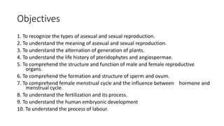
  • 1. Objectives 1. To recognize the types of asexual and sexual reproduction. 2. To understand the meaning of asexual and sexual reproduction. 3. To understand the alternation of generation of plants. 4. To understand the life history of pteridophytes and angiospermae. 5. To comprehend the structure and function of male and female reproductive organs. 6. To comprehend the formation and structure of sperm and ovum. 7. To comprehend female menstrual cycle and the influence between hormone and menstrual cycle. 8. To understand the fertilization and its process. 9. To understand the human embryonic development 10. To understand the process of labour.
  • 2. Main points • Double fertilization • Formation of sperm and ovule • Menstruation cycles and hormones • Fertilization and the development of zygote
  • 3. Difficult points • Importance of sexual reproduction • Alternative of generation in plants • Sexual reproduction in seed plants • Menstruation cycles and hormones
  • 4. Objectives 1. To understand the importance of reproduction. 2. To recognize the types of asexual reproduction. 3. To understand the meaning of asexual reproduction.
  • 6. Notice the sleeping movement of the plants
  • 7. Rewind… remember Junior High and Chapter 16 • How does a plant form more cells and grow? • How does a plant produce seeds? • Why does the sunflower plant needs to produce seed?
  • 8. Reproduction • Reproduction is the process of organisms producing offspring后代 • Continuation of species • Avoid extinction • Basic characteristic of living organisms.
  • 9. Types of reproduction • Asexual reproduction无性繁殖 • No fusion of reproductive cells / gametes配子. • produce a new individual directly from the parent body. • Sexual reproduction有性繁殖 • fusion of two gametes to form a zygote合子. • a new individual is form through the development of zygote.
  • 10. Gametes配子 • from Greek gamos ‘marriage’ • a mature haploid male or female germ cell which is able to unite with another of the opposite sex in sexual reproduction to form a zygote. Zygotes合子 • from Greek zugoun ‘to yoke’. • a diploid cell resulting from the fusion of two haploid gametes. yoke
  • 11.
  • 12. Quiz • Which of the following statements about asexual reproduction is NOT true? A. It does not improve genetic variation. B. It produces clones of the original cell. C. It does not require a mate for reproduction. D. It is a seldom used form of reproduction.
  • 14. Asexual reproduction • Binary fission • Budding • Sporulation • Fragmentation • Vegetative propagation
  • 15. Binary fission分裂生殖 • The parent cell divides to form genetically identical offspring with the same size and shape. • Simple fission二分裂 produces two offspring. • bacteria and amoeba • Multiple fission复分裂 produces many offspring. • Plasmodium疟原虫 裂殖体
  • 16.
  • 17. Budding出芽生殖 • A new individual is derived from an outgrowth (bud). • When the bud matures, the new individual separates from the body of the parent to form a new individual. • Sometimes the offspring do not separate from the parent before budding again. • Examples: yeast and hydra水螅.
  • 18. Sporulation孢子生殖 • Spores are stable, dormant cells that are produced from vegetative cells. • When the conditions are favourable, the spores germinate to form new individuals. • Example: most of the fungi and algae. • Some spores have flagella鞭毛that can swim - zoospores单胞藻
  • 19. Fragmentation断裂生殖 • Some parts of the organism break off and subsequently differentiate and develop into new individuals. • Example: spirogyra水绵, cnidarians腔肠动物 (e.g. hydra水螅) and annelids 环节动物. spirogyra
  • 20. Vegetative propagation 营养生殖 • New individuals develop from a part of nutritive organs (root, stem and leaves) of plants. • Example, tubers of potatoes, bulbs of onion
  • 21. Characters of asexual reproduction • Characters of the parents are maintained • New generations are genetically identical to the parents • Poorly adapt to new environment • Lack of diversity and genetic variation in the offspring population • Fast growing • Do not required complicated maturation process from zygote • Able to colonise an area quickly when the environment is favourable
  • 22. Quiz • Which of these statements is true of asexual reproduction? A. It produces offspring genetically identical to each other and requires one parent. B. It produces offspring genetically identical to each other and requires two parents. C. It produces offspring genetically different from each other and requires one parent. D. It produces offspring genetically different from each other and requires two parents.
  • 23. Quiz • When an organism undergoes a period of growth then splits in two separate organisms, it is called _____________ A. Budding B. Fission C. Fragmentation D. Parthenogenesis
  • 24.
  • 25. Quiz • Which of the following statements about gametes are true? I. In sexual reproduction, each parent provides genetic material for the formation of a gamete cell. II. Gametes are produced by mitosis. III. If a sexually reproducing organism is diploid, the gametes are haploid. A. I, II and III B. I and III C. II and III D. I and II
  • 27. Objectives 1. To recognize the types of sexual reproduction. 2. To understand the meaning of sexual reproduction. 3. To compare and contrast the characters of asexual and sexual reproduction.
  • 28. Sexual reproduction • Isogamy • Anisogamy • Oogamy
  • 29. Sexual reproduction有性生殖 • Two reproductive cells (gametes配子) (N + N) to form a zygote合子 (2N). • Zygotes develop into new individual. • Sexual reproduction can be divided into isogamy, anisogamy and oogamy according to their morphological appearance.
  • 30. Characters of sexual reproduction • More genetic variation in offsprings • Heredity characters of both parents • Crossovers • Better adaptive to changing environment
  • 31. lsogamy同配生殖 • Production and fusion of two gametes that are similar in size and structure. • Generally flagellated and mobile • Examples: lower animals and plants, such as certain protozoa 原生动物, algae (Chlamydomonas单 胞藻) and fungi.
  • 32. Anisogamy异配生殖 • The two gametes that fused to form zygotes are dissimilar in size. • Large gametes = female gametes = ova / egg cells 雌配子 • Small gametes = male gamates = sperm雄 配子 • Example: Pandorina空球藻.
  • 33. Oogamy卵式生殖 • Egg cell – female gametes • Sessile固定,不能运动 • Large • Provide nutrient for the zygotes受精卵,合子 • Sperm – male gametes • Mobile • Small • Search for the ova
  • 34.
  • 35. Parthenogenesis孤雌生殖/单性生殖 • Greek παρθένος, parthenos, 'virgin' + γένεσις, genesis, 'creation‘ • A natural form of asexual reproduction in which growth and development of embryos occur without fertilization. • In animals, parthenogenesis is the development of an embryo from an unfertilized egg cell. • In plants parthenogenesis is a component process of apomixis无融合生殖.
  • 37. Diploid parthenogenesis 双倍孤雌生殖 • In the spring / early summer • Lots of food • Produce 2N egg cells • Offspring born viviparity卵胎 生 • In the late summer / autum • Food is scarce • Produce offspring that have two sexes that will mate and produce fertilized eggs • Dormant in the winter aphid 蚜虫
  • 38.
  • 39.
  • 40.
  • 41. 17.2 Alternation of generation in plants
  • 42. Alternation of generation in plants • All plants undergo a life cycle that takes them through both haploid and diploid generations. • This fluctuation between these diploid and haploid stages that occurs in plants is called the alternation of generations世代交替.
  • 43. Sporophyte and gametophyte • Sporophyte孢子体 • grow from a zygote • diploid • produces spores through meiotic division. • Gametophyte配子体 • grow from a spore • haploid • produces gametes through mitosis
  • 44. Quiz: fill in the blanks Generalized Life Cycle Pattern For Animals & Plants 4 3 2 1 5
  • 45. Human Life Cycle • In the human life cycle (and the life cycles of most multicellular animals), the only cells that are haploid are the sperm and egg. • From the zygote to the diploid mother cells inside the sex organs, all the cells are diploid with two sets of chromosomes. • Humans are dioecious species with separate male and female individuals in the diploid population.
  • 47. general evolutionary trend • gradual increase in the diploid sporophyte phase • decrease in the haploid gametophyte phase
  • 48. Moss
  • 49.
  • 50. Moss Life Cycle • The dominant (conspicuous) part of the life cycle is the haploid, leafy N gametophyte. • The gametophytes are photosynthetic. • Most moss gametophytes are dioecious, with separate male and female individuals in the population. • Female sex organs = archegonia. • Male sex organ = antheridium • Fertilization involves a motile, biflagellate sperm that swims through water to reach the egg on female plants.
  • 51. Moss Life Cycle • The non-photosynthetic diploid sporophyte consists of a sporangium- bearing stalk that grows directly out of the gametophyte. • Spore mother cells within the sporangium undergo meiosis, producing numerous haploid spores that fall to the ground like tiny particles of dust. • Since the sporophyte is without chlorophyll, it is completely dependent on the gametophyte for its water, minerals and carbohydrate nutrition. • Consequently, the sporophyte of the moss is heterotrophic and parasitic on the gametophyte.
  • 52. Quiz
  • 53.
  • 56. Fern life cycle • The dominant (conspicuous) part of the life cycle is the diploid, leaf-bearing sporophyte. • The sporophytes of ferns are photosynthetic and autotrophic. • Spore mother cells within the sporangium undergo meiosis, producing numerous haploid spores. • The sporangia split open at maturity, releasing millions of spores that fall to the ground like tiny particles of dust.
  • 57. Fern life cycle • Each spore germinates and grows into a heart-shaped gametohyte (prothallus). • The gametophytes of ferns are photosynthetic and autotrophic. • male sex organs (antheridia) • female sex organs (archegonia) • Ferns are typically monoecious (=bisexual) with both male and female sex organs on the same gametophytes. • Fertilization involves a multiciliate sperm that swims through water to reach the egg. • Young sporophyte developed from the old grametophyte.
  • 58. Quiz Fertilization Mitosis (2n) Leaf of young sporophyte growing from gametophyte indusium sorus Meiosis Mitosis (n) Spore Gametophyte Sporophyte Embryo Sporangium
  • 59.
  • 60. Angiosperm • Also known as angiospermophyta or the flowering plant. • Greek angos –ανγοσ “box” + sperma –σπέρμα “seed” + phyto –φυτό
  • 61. Characters of life cycle of angiosperms • Gametophyte reduce • Gametophyte reply on sporophyte to survive • Does not need H20 to reproduce – pollen tube • Double fertilization • Embryo nourished with endosperm
  • 62. Life cycle of angiosperm • Sporophyte = the plant • Gametophyte • Megagametophyte = ovule • Microgametophyte = pollen grain • Forms in flower
  • 63. Structure of a flower 雌蕊 花药 花丝 雌蕊 柱头 花柱 子房 胚珠 花瓣 花萼 花托 花梗
  • 64. Quiz: label the flower. 1 2 3 4 5 6 7 8 9
  • 65. Formation of microspore • Stamen = anther + filament • Each anther has four pollen sacs with many 2N pollen mother cells. • 2N pollen mother cells meiosis to form four N pollen grains = microspores.
  • 66. Growth of microspore • The nucleus of each microspore undergo mitosis. • A N generative cell生殖细胞 without cell wall is formed within the N vegetative cell营 养细胞 • Generative cell will mitosis into two N sperm cells. • Vegetative cell provides nutrients and will form the tube cell.
  • 67. Mature pollen grain • N mature pollen grain = N male gametophyte • 1 vegetative cell • 1 generative cell OR 2 sperm cells • A pollen grain contain germ pore on the surface. • Pollen tube grows from these pores.
  • 68. A. Cross-section through an anther of Lilie (Lilium)sp.) with on the left and the right side two loculi each. In the loculi sporemothercells (SMCs) can be seen from which the four spores develop through meiosis I and II. Inbetween the loculi of each pair a thin layer of cells (arrow) is visible along which the loculus can burst open atmaturity and release the pollen grains. In the middle the cross-sectioned filament (Fi) to which the anther is attached is indicated. In the upper part the vascular bundle (v) of the loculus can be distinguished. B. Loculus. The lumen contains developing pollen. On the inner wall (w) of the loculus a layer constitued of block-shaped single cells is present, the tapetum (t). The tapetum feeds the developing spore and -later- pollen. C. Tetrad stage during pollen development. After the two meiotic divisions the four daughter cells are still interconnected and form a tetrad. They are still surrounded by the wall (arrow) of the original cell, the microspore mothercell (MMC). D. Mitotic division in the spore leading to the formation of a microgametophyte or pollen. Only the metaphase is shown here. The chromosomes lay in the equatorial plane of the cell. E. Nearly ripe pollen grain: visible are a vegetative cell with nucleus (VN), which later will form the pollen tube, and a generative cell with its own nucleus (GN), which later will divide into two sperm cells. F. Ripe pollen grain in which the texture of the outer cell wall, the exine, can be recognized. The grainy dark purple structure in the middle of the pollen grain is the vegetative nucleus. G. Diagram in 3 parts: Ripe pollen grain consisting of the vegetative cell (VC) and therein the smaller generative cell (GC). After landing on the stigma (St) the pollen grain germinates and forms a pollen tube. In the pollen tube the generative cell divides into two sperm cells (SC). The pollen tube grows to the embryo sac (ES) and delivers the two sperm cells that are involved in double fertilization.
  • 69.
  • 70. Structure of a pistil • Pistil = stigma + style + ovary • Inside the ovary, there may be one or numerous 2N ovule • Ovule = megasporangia 雌蕊 = 柱头 + 花柱 + 子房
  • 71. Structure of an ovary • Funicle: to support, projection and conduction • Nucellus: where development of female gametophyte occurs • Integument: to protect nucellus and embryo sac (megagametophyte) • Micropyle: for a passage for pollen tube to enter the ovule • Chalaza: transfer nutrients to nucellus
  • 72. Formation of a megaspore • The 2N megasporocyte (megaspore mother cell)大 孢子母细胞 undergoes meiosis • Four N megaspores大孢子 are form • Three megaspore that are closer to the micropyle degenerate • Only one megaspore珠孔 remaining functional micropyle
  • 73. Formation of an megagametophyte • After 3 mitosis • 7 N cells with 8 nucleus are form • = embryo sac胚囊 • = megagametophyte
  • 74. Megagametophyte = embryo sac • At the microphylar end • 1 egg cell卵细胞 • 2 synergids助细胞 • Middle • 1 central cell中央细胞 with 2 polar nuclei极核 • May fuse to form a 2N cell • At the chalazal end • 3 antipodals cells反足细胞
  • 75. Function of cells in the embryo sac • The central cell, after fertilization, develops into the endosperm胚乳, which produce nutrients to the zygote. • The synergids are thought to help the pollen nucleus reach the egg cell for fertilization. • The antipodal cells nourishes the embryo sac
  • 76.
  • 77.
  • 78. Pollination • Pollination is the act of transferring pollen grains from the male anther of a flower to the female stigma.
  • 79. Growth of pollen tube • A pollen grain germinate in response to a sugary fluid and lipids secreted by the mature stigma. • The vegetative cell then produces the pollen tube, a tubular protrusion from the pollen grain, which carries the sperm cells within its cytoplasm. • The germinated pollen tube must then drill its way through the nutrient-rich style and curl to the bottom of the ovary to reach the ovule. • Once the pollen tube reaches an ovule, it bursts to deliver the two sperm cells.
  • 80. Double fertilization • One of the sperm fertilizes the egg cell which develops into an 2N embryo胚, which will become the future plant. • The other sperm cell fuses with both polar nuclei of the central cell to form the 3N endosperm胚乳, which serves as the embryo's food supply.
  • 81. • The ovary will develop into a fruit. • The ovules will develop into seeds.
  • 82.
  • 84.
  • 85.
  • 86.
  • 87.
  • 89. • Human male reproductive system includes testes, epididymis, vas deferens (sperm duct), seminal vesicles, prostate gland, bulbourethral glands and penis etc.
  • 90. Testes • Human male reproductive system consists of two testes which both are enclosed by scrotum. • The functions of the testes are to produce both sperm and androgens, primarily testosterone.
  • 91. Tubules of testes • Within the testes are very fine coiled tubes called seminiferous tubules. • Primary cell types within the seminiferous tubules includes germ cells, sertoli and peritubular myoid cells. • Cells between the seminiferous tubules are called interstitial cells), primarily Leydig cells. • All the tubules in a testis are joined to a single tubes called epididymis. • Epididymis is the site for temporary storing of sperms and for the sperm to develop until mature. Lumen
  • 92. Primary cell types within the seminiferous tubules • The germ cells give rise to sperm through spermatogenesis. • The sertoli cells are the true epithelium of the seminiferous epithelium, and is critical for the support of germ cell development into spermatozoa. Sertoli cells also secrete an inhibitory hormone called inhibin. • Peritubular myoid cells are smooth muscle cells surrounding the seminiferous tubules.
  • 93. Primary cell types between the seminiferous tubules • The Leydig cells, which are generally refers as interstitial cells, secrete testosterone for sexual development and puberty, secondary sexual characteristics, supporting spermatogenesis and erectile function.
  • 94. Semen • Semen is a fluid that is a composition of sperms and other secretions. Gland/Site Features Testis/Epididymis Sperm / spermatozoa Seminal Vesicle Contains large amounts of fructose, which is used by the sperm mitochondria to generate ATP to allow movement through the female reproductive tract. Prostate Produce an alkaline, milky fluid that is critical to first coagulate and then decoagulate the semen following ejaculation. Bulbourethral Glands or Cowper’s glands Produce a thick, salty mucus that lubricates the end of the urethra and the vagina, and helps to clean urine residues from the penile urethra.
  • 95. Ejaculation • Ejaculation is the discharge of semen from the male reproductory tract. • When the male orgasm, small arteries of erectile tissue (spongy tissue) dilate to increase the blood supply, causes penis to become hard and erect.
  • 96. Ejaculation • Rhythmic contractions of muscles will produce waves of pressure within the urethra. • This enables sperm enter urethra and mixes with the fluid that secreted from seminal vesicles, prostate gland and bulbourethral gland to form semen. The resultant semen is forced out of the penis by powerful contractions of the urethra.
  • 99. Spermatogenesis • The process of the formation of sperm in the testes is called spermatogenesis. • Spermatogenesis occurs in the seminiferous tubules. • One production cycle, from spermatogonia through formed sperm, takes approximately 64 days.
  • 100. Spermatogenesis • Mitosis of a spermatogonium (stem cell) involves a single cell division that results in two identical, diploid daughter cells called primary spermatocyte. • Meiosis has two rounds of cell division: primary spermatocyte to secondary spermatocyte, and then secondary spermatocyte to spermatid. • A total of four spermatids are produced from each spermatogonium.
  • 101. Spermatogenesis • Although haploid, early spermatids look very similar to cells in the earlier stages of spermatogenesis, with a round shape, central nucleus, and large amount of cytoplasm. • These early spermatids reduced the cytoplasm to form sperm. • Eventually, the sperm are released into the lumen and are moved along a series of ducts in the testis toward a structure called the epididymis for the next step of sperm maturation.
  • 104. Three regions of a sperm • Sperm have a distinctive head, mid-piece, and tail region. • The head of the sperm contains the extremely compact haploid nucleus with very little cytoplasm. • An acrosome covers most of the head of the sperm cell as a “cap” that is filled with lysosomal enzymes important for preparing sperm to participate in fertilization. • Tightly packed mitochondria fill the mid-piece of the sperm. ATP produced by these mitochondria will power the flagellum. • The tail is a flagellum , which extends from the neck and the mid-piece through the tail of the sperm, enabling it to move the entire sperm cell.
  • 105. 17.3.1.3 Hormonal control of male reproductive system
  • 106. Hormonal Control of Male Reproduction • The hypothalamus monitors and causes the release of hormones from the pituitary gland using a negative feedback mechanism. • When the reproductive hormone is required, the hypothalamus sends a gonadotropin- releasing hormone (GnRH) to the anterior pituitary. • This causes the release of gonadotrophic hormones, i.e. follicle stimulating hormone (FSH) and luteinizing hormone (LH) from the anterior pituitary into the blood.
  • 107. Negative feedback loop of spermatogenesis • FSH enters the testes and stimulates the Sertoli cells to begin facilitating spermatogenesis. • The Sertoli cells produce the hormone inhibin, which is released into the blood when the sperm count is too high. • This inhibits the release of GnRH and FSH, which will cause spermatogenesis to slow down. • If the sperm count reaches 20 million/ml, the Sertoli cells cease the release of inhibin, and the sperm count increases.
  • 108. Negative feedback loop of testosterone • LH also enters the testes and stimulates the interstitial cells of Leydig besides the seminiferous tubule to make and release testosterone into the testes and the blood. • Testosterone, the hormone responsible for the secondary sexual characteristics that develop in the male during adolescence, also stimulates spermatogenesis. • A negative feedback system occurs in the male with rising levels of testosterone acting on the hypothalamus and anterior pituitary to inhibit the release of GnRH, FSH, and LH.
  • 109. Micrograph showing a cluster of Leydig cells (center of image). H&E stain. Histological section through testicular parenchyma of a boar. 1 Lumen of convoluted part of the seminiferous tubules, 2 spermatids, 3 spermatocytes, 4 spermatogonia, 5 Sertoli cell, 6 myofibroblasts, 7 Leydig cells, 8 capillaries
  • 110. Quiz • Which hormone causes Leydig cells to make testosterone? A. FSH B. LH C. inhibin D. estrogen
  • 111. Quiz • Which hormone causes FSH and LH to be released? A. testosterone B. estrogen C. GnRH D. progesterone
  • 115. The female reproductive system is located primarily inside the pelvic cavity.
  • 116. Vagina • Serves as the entrance to the reproductive tract. • Serves as the exit from the uterus during menses and childbirth. • The vagina is home to a normal population of microorganisms that help to protect against infection by pathogenic bacteria, yeast, or other organisms that can enter the vagina. • The vaginal bacteria secretes lactic acid, and thus protects the vagina by maintaining an acidic pH (below 4.5).
  • 117. Ovaries • The ovaries are the pair of female gonads. • The ovaries are supported by ovarian ligament that contains the ovarian blood and lymph vessels. • The cortex of ovaries is composed the germinal epithelium, that will eventually forms oocyte. • Beneath the cortex lies the inner ovarian medulla, the site of blood vessels, lymph vessels, and the nerves of the ovary.
  • 118. Ovaries • The ovaries are the pair of female gonads. • The ovaries are supported by ovarian ligament that contains the ovarian blood and lymph vessels. • The cortex of ovaries is composed the germinal epithelium, that will eventually forms oocyte. • Beneath the cortex lies the inner ovarian medulla, the site of blood vessels, lymph vessels, and the nerves of the ovary.
  • 119. Oviduct / Fallopian tubes • The oviducts serve as the conduit of the oocyte from the ovary to the uterus. • Each of the two uterine tubes is close to, but not directly connected to, the ovary. • The distal end (closer to the ovaries) flares out with slender, finger-like projections called fimbriae. • The inner layer of the oviduct contains ciliated cells that beat in the direction of the uterus, producing a current that move the oocyte.
  • 120. The Uterus and Cervix • The uterus is the muscular organ that nourishes and supports the growing embryo. • The cervix is the narrow inferior portion of the uterus that projects into the vagina. • The cervix produces mucus secretions that become thin and stringy under the influence of high systemic plasma estrogen concentrations, and these secretions can facilitate sperm movement through the reproductive tract. • The innermost layer of the uterus is called the endometrium. • Structurally, the endometrium consists of two layers: the stratum basalis and the stratum functionalis (the basal and functional layers). The stratum basalis layer does not shed during menses. • In contrast, the thicker stratum functionalis grows and thickens in response to increased levels of estrogen and progesterone. This inner functional layer provides the proper site of implantation for the fertilized egg, and—should fertilization not occur—it is only the stratum functionalis layer of the endometrium that sheds during menstruation.
  • 122. Before birth • The ovarian stem cells, or oogonia (sg. oogonium), are formed during fetal development, and divide via mitosis, from the germinal epithelium. • Oogonia form primary oocytes in the fetal ovary prior to birth. • These primary oocytes are then arrested in prophase I of meiosis I.
  • 123. After puberty • Meiosis resumes. • The cytoplasm is divided unequally, and one daughter cell is much larger than the other. • This larger cell is the secondary oocyte. • The secondary oocyte arrests at metaphase II. • The smaller cell, called the first polar body, eventually disintegrates.
  • 124. After puberty • Meiosis of a secondary oocyte is completed only if a sperm succeeds in penetrating its barriers. • Meiosis II then resumes, producing one haploid ovum that, at the instant of fertilization by a (haploid) sperm, becomes the first diploid cell of the new offspring (a zygote). • Thus, the ovum can be thought of as a brief, transitional, haploid stage between the diploid oocyte and diploid zygote.
  • 125. Follicle development • Ovarian follicles are oocytes and their supporting cells. • Primordial follicles are resting follicle with a single flat layer of support follicular cells, that surround the oocyte, and they can stay in this resting state for years—some until right before menopause. • After puberty, a few primordial follicles will join a pool of immature growing follicles called primary follicles, as the follicular cells increase in size and proliferate. • Secondary follicles increase in diameter, adding a new outer layer of connective tissue, blood vessels, and starts to produce estrogens. • Tertiary follicle has a thick fluid, called follicular fluid, collected in a large pool.
  • 126. Ovulation • Several follicles reach the tertiary stage at the same time, and most of these will undergo atresia (die). • The one that does not die will continue to grow and develop until ovulation, when it will expel its secondary oocyte surrounded by several layers of follicular cells from the ovary.
  • 127. In this electron micrograph of a secondary follicle, the oocyte, theca cells (thecae folliculi), and developing antrum are clearly visible. EM × 1100. (Micrograph provided by the Regents of University of Michigan Medical School © 2012)
  • 128. Ovulation • Several follicles reach the tertiary stage at the same time, and most of these will undergo atresia (die). • The one that does not die will continue to grow and develop until ovulation, when it will expel its secondary oocyte surrounded by several layers of follicular cells from the ovary.
  • 129. Afterwards • The collapsed follicle is transformed into a new endocrine structure called the corpus luteum. • Corpus luteum produces large amounts of the sex steroid hormone progesterone, a hormone that is critical for the establishment and maintenance of pregnancy and to prevent development of new dominant follicles develop at this time. • If pregnancy does not occur within 10 to 12 days, the corpus luteum will stop secreting progesterone and degrade into the corpus albicans, a nonfunctional “whitish body” that will disintegrate in the ovary over a period of several months.
  • 130. The ovarian cycle • The ovarian cycle refers to the series of changes in the ovary during which the follicle matures, the ovum is shed, and the corpus luteum develops. • The follicular phase describes the development of the follicle in response to follicle stimulation hormone ( FSH ). • As luteinizing hormone ( LH ) and FSH levels increase they stimulate ovulation, or the release of a mature oocyte into the fallopian tubes. • In the luteal phase, the corpus luteum forms on the ovary and secretes many hormones, most significantly progesterone, which makes the endometrium of the uterus ready for implantation of an embryo. • If implantation does not occur, the corpus luteum will be degraded, resulting in menstruation. • If implantation occurs the corpus luteum is maintained.
  • 132. Ovum • Ovum is a rounded and non-motile structure. • The cytoplasm of the egg is called ooplasm. • It contains a very little amount of yolk in man. • In animals where huge amount of yolk is present, the cytoplasm of egg consists of lipoproteins, pigment granules, water, along with other cytoplasmic organelles. • The peripheral layer of ooplasm is known as cortex and it contains many microvilli and cortical granules. • The cortical granules are most associated with polyspermy prevention after the event of fertilization. • Microvilli are tubular outrushing of plasmalemma for transportation of substances into and out of egg cytoplasm.
  • 133. Ovum • In man the ovum is covered over by a thin vitelline membrane (made up of protein fibre) which is further covered over by another primary membrane known as zona pellucida. • During discharge of ovum from the graafian follicle, several layer’ of follicular cells adhere to the outer surface of zona pellucida and are arranged radially forming corona radiata.
  • 135. Menstrual cycle • Menstrual cycle is the series of changes in which the uterine lining is shed, rebuilds, and prepares for implantation. • The cycle length is determined by counting the days between the onset of bleeding in two subsequent cycles. • The average length of a woman’s menstrual cycle is 28 days, but the length of the menstrual cycle varies among women, and even in the same woman from one cycle to the next, typically from 21 to 32 days.
  • 136. Phases of the menstrual cycle • The three distinct phases of the menstrual cycle. • The menses phase of the menstrual cycle is the phase during which the lining is shed. • The endometrium begins to proliferate again in the proliferative phase. • The secretory phase of the menstrual cycle refers the maintenance of the thickness of the endometrium as the endometrial lining prepares for implantation.
  • 137. Menses phase • The menses phase occurs during the early days of the follicular phase of the ovarian cycle, when progesterone, FSH, and LH levels are low. • Progesterone concentrations decline as a result of the degradation of the corpus luteum, marking the end of the luteal phase. • This decline in progesterone triggers the shedding of the endometrium.
  • 138. Proliferative phase • The tertiary follicles begin to produce increased amounts of estrogen that stimulate the endometrial lining to rebuild. • The high estrogen concentrations will lead to a decrease in FSH as a result of negative feedback, resulting in atresia of all but one of the developing tertiary follicles. • The switch to positive feedback—which occurs with the elevated estrogen production from the dominant follicle— then stimulates the LH surge that will trigger ovulation. • In a typical 28-day menstrual cycle, ovulation occurs on day 14. • Ovulation marks the end of the proliferative phase as well as the end of the follicular phase.
  • 139. Secretory Phase • In the ovary, the collapsed follicle forms the progesterone-producing corpus luteum, marking the beginning of the luteal phase of the ovarian cycle. • In the uterus, progesterone from the corpus luteum begins the secretory phase of the menstrual cycle, in which the endometrial lining prepares for implantation. • Over the next 10 to 12 days, the endometrial glands secrete a fluid rich in glycogen that can nourish developing zygote. At the same time, the spiral arteries develop to provide blood to the thickened endometrium.
  • 140. Secretory Phase • If no pregnancy occurs within approximately 10 to 12 days, the corpus luteum will degrade into the corpus albicans. • Levels of both estrogen and progesterone will fall, and the endometrium will grow thinner. • Prostaglandins will be secreted that cause constriction of the spiral arteries, reducing oxygen supply. • The endometrial tissue will die, resulting in menses—or the first day of the next cycle.
  • 141. Summary • The follicular phase begins with an increase in follicle -stimulation hormone (FSH), which causes increases in luteinizing hormone (LH) and gonadotropin-releasing hormone (GnRH). Concurrent increases in estrogen levels cause increases in progesterone, stimulating proliferation of the endometrium. • A spike in LH and FSH (“LH surge”) causes ovulation, following a suppression of GnRH. • Estrogen levels continue to rise following ovulation and the corpus luteum forms, which secretes progesterone in significant levels and causes decreases in LH and FSH levels. • Without implantation, estrogen and progesterone levels will fall and the corpus luteum will degrade.
  • 142. 17.3.3.2 Hormonal control in the female reproductive system
  • 143. Follicular phase • The hypothalamus produces GnRH, a hormone that signals the anterior pituitary gland to produce the gonadotropins FSH and LH. • FSH stimulates the follicles to grow, and the five or six tertiary follicles expand in diameter. • The release of LH also stimulates the follicles to produce the sex steroid hormone estradiol, a type of estrogen.
  • 144. Follicular phase • The larger a follicle is, the more estrogen it will produce in response to LH stimulation. • As a result of these large follicles producing large amounts of estrogen, systemic plasma estrogen concentrations increase. • Following a classic negative feedback loop, the high concentrations of estrogen will stimulate the hypothalamus and pituitary to reduce the production of GnRH, LH, and FSH. • Because the large tertiary follicles require FSH to grow and survive at this point, this decline in FSH caused by negative feedback leads most of them to die (atresia). • Typically only one follicle, now called the dominant follicle, will survive this reduction in FSH, and this follicle will be the one that releases an oocyte.
  • 145. Ovulation • The remaining follicle secrets a high concentrations of estrogen • This trigger the anterior pituitary to responds by secreting large amounts of LH and FSH into the bloodstream. • It is this large burst of LH (called the LH surge) that leads to ovulation of the dominant follicle.
  • 146. Luteal phase • The surge of LH also stimulates the luteinization of collapsed follicle into a new endocrine structure called the corpus luteum. • The corpus luteum begin to produce large amounts of the sex steroid hormone progesterone. • Progesterone triggers negative feedback at the hypothalamus and pituitary, which keeps GnRH, LH, and FSH secretions low, so no new dominant follicles develop at this time.
  • 147. 17.3.4 Development of human embryo
  • 148. Gestation • From Latin gestare ‘carry, carry in the womb’ • Gestation is the period of time required for full development of a fetus in utero. • Prenatal development can be subdivided into distinct gestational periods: • Pre-Embryonic / Germinal stage – first two weeks • Embryonic stage – weeks 3 -8 • Fetus stage – week 9 until birth
  • 149. Pre-implantation Embryonic Development • Following fertilization, the zygote and its associated membranes (= conceptus), continue to be projected toward the uterus by peristalsis and beating cilia. • During its journey to the uterus, the zygote undergoes five or six rapid mitotic cell divisions in the process called cleavage. • Although each cleavage results in more cells, it does not increase the total volume of the cells. • Each daughter cell produced by cleavage is called a blastomere (blastos = “germ,” in the sense of a seed or sprout).
  • 150. Pre-implantation Embryonic Development • Approximately 3 days after fertilization, a 16-cell conceptus reaches the uterus. • The cells that had been loosely grouped are now compacted and look more like a solid mass. • The name given to this structure is the morula (morula = “little mulberry”).
  • 151. Pre-implantation Embryonic Development • Once inside the uterus, the conceptus continues to divide, creating a ball of approximately 100 cells, and consuming nutritive endometrial secretions called uterine milk while the uterine lining thickens. • The ball of now tightly bound cells starts to secrete fluid and organize themselves around a fluid-filled cavity, the blastocoel. • At this developmental stage, the conceptus is referred to as a blastocyst.
  • 152. Pre-implantation Embryonic Development • Within this structure, a group of cells forms into an inner cell mass, which is fated to become the embryo. • The cells that form the outer shell are called trophoblasts (trophe = “to feed” or “to nourish”). • As the blastocyst forms, the trophoblast excretes enzymes that begin to degrade the zona pellucida in a process called “hatching,” • The conceptus breaks free of the zona pellucida in preparation for implantation. • The trophoblasts will develop into the extra- embryonic structures.
  • 153. Day 3 to 6
  • 154. Implantation • At the end of the first week, the blastocyst comes in contact with the uterine wall and adheres to it, embedding itself in the uterine lining via the trophoblast cells. • The trophoblast is invasive, secreting proteolytic enzymes that allow the blastocyst to penetrate into the endometrium.
  • 155. Disorders from incorrect implantation • In one to two percent of cases, the embryo implants either outside the uterus (an ectopic pregnancy) or in a region of uterus that can create complications for the pregnancy.
  • 156. Disorders from incorrect implantation • If the embryo implants in the inferior portion of the uterus, the placenta can potentially grow over the opening of the cervix, a condition call placenta previa.
  • 157. Embryonic Membranes • During the second week of development, with the embryo implanted in the uterus, cells within the blastocyst start to organize into layers. • Some grow to form the extra-embryonic membranes needed to support and protect the growing embryo: the amnion, the yolk sac, the allantois, and the chorion.
  • 158.
  • 159. Amnion • At the beginning of the second week, the cells of the inner cell mass form into a two-layered disc of embryonic cells, and a space—the amniotic cavity— opens up between it and the trophoblast. • Cells from the upper layer of the disc (the epiblast) extend around the amniotic cavity, creating a membranous sac that forms into the amnion by the end of the second week. • The amnion fills with amniotic fluid and eventually grows to surround the embryo.
  • 160. Amniotic fluid • Floating within the amniotic fluid, the embryo—and later, the fetus—is protected from trauma and rapid temperature changes. • During delivery, the amniotic fluid also lubricates the vagina.
  • 161. Yolk sac • On the ventral side of the embryonic disc, opposite the amnion, cells in the lower layer of the embryonic disk (the hypoblast) extend into the blastocyst cavity and form a yolk sac. • The yolk sac supplies some nutrients absorbed from the trophoblast and also provides primitive blood circulation to the developing embryo for the second and third week of development. • When the placenta takes over nourishing the embryo at approximately week 4, the yolk sac has been greatly reduced in size and its main function is to serve as the source of blood cells and germ cells (cells that will give rise to gametes).
  • 162. Allantois • During week 3, a finger-like outpocketing of the yolk sac develops into the allantois, a primitive excretory duct of the embryo that will become part of the urinary bladder. • Together, the stalks of the yolk sac and allantois establish the outer structure of the umbilical cord.
  • 163. Chorion • The last of the extra- embryonic membranes is the chorion, which is the one membrane that surrounds all others. • The chorionic membrane forms finger-like structures called chorionic villi that burrow into the endometrium like tree roots, making up the fetal portion of the placenta.
  • 164. Placenta • The placenta is an organ that connects the developing fetus to the uterine wall to allow nutrient uptake, thermo- regulation, waste elimination, and gas exchange via the mother's blood supply; to fight against internal infection; and to produce hormones which support pregnancy. • The placenta attaches to the wall of the uterus, and the fetus's umbilical cord develops from the placenta.
  • 165. Exchange of substances • Some substances such as oxygen, carbon dioxide, and any other lipid- soluble substances move across the placenta by simple diffusion. • Other substances such as water- soluble glucose move across by facilitated diffusion. • The fetus has a high demand for amino acids and iron, and those substances are moved across the placenta by active transport.
  • 166. Placenta blood barrier • Maternal and fetal blood does not commingle because blood cells cannot move across the placenta. • This separation prevents the mother’s cytotoxic T cells from reaching and subsequently destroying the fetus, which bears “non-self” antigens. • Further, it ensures the fetal red blood cells do not enter the mother’s circulation and trigger antibody development (if they carry “non-self” antigens)—at least until the final stages of pregnancy or birth.
  • 167. Functions of the Placenta Nutrition and digestion Respiration Endocrine function •Mediates diffusion of maternal glucose, amino acids, fatty acids, vitamins, and minerals •Stores nutrients during early pregnancy to accommodate increased fetal demand later in pregnancy •Excretes and filters fetal nitrogenous wastes into maternal blood •Mediates maternal-to-fetal oxygen transport and fetal- to-maternal carbon dioxide transport •Secretes several hormones, including hCG, estrogens, and progesterone, to maintain the pregnancy and stimulate maternal and fetal development •Mediates the transmission of maternal hormones into fetal blood and vice versa
  • 168.
  • 169. Umbilical cord • The umbilical cord is a conduit between the developing embryo or fetus and the placenta. • The umbilical cord develops from and contains remnants of the yolk sac and allantois. • The umbilical cord is physiologically normally contains two arteries (the umbilical arteries) and one vein (the umbilical vein. • The umbilical vein supplies the fetus with oxygenated, nutrient- rich blood from the placenta. • The fetal heart pumps deoxygenated, nutrient-depleted blood through the umbilical arteries back to the placenta.
  • 170. Germs layers • As the third week of development begins, the two-layered disc of cells becomes a three-layered disc. • The first layer is the endoderm, a sheet of cells that displaces the hypoblast and lies adjacent to the yolk sac. • The second layer of cells fills in as the middle layer, or mesoderm. • The ectoderm is the layer adjacent to the amniotic cavity.
  • 171. Germs layers • Each of these germ layers will develop into specific structures in the embryo.
  • 172. Germs layers • The ectoderm gives rise to cell lineages that differentiate to become the central and peripheral nervous systems, sensory organs, epidermis, hair, and nails. • Mesodermal cells ultimately become the skeleton, muscles, connective tissue, heart, blood vessels, and kidneys. • The endoderm goes on to form the epithelial lining of the gastrointestinal tract, liver, and pancreas, as well as the lungs.
  • 173. Growth of embryo • A 4-week embryo is about 6 mm long and 0.01 gram in mass and the embryo heart beat is starts. • During 5th to 6th weeks, hands and legs grow out from the limb bud. • During the 8th weeks, embryo’s head, face and four limbs etc. will appear and look like a man, it is now known as a fetus. •
  • 174. Growth of fetus • When the fetus develops until 12th weeks, all the internal organs and systems are formed. • It focuses on the growth and the differentiation of main organs and tissues for the future development. • The head of the fetus is normally tilted downwards after 7 months of pregnancy. • During the end of 9th months, the fetus is mature.
  • 175. Birth • Birth is the act or process of bearing or bringing forth offspring. • The normal process of childbirth takes several hours and has three stages.
  • 176. The first stage • The first stage starts with a series of involuntary contractions of the muscular walls of the uterus and gradual dilation of the cervix. • The active phase of the first stage starts when the cervix is dilated more than about 4 cm in diameter and is when the contractions become stronger and regular. • The head (or the buttocks in a breech birth) of the baby is pushed against the cervix, which gradually dilates until is fully dilated at 10 cm diameter. • At some time, the amniotic sac bursts and the amniotic fluid escapes (also known as rupture of membranes or breaking the water).
  • 177. The second stage • In stage two, starting when the cervix is fully dilated, strong contractions of the uterus and active pushing by the mother expels the baby out through the vagina, which during this stage of labour is called a birth canal as this passage contains a baby, and the baby is born with umbilical cord attached. • After birth, doctors will cut the umbilical cord and apply a layer of medicine onto the wound.
  • 178. The third stage • In stage three, which begins after the birth of the baby, further contractions expel the placenta, amniotic sac, and the remaining portion of the umbilical cord usually within a few minutes. • About 10 minutes later, placenta and ectoderm of the fetus will be expelled from the mother body. The whole process of birth is completed.

Hinweis der Redaktion

  1. https://dr282zn36sxxg.cloudfront.net/datastreams/f-d%3Af9b681a7c59c2a09aeb8eb512f2a52a6d6996d09c789a596c616229b%2BIMAGE_TINY%2BIMAGE_TINY.1
  2. https://www.youtube.com/watch?v=zst08tm9s6M
  3. https://static.diffen.com/uploadz/thumb/8/8f/Asexual-Sexual-Reproduction.png/300px-Asexual-Sexual-Reproduction.png
  4. https://askgramps.org/files/2017/11/yoke-and-oxen.jpg https://qph.fs.quoracdn.net/main-qimg-978f3c936c0d2523a141b4edfb6f04e1-c
  5. https://i.pinimg.com/736x/75/09/cb/7509cb7b5e96deed1ea7f3f0d73632ad--reproduction-a-photo.jpg
  6. https://s3mn.mnimgs.com/img/shared/ck-files/ck_58bb07393ba6c.png https://qph.fs.quoracdn.net/main-qimg-c641567e9aa3ccde4186dd5562630e74-c
  7. https://dr282zn36sxxg.cloudfront.net/datastreams/f-d%3A62148f13905f82ac40c55dbe9cf2db3bf148a9daab4f3820aa319aad%2BIMAGE_THUMB_POSTCARD_TINY%2BIMAGE_THUMB_POSTCARD_TINY.1
  8. http://album.udn.com/community/img/PSN_PHOTO/Gabriel33/f_5025397_1.jpg https://www.researchgate.net/profile/Rasha_Aref2/publication/308144762/figure/fig1/AS:456616182063104@1485877189399/Scanning-electron-microscopy-image-of-Saccharomyces-cerevisiae-The-budding-yeast-cells.ppm http://www.biologydiscussion.com/invertebrate-zoology/phylum-coelenterata/hydra-history-habitat-and-locomotion-with-diagram/28686
  9. http://www.biologydiscussion.com/plants/plant-kingdom/list-of-35-thallophytes-fungi/31503 http://www.kepu.net.cn/gb/lives/plantae/pic/Fl201001060959109989.jpg https://samagra.itschool.gov.in/uploads/12/botony/916/1716/12_Ch916_12151/R_Zoospres%20in%20Chlamydomonas.jpg
  10. http://ctkcole16.weebly.com/uploads/5/7/8/1/57814483/8414642_orig.jpg https://image.tutorvista.com/content/feed/tvcs/spirogyra-fragmentation.jpeg
  11. http://cdn.biologydiscussion.com/wp-content/uploads/2014/12/clip_image002_thumb153.jpg
  12. https://s3-ap-southeast-1.amazonaws.com/learnhive/lcards/Types-of-Asexual-Reproduction-52d9233436401.png
  13. http://2.bp.blogspot.com/-PA2r_RDsi4w/UNh6eUNEOQI/AAAAAAAAEQc/CTrX7Z4pFH8/s1600/chlamydomonasrepro.jpg http://wsm.hebeijiaoyu.com.cn/html/cp/whys_100k/swgwsm/port1/zao/doc-18020.html
  14. http://fmp.conncoll.edu/Silicasecchidisk/Pics/Other%20Algae/Green_jpegs/Pandorina1.jpg
  15. http://wsm.hebeijiaoyu.com.cn/html/cp/whys_100k/swgwsm/port1/zao/doc-18020.html http://www.biologydiscussion.com/algae/life-cycle-of-chlamydomonas-with-diagram/53699
  16. http://pediaa.com/difference-between-anisogamy-isogamy-and-oogamy/
  17. https://cdn.britannica.com/09/189109-004-DEEDDB7C.jpg
  18. https://qph.fs.quoracdn.net/main-qimg-eb6e0392e8f5651ad1d09e0290f0e56a
  19. http://www.g3journal.org/content/4/4/657
  20. http://vickypang.weebly.com/uploads/2/5/4/7/25474594/87918.jpg?375
  21. https://farm5.staticflickr.com/4396/36329702734_269702f04a_o.png
  22. https://www.youtube.com/watch?v=fcGDUcGjcyk
  23. https://upload.wikimedia.org/wikipedia/commons/thumb/7/78/Sporic_meiosis.svg/400px-Sporic_meiosis.svg.png
  24. https://upload.wikimedia.org/wikipedia/commons/thumb/7/78/Sporic_meiosis.svg/400px-Sporic_meiosis.svg.png
  25. https://www2.palomar.edu/users/warmstrong/lmexer8.htm
  26. https://www.zhihu.com/question/35260169/answer/165686308
  27. https://www2.palomar.edu/users/warmstrong/images/alcycle1.jpg
  28. http://image.slidesharecdn.com/bio22plants-090509102713-phpapp02/95/chapter-22-lecture-plants-35-728.jpg?cb=1241864889 http://biostudy4u.com/wp-content/uploads/2012/11/bryophytes.jpg http://studiootb.com/wp-content/uploads/label-the-structures-this-diagram-moss-mossanat-delightful-photoshot-moss-structure.gif
  29. http://cronodon.com/sitebuilder/images/Moss_fig_a_labeled-685x864.jpg http://studiootb.com/wp-content/uploads/label-the-structures-this-diagram-moss-mossanat-delightful-photoshot-moss-structure.gif
  30. https://www2.palomar.edu/users/warmstrong/lmexer8.htm
  31. https://www2.palomar.edu/users/warmstrong/lmexer8.htm
  32. https://d2vlcm61l7u1fs.cloudfront.net/media%2F1e0%2F1e06084f-f6d0-4158-b531-cbd6d6f34070%2Fphpbcdkc0.png
  33. https://www.youtube.com/watch?v=ihj3UfSxRqA
  34. http://3.bp.blogspot.com/_ardEXc55Vv4/S8JwWU7xbQI/AAAAAAAAAH4/FtPJLKeifBo/s1600/Fern_labeled_MC_%5B1%5D.jpg https://www.uwgb.edu/biodiversity/herbarium/pteridophytes/fern_sterile_and_fertile_fronds01.jpg http://www.happybotanist.com/wp-content/uploads/2016/10/sporan3-300x240.jpg https://www.uwgb.edu/biodiversity/herbarium/pteridophytes/fern_intro01.htm https://www.uwgb.edu/biodiversity/herbarium/pteridophytes/fern_indusium01.jpg
  35. https://lh3.googleusercontent.com/QRIld2u0PKfGBTeki2a3qy_gaUsxD22uoQgRUJ69mKzlk2Bwyj4Bi5O3R-FGjiF3nO4beHQ0lKyhpoyA8-wwqNjxuK9od-98-rghccmHb-9GUJgmcQwbkOYM8vlP_ztVTCDTKGT4xGM
  36. https://www2.palomar.edu/users/warmstrong/lmexer8.htm
  37. https://www2.palomar.edu/users/warmstrong/lmexer8.htm
  38. https://d2vlcm61l7u1fs.cloudfront.net/media%2Fd2a%2Fd2ab34da-d029-478f-8db6-34be39678b2f%2FphpvVgYJf.png
  39. https://www.youtube.com/watch?v=LlyqzOpyM9U
  40. https://qph.fs.quoracdn.net/main-qimg-f52ced77c18a2785f06a5466b21832fd-c
  41. https://www.exploringnature.org/graphics/life_cycle_angiosperm72.jpg
  42. https://www2.palomar.edu/users/warmstrong/lmexer8.htm
  43. https://image.slidesharecdn.com/flower-170310043940/95/flower-structure-and-pollination-mechanisms-3-638.jpg?cb=1489120858
  44. https://i.pinimg.com/originals/94/25/b7/9425b708310ed521be6898fab4e488c1.jpg
  45. https://images.tutorvista.com/cms/images/123/anther-structure-androecium.jpeg
  46. https://www2.le.ac.uk/departments/genetics/people/twell/lab/images/pollis.gif http://www.biologydiscussion.com/angiosperm/modes-of-reproduction-in-angiosperms-with-diagrams-botany/20844
  47. https://qph.fs.quoracdn.net/main-qimg-8681292ab99d0be78f533f8ef7689f17 http://www.biologydiscussion.com/angiosperm/modes-of-reproduction-in-angiosperms-with-diagrams-botany/20844 http://125.75.235.126:1517/xdnyzy/ZYKC201330-A04-%E6%A4%8D%E7%89%A9%E4%B8%8E%E6%A4%8D%E7%89%A9%E7%94%9F%E7%90%86-%E7%BD%91%E7%BB%9C%E8%AF%BE%E7%A8%8B-%E6%88%90%E6%9E%9C/ZYKC201330-A04-%E6%A4%8D%E7%89%A9%E4%B8%8E%E6%A4%8D%E7%89%A9%E7%94%9F%E7%90%86-%E6%99%AE%E9%80%9A%E7%BD%91%E7%BB%9C%E8%AF%BE%E7%A8%8B/scos/ZYKC201330_A04_7_2_2/images/7-2-4.jpg
  48. http://www.vcbio.science.ru.nl/en/virtuallessons/pollendevelopment/
  49. https://www.researchgate.net/profile/Ettore_Pacini/publication/240538033/figure/fig2/AS:318561345130497@1452962351929/Male-gametophyte-development-in-angiosperms-Pollen-grains-develop-in-the-stamen-which.png
  50. http://www-plb.ucdavis.edu/labs/rost/cotton/Reproduction/flwrcut5.gif
  51. https://www.emedicalprep.com/wp-content/uploads/5-Structure-of-Ovule.png
  52. http://www.sureden.com/images/notes/12/PMT/Biology/Sexual%20Reproduction%20in%20Flowering%20Plants/Ovule/image2.png
  53. http://leavingcertbiology.net/uploads/3/4/3/2/34323540/1413839570.jpg
  54. https://qph.fs.quoracdn.net/main-qimg-658569be48395e4edc0b6d6f682d75eb-c https://upload.wikimedia.org/wikipedia/commons/thumb/c/cb/Embryosac.png/220px-Embryosac.png
  55. https://www.researchgate.net/profile/Marcelina_Garcia-Aguilar/publication/280637740/figure/fig2/AS:433516572876801@1480369813331/Schematic-representation-of-a-Polygonum-type-mature-embryo-sac-In-the-ovary-a-mature.png
  56. https://image.slidesharecdn.com/bio130ovuledev-130709083015-phpapp02/95/bio-130-ovule-dev-ist-sem-2013-2014-21-638.jpg?cb=1373358753 http://www.k-state.edu/organismic/images/lily_ovary.jpg
  57. http://www.biology-pages.info/A/angiosperm.jpg http://plantphys.info/plants_human/pollemb.gif
  58. https://cdn.britannica.com/700x450/88/95388-004-6E2508A9.jpg https://en.wikipedia.org/wiki/File:Stigma3475.jpg https://www.google.com/imgres?imgurl=https%3A%2F%2Fupload.wikimedia.org%2Fwikipedia%2Fcommons%2F5%2F55%2FBumblebee-2009-04-19-01.jpg&imgrefurl=http%3A%2F%2Fthatslifesci.com%2F2017-01-05-Pollination-101-AGuirre%2F&docid=Xrh2O3uWtcLn1M&tbnid=CA3aIhguy6IAoM%3A&vet=10ahUKEwiEn4W588rcAhVDVH0KHTXoDJkQMwiRAigbMBs..i&w=3606&h=2404&bih=767&biw=1440&q=pollination&ved=0ahUKEwiEn4W588rcAhVDVH0KHTXoDJkQMwiRAigbMBs&iact=mrc&uact=8 https://www.google.com/imgres?imgurl=https%3A%2F%2Fwww.gotscience.org%2Fwp-content%2Fuploads%2F2016%2F11%2FConverted_file_44a64a37-e1480351829234.jpg&imgrefurl=https%3A%2F%2Fwww.gotscience.org%2F2016%2F12%2Fflower-power-physics-pollination%2F&docid=J97CQm8UMGO76M&tbnid=mUb9jGNOyufMYM%3A&vet=10ahUKEwiEn4W588rcAhVDVH0KHTXoDJkQMwj7ASgPMA8..i&w=686&h=382&bih=767&biw=1440&q=pollination&ved=0ahUKEwiEn4W588rcAhVDVH0KHTXoDJkQMwj7ASgPMA8&iact=mrc&uact=8
  59. https://www2.palomar.edu/users/warmstrong/images/pollen2a.gif https://en.wikipedia.org/wiki/File:LilySEM.jpg
  60. https://peda.net/kenya/css/subjects/biology/form-three/ripaa/fertilization/df2:file/photo/24e629c263a55d0ea8bdc1ab4dfad6be184e6dc6/DOUBLE%20FERTILISATION.jpg
  61. https://image.slideserve.com/1194453/development-of-fruit-and-seeds-from-flower-parts-n.jpg https://s3-us-west-2.amazonaws.com/courses-images/wp-content/uploads/sites/1223/2017/01/31234637/Figure_32_02_09.png
  62. http://www.geochembio.com/IMG/apple-fruit-development.jpg
  63. http://www.plantcell.org/content/25/6/1960/tab-figures-data
  64. https://www.youtube.com/watch?v=Clpq9fwXl4A
  65. https://d2vlcm61l7u1fs.cloudfront.net/media%2F24d%2F24d2aee9-2c95-4bf0-98cd-1dc9686c6d4e%2Fphp0PlGhJ.png
  66. https://www.google.com/search?q=difference+between+gametophyte+and+sporophyte&tbm=isch&source=iu&ictx=1&tbs=simg:CAESzQIJ-b3q7fQjy5QawQILEKjU2AQaBAgVCAoMCxCwjKcIGmIKYAgDEijcHd0dthOfFNYClhSpE5kU3h2kFOY35TfnN5op3CnoN9sp1CneN989GjBTfvkG9uZztJboo9CgtCpnKErdkYbiGsPAy7u5Rb_12qITvYvjTcEJx3LONI8UVoS8gBAwLEI6u_1ggaCgoICAESBBlOVFUMCxCd7cEJGqwBCiIKDmZpZWxkIG1hcmlnb2xk2qWI9gMMCgovbS8wNTY2MHQ2ChsKB2FsZXRyaXPapYj2AwwKCi9tLzAyNjBzZ3IKHgoLY2Flc2FscGluaWHapYj2AwsKCS9tLzA4NG5xMwooChRhY2lhbnRoZXJhIHBlY3RpbmF0YdqliPYDDAoKL20vMDY1enZ3dgofCgtoYWlyeSBhc3RlctqliPYDDAoKL20vMDY1ejRoYww&fir=8EmuocvQLS25FM%253A%252CUdIgXQqKQUE7RM%252C_&usg=__fM4xai24iBVzfi9BecpprmJFouY%3D&sa=X&ved=0ahUKEwjv-OPz8ZXcAhVLfisKHcXlB0EQ9QEIOzAC#imgrc=MyTrOChViWF4KM:
  67. http://image.sciencenet.cn/album/201408/04/032224vszl88lvmr8d5xvs.jpg
  68. http://www.clevelandclinic.org/healthinfo/ShowImage.ashx?PIC=4106&
  69. https://cnx.org/resources/812d4f0375ca23ad5352e2a16b013f50b5a08d83/Figure_28_01_02.JPG
  70. https://upload.medbullets.com/topic/116006/images/05172017vldstep1reprotestis.jpg http://legacy.owensboro.kctcs.edu/gcaplan/anat2/notes/seminiferous_tubule.jpg
  71. https://www.dartmouth.edu/~anatomy/Histo/lab_6/male/DMS186/09.gif http://faculty.une.edu/com/abell/histo/semtubule1w.jpg
  72. https://www.researchgate.net/profile/Lalan_Fernando/publication/302987409/figure/fig4/AS:360956816314372@1463070219815/A-cross-section-of-the-testis-showing-Sertoli-cells-and-Leydig-cells.png
  73. https://239f21.medialib.edu.glogster.com/LrTzxKxlNVvBj58zrBsx/media/c8/c8126cf0f5e1de3ddf0a049bb8c8fccc85dacd8c/male-sdfsdc.png
  74. http://www.kirkkojyvaskyla.net/wp-content/uploads/2018/05/anatomy-of-an-erection-three-columns-of-erectile-tissue-make-up-most-of-the-volume-of-the.jpg
  75. http://www.kirkkojyvaskyla.net/wp-content/uploads/2018/05/anatomy-of-an-erection-three-columns-of-erectile-tissue-make-up-most-of-the-volume-of-the.jpg
  76. https://humananatomycharty.com/wp-content/uploads/2017/12/male-reproductive-system-glands-male-fertility-parts-functions-of-the-male-reproductive-system.png
  77. https://opentextbc.ca/anatomyandphysiology/chapter/27-1-anatomy-and-physiology-of-the-male-reproductive-system/
  78. http://www.drgpbiology.com/wp-content/uploads/2017/04/Spermatogenesis.png
  79. http://www.drgpbiology.com/wp-content/uploads/2017/04/Spermatogenesis.png
  80. https://cdn.britannica.com/49/135449-004-F2AB0A3F.jpg
  81. https://opentextbc.ca/anatomyandphysiology/chapter/27-1-anatomy-and-physiology-of-the-male-reproductive-system/
  82. http://g.fmanager.net/image/hormon_mucizesi/male_hormone_en.jpg
  83. https://files.mtstatic.com/site_4463/3956/0?Expires=1534209368&Signature=ERVhwUV15Su-Yr4getqk9vbhwY~5OuFOkuhtTQAayWMEbYaJtH7qQcEfKsOywWhwR5-oC2oNiBX4TFStU1YSLvdYHCut2MKpMRfLTtY-9bG8VArPqngq7mCFDNsm7Y-gQFiid13CLMMRWf~TuSyEvRoI~jziU4SD8g0ITfFqMkg_&Key-Pair-Id=APKAJ5Y6AV4GI7A555NA
  84. https://files.mtstatic.com/site_4463/3956/0?Expires=1534209368&Signature=ERVhwUV15Su-Yr4getqk9vbhwY~5OuFOkuhtTQAayWMEbYaJtH7qQcEfKsOywWhwR5-oC2oNiBX4TFStU1YSLvdYHCut2MKpMRfLTtY-9bG8VArPqngq7mCFDNsm7Y-gQFiid13CLMMRWf~TuSyEvRoI~jziU4SD8g0ITfFqMkg_&Key-Pair-Id=APKAJ5Y6AV4GI7A555NA
  85. https://opentextbc.ca/biology/chapter/24-4-hormonal-control-of-human-reproduction/
  86. http://ib.bioninja.com.au/_Media/female-front-labelled_med.jpeg
  87. http://ib.bioninja.com.au/_Media/female-front-labelled_med.jpeg
  88. http://ib.bioninja.com.au/_Media/female-front-labelled_med.jpeg
  89. http://ib.bioninja.com.au/_Media/female-front-labelled_med.jpeg
  90. http://ib.bioninja.com.au/_Media/female-front-labelled_med.jpeg
  91. https://opentextbc.ca/anatomyandphysiology/chapter/27-2-anatomy-and-physiology-of-the-female-reproductive-system/
  92. https://opentextbc.ca/anatomyandphysiology/chapter/27-2-anatomy-and-physiology-of-the-female-reproductive-system/
  93. https://opentextbc.ca/anatomyandphysiology/chapter/27-2-anatomy-and-physiology-of-the-female-reproductive-system/
  94. https://www.repropedia.org/sites/repropedia/files/primordial-follicle-final.jpg
  95. https://www.repropedia.org/sites/repropedia/files/primordial-follicle-final.jpg
  96. https://opentextbc.ca/anatomyandphysiology/chapter/27-2-anatomy-and-physiology-of-the-female-reproductive-system/
  97. https://www.repropedia.org/sites/repropedia/files/primordial-follicle-final.jpg
  98. https://www.repropedia.org/sites/repropedia/files/primordial-follicle-final.jpg
  99. https://opentextbc.ca/anatomyandphysiology/chapter/27-2-anatomy-and-physiology-of-the-female-reproductive-system/ https://courses.lumenlearning.com/boundless-ap/chapter/physiology-of-the-female-reproductive-system/
  100. https://qph.fs.quoracdn.net/main-qimg-ad5348d5861c7252b0df330fa6f1a2a5-c
  101. https://opentextbc.ca/anatomyandphysiology/chapter/27-2-anatomy-and-physiology-of-the-female-reproductive-system/
  102. https://opentextbc.ca/anatomyandphysiology/chapter/27-2-anatomy-and-physiology-of-the-female-reproductive-system/
  103. https://opentextbc.ca/anatomyandphysiology/chapter/27-2-anatomy-and-physiology-of-the-female-reproductive-system/
  104. https://opentextbc.ca/anatomyandphysiology/chapter/27-2-anatomy-and-physiology-of-the-female-reproductive-system/
  105. https://opentextbc.ca/anatomyandphysiology/chapter/27-2-anatomy-and-physiology-of-the-female-reproductive-system/
  106. https://courses.lumenlearning.com/boundless-ap/chapter/physiology-of-the-female-reproductive-system/
  107. https://opentextbc.ca/anatomyandphysiology/chapter/27-2-anatomy-and-physiology-of-the-female-reproductive-system/
  108. https://opentextbc.ca/anatomyandphysiology/chapter/27-2-anatomy-and-physiology-of-the-female-reproductive-system/
  109. https://opentextbc.ca/anatomyandphysiology/chapter/27-2-anatomy-and-physiology-of-the-female-reproductive-system/
  110. https://opentextbc.ca/anatomyandphysiology/chapter/27-2-anatomy-and-physiology-of-the-female-reproductive-system/
  111. https://www.verywellmind.com/thmb/JPnYNgwANy_iHnO9ihQ5q0Vflek=/768x0/filters:no_upscale():max_bytes(150000):strip_icc()/2795073-stages-of-prenatal-development-01-5a3040f6eb4d5200362d5553.png
  112. https://opentextbc.ca/anatomyandphysiology/chapter/28-2-embryonic-development/
  113. https://opentextbc.ca/anatomyandphysiology/chapter/28-2-embryonic-development/
  114. https://opentextbc.ca/anatomyandphysiology/chapter/28-2-embryonic-development/
  115. https://courses.washington.edu/conj/bess/implantation/implantation.htm
  116. https://opentextbc.ca/anatomyandphysiology/chapter/28-2-embryonic-development/
  117. https://opentextbc.ca/anatomyandphysiology/chapter/28-2-embryonic-development/
  118. https://upload.wikimedia.org/wikipedia/commons/thumb/4/4c/Ectopic_Pregnancy.png/440px-Ectopic_Pregnancy.png
  119. http://cdn1.teachmeseries.com/tmobgyn/wp-content/uploads/2017/07/22092440/Placenta-Praevia-Antepartum-Haemorrhage.png
  120. https://opentextbc.ca/anatomyandphysiology/chapter/28-2-embryonic-development/
  121. https://opentextbc.ca/anatomyandphysiology/chapter/28-2-embryonic-development/
  122. https://opentextbc.ca/anatomyandphysiology/chapter/28-2-embryonic-development/
  123. https://opentextbc.ca/anatomyandphysiology/chapter/28-2-embryonic-development/
  124. https://opentextbc.ca/anatomyandphysiology/chapter/28-2-embryonic-development/
  125. https://opentextbc.ca/anatomyandphysiology/chapter/28-2-embryonic-development/
  126. https://en.wikipedia.org/wiki/Placenta#/media/File:Placenta.svg
  127. https://session.masteringbiology.com/problemAsset/1187284/21/MB_1187284_011.jpg
  128. https://biologydictionary.net/wp-content/uploads/2017/07/Placenta.jpg
  129. https://www.researchgate.net/profile/Gernot_Desoye/publication/257837399/figure/fig1/AS:297374665723904@1447911053490/Illustration-of-the-placental-barrier-that-separates-between-fetal-and-maternal.png
  130. http://schoolbag.info/biology/mcat/mcat.files/image043.jpg
  131. https://opentextbc.ca/anatomyandphysiology/chapter/28-2-embryonic-development/ https://ogdensd.instructure.com/courses/11660/pages/the-menstrual-cycle-2 https://opentextbc.ca/anatomyandphysiology/chapter/27-2-anatomy-and-physiology-of-the-female-reproductive-system/