VisualSonics University Pamphlet North America 2011
•
0 likes•316 views

Courses offered for the 2011 year.

Recommended
Recommended
More Related Content
Viewers also liked
Viewers also liked (20)
More from FUJIFILM VisualSonics Inc.
More from FUJIFILM VisualSonics Inc. (20)
Recently uploaded
Recently uploaded (20)
VisualSonics University Pamphlet North America 2011
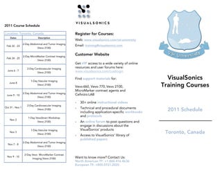
- 1. 2011 Course Schedule Location: Toronto, Canada Register for Courses: Dates Description Web: www.visualsonics.com/vsi-university 2-Day Abdominal and Tumor Imaging Feb 22 - 23 Email: training@visualsonics.com (Vevo 2100) 2-Day MicroMarker Contrast Imaging Customer Website Feb 24 - 25 (Vevo 2100) Get VIP access to a wide variety of online 2-Day Cardiovascular Imaging resources and user forums here: June 6 - 7 www.visualsonics.com/custlogin (Vevo 2100) 1-Day Vascular Imaging Find support materials for: VisualSonics Training Courses June 8 (Vevo 2100) Vevo 660, Vevo 770, Vevo 2100, MicroMarker contrast agents and 2-Day Abdominal and Tumor Imaging June 9 - 10 Cellvizio LAB (Vevo 2100) • 30+ online instructional videos 2-Day Cardiovascular Imaging Oct 31 - Nov 1 • Technical and procedural documents (Vevo 2100) including application-specific workbooks 2011 Schedule 1-Day VevoStrain Workshop and protocols Nov 2 (Vevo 2100) • An online forum to post questions and engage in discussions about the 1-Day Vascular Imaging VisualSonics’ products Nov 3 (Vevo 2100) Toronto, Canada • Access to VisualSonics’ library of published papers 2-Day Abdominal and Tumor Imaging Nov 7 - 8 (Vevo 2100) 2-Day Vevo MicroMarker Contrast Nov 9 - 10 Want to know more? Contact Us: Imaging (Vevo 2100) North American TF: +1.866.416.4636 European TF: +800.0751.2020
- 2. Course Descriptions Vascular and Doppler ® Our Vevo courses offer hands-on One-Day Training Our Vascular and Doppler Training Program will use the mouse instruction at our laboratories in Toronto or Abdominal and 3-Dimensional model to help users optimize their use of the Vevo high resolution Two-Day Training imaging system for cardio and vascular phenotyping. Amsterdam. You can sign up for 1 or all 5 Our Abdominal and 3D Techniques Training Program will use the courses. You will receive personal mouse model to help users optimize their use of the Vevo high • Overview of basic Doppler principles for quantification of blood resolution imaging system for phenotyping of cancer models instruction from our skilled applications and/or abdominal imaging. flow • Locating and obtaining 2D images of various vessels in the team, who have trained researchers at • Obtaining 2D views of the liver, spleen, pancreas, gallbladder, body, such as the carotid arteries, femoral arteries, renal several institutions worldwide. kidney and adrenals arteries, splenic arteries, abdominal aorta and branches off of the vessel, i.e. mesenteric arteries • Use of Pulsed Wave Doppler to obtain flow measurements of • Obtaining Pulsed Wave Doppler of these vessels and the renal artery and vein, portal vein, hepatic vessels, splenic implementing flow measurement and quantification tools in the vessels, abdominal aorta and IVC software such as peak velocity, VTI, resistive and pulsatility • Implementation of standard measurements used in the indices… abdomen • How to use Color Doppler (for Vevo 2100 only) • Creation of a 3D volumetric measurement in vivo in a • Use of M-mode to aid in wall measurements of vessels subcutaneous tumor model • Workflow optimization • How to use Color Doppler (for Vevo 2100 only) • Use of Power Doppler to quantify percent vascularity • Workflow optimization VevoStrain One-Day Workshop Cardiovascular This workshop is intended for Vevo 2100 user. If you are a Two-Day Training Vevo 770 user and would like to see how this software works, Our Cardiovascular Training Program will use the mouse model to you may certainly attend. However, this software is only available help users optimize their use of the Vevo high resolution imaging on the Vevo 2100 system. system for cardiovascular phenotyping. Our VevoStrain Workshop will use the mouse model to help users • How to do a mouse echocardiogram, including imaging the optimize the use of the VevoStrain software for global wall heart in long axis, short axis and apical views. motion tracking in the left ventricle. Courses Offered: • How to obtain images of the aortic valve, pulmonary valve, mitral and tricuspid valves • Normal and infarcted images for analysis will be provided • How to use Color Doppler (for Vevo 2100 only) • Overview of regional and global wall motion tracking • Abdominal and 3-Dimensional • How to obtain and measure Pulsed Wave Doppler and M-mode • How to quantify: velocity/displacement/strain/strain rate time to spectrums Two-Day Training • Diastolic function measurements peak • Quantification with a variety of graphs and parametric displays • Left ventricular analysis • How to export and understand the values • Cardiovascular • Workflow optimization • Image guided injection into the myocardium Two-Day Training This is a post processing feature. Computers for analysis will be provided for this hands-on session. There is no animal handling MicroMarker Contrast Imaging with this course • MicroMarker™ Contrast Imaging Two-Day Training Two-Day Training Our MicroMarker Training Program will use the mouse model to help users optimize their use of the Vevo high resolution imaging system for contrast-enhanced and molecular imaging, using • Vascular and Doppler MicroMarker contrast agents. One-Day Training • Overview of available MicroMarker contrast agents for various • VevoStrain™ applications • Introduction of tail vein cannulation techniques (for agent One-Day Workshop introduction) • Hands-on practice sessions will involve both targeted and non- targeted imaging with MicroMarker contrast agents and software analysis tools • Models may include subcutaneous tumors, normal abdominal organs, hind limb inflammatory model and normal eye and heart models • Workflow optimization