Basal Cell Carcinoma of Scalp Diagnosed on Biopsy
•Als PPTX, PDF herunterladen•
3 gefällt mir•4,577 views

CPC Dermatopathology Session on 28/6/2018

Empfohlen
Weitere ähnliche Inhalte
Was ist angesagt?
Was ist angesagt? (20)
Ähnlich wie Basal Cell Carcinoma of Scalp Diagnosed on Biopsy
Ähnlich wie Basal Cell Carcinoma of Scalp Diagnosed on Biopsy (20)
Mehr von Dr. Varughese George
Mehr von Dr. Varughese George (20)
Kürzlich hochgeladen
Kürzlich hochgeladen (20)
Basal Cell Carcinoma of Scalp Diagnosed on Biopsy
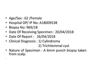
- 1. • Age/Sex : 62 /Female • Hospital OP/ IP No: A18009538 • Biopsy No: 969/18 • Date Of Receiving Specimen : 20/04/2018 • Date Of Report : 26/04/2018 • Clinical Diagnosis : 1) Cylindroma 2) Trichilemmal cyst • Nature of Specimen : A 6mm punch biopsy taken from scalp. .
- 2. Gross Examination Container labelled skin biopsy from scalp Received single nodular grey white skin attached soft tissue bit measuring 0.5x0.5x0.5cm. External surface show hair follicles. Cut surface shows grey white areas. All embedded in one block. .
- 3. Microscopy 4x A localized basal cell proliferative cup-shaped lesion with atrophic overlying epidermis.
- 4. Microscopy 10x Tumor cells are arranged in the form of islands, cords and groups with focal areas of sclerosis.
- 5. Microscopy 40x Clefts and peripheral palisading of basaloid cells of the nests
- 6. Microscopy Section studied shows • A localized basal cell proliferative lesion of the epidermis arranged in the form of islands, cords and groups with focal areas of sclerosis. • The entire lesion is cup shaped with atrophic overlying epidermis. • One focus shows a transition from the basal pigment layer to the basal cell carcinoma.
- 7. Impression Biopsy of scalp shows features of basal cell carcinoma. The edges of the biopsy are free of tumor.
- 8. Differential Diagnosis ● Well-circumscribed dermal nodules composed of islands of epithelial cells that fit together like pieces of jigsaw puzzle and are separated from each other only by thick hyaline sheaths. ● Two types of cells are present in the epithelial islands : - – Cells with small, dark-staining nuclei at the periphery of the islands. – Cells with large light-staining nuclei in the center of the islands. ● Tubular lumina lined by ductal cells and filled with amorphous material are often present. ● Drops of eosinophilic hyaline material can be present within the epithelial islands. Cylindroma
- 9. Differential Diagnosis Trichilemmal cyst ● Calcifications are frequently found. ● Proliferating trichilemmal cystic neoplasm: a low-grade neoplasm characterized by lobules of eosinophilic epithelial cells (isthmic) and infiltrative growth pattern ● Benign cyst occurring most commonly on the scalp as multiple cystic nodules. ● Cyst contents consist of compact keratin, and the lining resembles the isthmus of hair follicle; abrupt keratinization with absent granular layer is characteristic.
- 11. Discussion
- 12. Basal Cell Carcinoma • Typically affects older individuals • Predilection for sun- exposed skin (face, hands) • Small, well-circumscribed, pearly tan-gray papule devoid of scale • Lesions enlarge with time and tend to ulcerate • (rodent ulcers)
- 13. Basal Cell Carcinoma Histopathology • Nests and islands of basaloid cells attached to the undersurface of epidermis and extending into the dermis • Peripheral palisading of basaloid cells of the nests • Basaloid cells are typically uniform with frequent mitotic activity and abundant apoptotic cells
- 14. Basal Cell Carcinoma Histopathology • Characteristic retraction artifact between the palisading cells and the normal stroma. • Areas of squamous differentiation and perineural invasion are seen in aggressive (infiltrative) forms • Variants of basal cell carcinoma: – pigmented, – morphea-like or sclerosing, – superficial, – nodular, – keratotic, – adenoid, – micronodular – fibroepithelial
- 15. Basal Cell Carcinoma Differential Diagnosis Trichoepithelioma • Nests of basaloid cells usually without mitotic activity, individual cell necrosis, or separation artifacts. • Abundant fibrotic stroma. • Retraction artifacts within a cellular stroma rather than around the epithelial nests. • Evidence of follicular differentiation in the form of germs, bulbs, and papillae is more common. • CD10-positive stroma.
- 16. • Basal cell nevus syndrome: multiple basaloid hamartomas on the cutaneous surface associated with palmar keratotic pits, jaw cysts, and basal cell carcinomas in non–sun- exposed locations. • BCCs rarely metastasize; when they do, the primary lesion is usually advanced