SlideShare ist ein Scribd-Unternehmen logo
1 von 22
Downloaden Sie, um offline zu lesen
The Department of Radiation Oncology at the
University of Alabama at Birmingham (UAB) has
partnered with Varian Medical Systems to
become a showcase site for excellence in cancer
care. UAB Radiation Oncology is proud to
provide other cancer centers the opportunity to
preview Varian equipment in a clinical setting.
Tours of Excellence
EXTENSIVE EXPERIENCE with delivering the most
advanced radiation therapies available goes hand in hand
with compassionate patient care at the UAB Department of
Radiation Oncology.
About UAB
The University of Alabama at Birmingham (UAB) Health
System is a network of services that provides a complete
continuum of care, including comprehensive cancer care, for
patients from Alabama and all over the world. As one of the
original National Cancer Institute-designated comprehensive
centers created, the UAB Comprehensive Cancer Center has
maintained the designation for 40 years. As a part of the
UAB Comprehensive Cancer Center, the UAB Department of
Radiation Oncology is a world leader in the delivery of cancer
care with highly experienced clinical faculty, utilizing the
most advanced treatment modalities and technologies available
in radiation therapy.
Depth of Expertise
The UAB Department of Radiation Oncology has achieved an
international reputation for leadership through:
• A multidisciplinary approach to diagnosis and treatment
• An extensive and highly experienced clinical faculty that
sub-specializes in the breadth of tumor types
• A clinical care team of dosimetrists, nurses, and radiation therapists
who are trained and certified to the highest standards.
Twelve faculty physicians are board-certified in radiation oncol-
ogy. Each clinical faculty physician sub-specializes by tumor type.
Many hold PhD degrees in addition to their MD. Physicians stay
abreast of the latest breakthroughs and technologies through their
research into novel treatment modalities and methods to improve
radiation delivery. Because UAB is a teaching institution, its physi-
cians are training the radiation oncologists of tomorrow.
The department physics team is the largest in Alabama. It
includes seven PhD, board-certified, medical physicists. Faculty
members support quality patient care, perform research to
enhance cancer care via the use of radiation physics, and teach in
the UAB physics and resident physician program.
Leadership and Innovation
The UAB Department of Radiation Oncology is proud of its
contributions to advancing the use of radiation to treat cancer.
Notable achievements include:
• First in the world to use Intrafraction Motion Review tech-
nology from Varian to continually monitor tumor location
during radiosurgery for lung cancer (2011)
James A. Bonner, MD
Specialty: Lung,
Head & Neck
Residency: University
of Michigan
MD: Wayne State
University
James A Bonner M John Fiveash, MD
Specialty: CNS,
GU, Ocular
Melanoma, Sarcoma
Residency: Medical
College of Georgia
MD: Medical
College of Georgia
John Fi eash MDJennifer De Los
Santos, MD
Specialty: Breast,
GYN
Residency: University
of Texas-MD
Anderson Cancer
Center
MD: University of
Florida
Jennifer De Los Rojymon Jacob, MD
Specialty: GI, GU,
Sarcoma, Benign
Disease
Fellowship: Royal
College of
Radiologists, London
MD: Kerala University,
India
Roj mon Jacob MDMichael Dobelbower,
MD, PhD
Specialty: CNS, GI,
GU, Head & Neck,
Lung, Benign Disease
Residency: University
of Alabama at
Birmingham
MD: Medical College
of Ohio
PhD: Ohio State
University
Michael Dobelbo e
UAB Department of Radiation Oncology: Clinical Faculty
Advanced treatment.
Compassionate care.
The Kirklin Clinic at Acton Road
O. L. Burnett III, MD
Specialty: GU,
Lymphoma,
Pediatrics, Breast,
Sarcoma, GI
MD: Emory
University
O L B rnett III MD
• Among the first to use the TrueBeam™
system to treat lung,
liver, pancreas, head & neck, brain, and spine cancers (2010)
• First in the United States to treat cancer patients with
RapidArc®
radiotherapy (2008)
• First in the region to perform intensity-modulated radiation
therapy (IMRT) (1999)
• Among the first to perform linear accelerator-based radiosurgery
• First to clinically use ultrasound-based image guidance (1974).
• First department in the region to separate therapeutic radiology
from diagnostic radiology (1969).
Varian Technology at UAB
The UAB Department of Radiation Oncology delivers the
following advanced modalities with Varian equipment: IMRT;
image-guided radiation therapy (IGRT); and stereotactic body
radiation therapy (SBRT). Professionals visiting UAB will be able
to view Varian products in action in a high volume department
that handles 30,000 treatment visits annually. In addition,
department staff are available to answer questions about security,
integration and interfaces, support, and many other issues
common to large academic radiation oncology departments.
During the tour, visitors can see the following Varian
technology in use:
• TrueBeam™
STx system
• Clinac®
iX linear accelerators
• On-Board Imager®
(OBI) kV imaging system for treatment
localization
- OBI radiographic: kV-kV anatomy matching & OBI CBCT
• RapidArc®
radiotherapy technology
• Varian Real-time Position Management™
(RPM) system for
respiratory gating
• VariSource™
high dose rate afterloader
• Varian Acuity™
treatment planning, simulation, and verifica-
tion system for fluoroscopic simulation
• ARIA®
oncology information system
• Eclipse™
treatment planning system for RapidArc, IMRT,
IGRT, and SBRT
Hazelrig-Salter Radiation Oncology Center
Robert Kim, MD
Specialty: GU, GYN,
Ocular Melanoma,
Orbital tumors
Residency: University
of Alabama at
Birmingham
MD: Yonsei University,
Korea
Robert Kim MD Christopher Willey,
MD, PhD
Specialty: CNS, Head
& Neck, Lung,
Pancreas
Residency: Vanderbilt
University Medical
Center
MD: Medical University
of South Carolina
PhD: Medical
University of South
Carolina
Christopher Wille Eddy Yang, MD, PhD
Specialty: Prostate,
Breast, Head & Neck,
Lung
Residency:
Vanderbilt University
School of Medicine
MD: University of
Miami School of
Medicine
PhD: University of
Miami School of
Medicine
Edd Yang MD PhRuby Meredith, MD,
PhD
Specialty: Breast,
CNS, Lung, Lymphoma
Residency: Medical
College of Virginia
MD: Ohio State
University
PhD: Indiana
University
R b Meredith MDKimberly Keene, MD
Specialty: Breast, GI,
Head & Neck,
Pediatrics, Skin
Residency: University
of Virginia
MD: University of
Florida
Kimberl Keene MD Sharon Spencer, MD
Specialty: Head &
Neck, Lung,
Lymphoma, Pediatrics,
Sarcoma, Skin
Residency: The
University of Alabama
at Birmingham
MD: University of
Alabama at
Birmingham
Sharon Spencer M
Joint venture clinic
3100 Hansen Way, M/S MGM, Palo Alto, CA 94304
For more information, contact your Varian representative.
RAD 5832A © 2010, 2012 Varian Medical Systems, Inc. Printed in USA 5/12 (350)
UAB Department of Radiation Oncology: Physics Faculty
Ivan Brezovich, PhD
Professor and Director
PhD: Physics,
University of Alabama
at Birmingham
MS: Physics, University
of Alabama at
Birmingham
I an Bre o ich Ph
Richard Popple, PhD
Associate Professor
Postdoctoral
Fellowship: University
of Texas-MD Anderson
Cancer Center
Postdoctoral
Fellowship: Rice
University
Richard Popple Ph
Jun Duan, PhD
Associate Professor
PhD: Physics, Florida
State University
MS: Medical Physics,
University of Florida
J n D an PhD Prem Pareek, PhD
Adjunct Professor
Post-Doctoral
Fellowship: Allegheny
General Hospital
PhD: University of
Nebraska
Prem Pareek PhD Rex Cardan, PhD
Assistant Professor
Medical Physics
Residency: James
G. Brown Cancer
Center, University of
Louisville
PhD: University of
Texas at San Antonio
Re Cardan PhD
Xingen Wu, PhD
Assistant Professor
Postdoctoral
Fellowship: St. Jude
Children’s Research
Hospital
Postdoctoral
Fellowship: Zhejiang
University, China
Xingen W PhDSui Shen, PhD
Associate Professor
Postdoctoral
Fellowship: University
of California at Davis
Medical Center
PhD: University of
California at Davis
S i Shen PhD
Advanced treatment.
Compassionate care.
1700 6th Avenue South, Birmingham, AL 35249
The Tour Experience
With approximately 250,000 patient visits over the past
10 years, the UAB Department of Radiation Oncology offers a
unique experience for professionals who want to learn about
Varian technology. In 2010 and 2011, the UAB Department
of Radiation Oncology hosted 32 visiting delegations. They
represented community hospitals and university medical
centers in 15 states, Canada, Mexico, and Brazil. UAB seeks
to customize each tour to the expressed interests of our visitors
in technology and types of cases treated. Radiation oncologists,
neurosurgeons, physicists, dosimetrists, radiation therapists,
and administrators have the opportunity to meet with UAB
counterparts to exchange information and perspectives.
The tour is often the beginning of continuing collaborations.
2012 UAB Radiosurgery Program Outcomes
UAB Radiosurgery Program
Hazelrig-Salter Radiation Oncology Center
HSROC 2248 • 1700 6th Avenue South
619 19TH ST S
BIRMINGHAM AL 35249-6832
Non-Profit Org.
U.S. Postage
PAID
Permit No. 1256
Birmingham, AL
The UAB Comprehensive Cancer Center
To refer a patient to the UAB Radiosurgery Program or schedule
appointments, contact UAB MIST at 1.800.822.6478.
For information about the UAB Radiosurgery Program, visit
uabmedicine.org/radiosurgery or uab.edu/radonc
The 2012 UAB Radiosurgery Program Outcomes booklet continues our effort to
communicate the strides our team has made in providing the best possible care to
the citizens of Birmingham and beyond.
We believe that the innovative techniques being harnessed within our program
make UAB a standout in patient care, research, and education. Advancing
treatments for optimal patient care and outcomes, as well as contributing to the
body of radiosurgery knowledge, is helping us work toward our ultimate goal of
developing better cancer therapeutics.
Two of our most exciting and cutting-edge treatments are discussed in this
booklet: triggered imaging technique for thoracic radiosurgery and Gamma
Knife radiosurgery for pituitary tumors. Each of these care tactics builds on the
Radiosurgery Program’s culture of collaboration and aim of providing our patients
with care that is as individualized as they are.
Triggered imaging for thoracic surgery is the newest development in motion
management at UAB and allows our care teams to even more accurately deliver
treatment to our patients. Thoracic radiosurgery is a technically complex
procedure that requires advanced technologies and multidisciplinary care, which
in Alabama are available uniquely at UAB.
For the treatment of pituitary adenomas that require salvage treatment, Gamma
Knife radiosurgery offers a precise, successful treatment modality. This salvage
therapy offers a high rate of controlling the tumor while minimizing potential
radiation-induced damage to adjacent normal tissue—an advantage that
decreases the risk of neurocognitive impairment and secondary malignancy.
These strides in patient treatment combined with our comprehensive team
approach are a hallmark of our radiosurgery program. We strive to deliver these
treatments with a patient-centered approach that allows for compassionate and
superior care for each and every patient.
We welcome any questions and comments you may have. If you would like to
learn more about the progress of our program, you may contact the Department
of Radiation Oncology at 205.934.5670.
John B. Fiveash, MDJames A. Bonner, MD
James M. Markert, Jr,
MD, MPH
Kirby I. Bland, MD
At UAB, a new technique called triggered imaging is being
used to monitor tumor position in real-time during thoracic
radiosurgery. Triggered imaging is improving the accuracy and
precision of radiosurgery.
Radiosurgery is becoming an increasingly important tool for
managing lung cancer in non or marginally operable patients,
with outcomes comparable to surgery [8]. Numerous multi-
institutional clinical trials are ongoing, with early results showing
that this approach is safe and can result in cancer-free survivals
at three years similar to surgery with less morbidity in the short
term [8,10-13]. Reported control rates for thoracic tumors treated
with radiosurgery have reached more than 90 percent [14].
Thoracic radiosurgery is technically challenging, requiring accurate
targeting of the radiation beam so that the tumor receives the full,
ablative radiation dose while dose to healthy tissue is minimized. To
assure the best possible outcome, radiosurgery at UAB is performed
by a multidisciplinary team comprised of thoracic surgeons, radiation
oncologists, and medical physicists. Team members work in close
collaboration throughout the entire treatment process, from initial
consultation to the radiosurgical procedure, to patient follow up.
A particularly complex technical challenge facing thoracic
radiosurgery is respiratory motion of the tumor. Tumor motion is
highly variable; Tumors at the apex of the lung typically remain
stationary, while diaphragmatic tumors can move as much as
4 cm (K. M. Langen., And D. T. L. Jones, “Organ motion and its
management,” Int. J. Radiation Oncology Biol. Phys., Vol. 50, No. 1,
pp. 265–278, 2001). At UAB, management of tumor motion begins
with the thoracic surgeon. Using navigational bronchoscopy, the
surgeon implants fiducial markers in the tumor. Conventional
bronchoscopic techniques cannot reach many tumors, for which
the only other option for implanting fiducial markers is trans-
thoracically. The trans-thoracic approach has a pneumothorax rate
as high as 30% ( Yousefi S, Collins BT, Reichner CA, Anderson ED,
Jamis-Dow C, Gagnon G, Malik S, Marshall B, Chang T, Banovac F.
Complications of thoracic computed tomography-guided fiducial
placement for the purpose of stereotactic body radiation therapy.
Clin Lung Cancer. 2007 Jan;8(4):252-6.), compared to less than
6% for navigational bronchoscopy (Schroeder C, Hejal R, Linden
PA. Coil spring fiducial markers placed safely using navigation
bronchoscopy in inoperable patients allows accurate delivery of
CyberKnife stereotactic radio surgery. J Thorac Cardiovasc Surg.
Triggered Imaging Technique for Thoracic Radiosurgery
A Message From the Chairs
Triggered Imaging Technique for
Thoracic Radiosurgery............... 2-3
Gamma Knife Radiosurgery for
Pituitary Tumors..................... 4-5
Quality and Outcome Measures...6-7
Publications...............................8
Educational Site Visits.................9
Clinical Faculty.........................10
Contents
2
Merle M. Salter Professor and Chair
UAB Department of Radiation Oncology
Fay Fletcher Kerner Professor and Chair
UAB Department of Surgery
Robert Y. Kim Endowed Chair, Professor and Vice Chair
UAB Department of Radiation Oncology

Interim Associate Director for Clinical Research
UAB Comprehensive Cancer Center
James Garber Galbraith Endowed Chair,
Professor and Director
UAB Division of Neurosurgery Cover photo: Dr. Sharon Spencer, Dr. Barton Gurthrie, and Dr. Kristen Riley
Participating Faculty
James A. Bonner, MD
Kirby I. Bland, MD
Michael C. Dobelbower,
MD, PhD
John B. Fiveash, MD
Barton L. Guthrie, MD
Douglas J. Minnich, MD
Richard A. Popple, PhD
Kristen Riley, MD
Sharon A. Spencer, MD
Editorial Support
John Brinkerhoff
Valeria Pacheco-Rubi
Joey Slatsky
Fresia Vega
Above: Dr. Richard Popple
2010 Nov;140(5):1137-42. Epub 2010 Sep 20.; Harley DP, Krimsky WS,
Sarkar S, Highfield D, Aygun C, Gurses B. Fiducial marker placement
using endobronchial ultrasound and navigational bronchoscopy for
stereotactic radiosurgery: an alternative strategy. Ann Thorac Surg.
2010 Feb;89(2):368-73; discussion 373-4). At UAB, we have had no
pneumothoraces with fiducial placement. The markers are typically
implanted during a diagnostic bronchoscopy, so the patient does not
need to undergo an additional procedure.
After bronchoscopy, the radiation oncologist and the thoracic
surgeon consult to determine the best treatment strategy. Once the
decision has been made to use radiosurgery, the patient receives a CT
scan to identify the tumor and nearby healthy structures that need
to be protected from the radiation. The CT scan is the next stage
in the management of tumor motion. During the scan, an optical
technique is used to measure the chest motion. The scan is a special
type, called a 4D CT, composed of 10 complete 3-dimensional CT
image sets. Each CT corresponds to a snapshot at a different point in
the respiratory cycle, which is correlated with the chest motion.
One method to ensure that the tumor remains within the radiation
beam is to simply treat the entire volume encompassed by tumor
motion. However, this approach results in a relatively large volume
of lung receiving a high radiation dose (Wu J, Li H, Shekhar R,
Suntharalingam M, D’Souza W., “An evaluation of planning techniques
for stereotactic body radiation therapy in lung tumors,” Radiother
Oncol. 2008 Apr;87(1):35-43. Epub 2008 Mar 24). This approach is
particularly undesirable in the context of the high, ablative radiation
dose delivered by radiosurgery. An alternative approach preferred
at UAB is to gate the radiation beam, turning it on only at the end
of expiration, when the lung is at rest and the tumor is relatively
stationary. The scans are evaluated for tumor motion by the medical
physicist, who determines the optimal point in breathing cycle
to turn the radiation beam on and off. The medical physicist also
locates the fiducial markers in the CT images. The radiosurgery team
then develops and tests an individualized treatment plan. The dose
distribution is sculpted to tightly conform to the tumor and limit
radiation dose to the lung, chest wall, and other healthy tissues.
The final and most critical step in motion management is treatment,
usually one to five treatments over one to two weeks. Prior to
starting radiation delivery, x-rays are taken to ensure that the tumor
is in the correct position. The fiducial markers are easily seen in the
x-rays and are compared with outlines of the expected position,
derived from the 4D CT scan and the preparation by the medical
physicist. If the outline and the image on the x-ray do not coincide,
the patient is shifted until they do. When the patient is in the correct
position and the tumor is centered in the radiation beam, the beam
is turned on. During treatment, the same optical technique used
during the CT scan is used to track the patient’s breathing. The
optical system instructs the radiation beam to turn on at the end of
expiration and to turn off as inspiration begins.
The newest development in motion
management at UAB is triggered
imaging. Using triggered imaging,
we observe the fiducial marker
during treatment delivery. At the
beginning of each expiratory cycle,
immediately before the radiation
beam comes on, an x-ray image is
taken. The image is displayed along
with a circle around the expected
position of the fiducial marker. The radiation oncologist and thoracic
surgeon are thus able to monitor the position of the tumor in
real time as the treatment progresses. If the patient moves or the
breathing pattern changes, treatment is suspended, the position
corrected, and treatment resumed.
Thoracic radiosurgery is a technically complex procedure requiring
advanced technologies and multidisciplinary care, which in Alabama
are available uniquely at UAB. The experienced team at UAB will
continue to remain at the forefront of innovation as the technologies
for thoracic radiosurgery continue to evolve.
Pituitary adenomas represent one of the most common intracranial neoplasms.
Found in 10-15% of the population, these benign tumors often pose complex
management situations. While the majority of pituitary tumors can be treated with
medication or surgery alone, a significant proportion require salvage treatment.
Pituitary tumors that generally require additional treatment include functional tumors
not controlled with surgery or medication and nonfunctional tumors that recur
following surgery.
Gamma Knife radiosurgery offers a precise, successful treatment modality for pituitary
adenomas. Tumor growth can be controlled in 90% of patients treated, frequently with
reduction in tumor volume. Radiosurgery effects on biochemical cure vary depending
on tumor type. (Sheehan et al 2011). Patient selection for radiosurgery depends on
endocrine evaluation, tumor size, location, growth pattern, and pathology.
Pituitary adenomas are classified according to size and endocrine profile.
Microadenomas, defined as smaller than 10mm in size, rarely cause clinical concern
due to size, but may require treatment if they are functionally active. Macroadenomas,
larger than 10mm, may cause visual difficulty if the optic pathways become
compressed by the tumor. Located at the base of the skull, pituitary tumors occur
adjacent to many critical structures such as the optic nerves, optic chiasm, cranial
nerves within the cavernous sinuses, carotid artery, and brainstem. The location of
these tumors requires specialized knowledge and techniques for management.
Additionally, pituitary adenomas often have either hormone overproduction
or deficiency. All management decisions regarding these tumors require a
multidisciplinary approach. While many tumors require only observation, a significant
number have endocrine and anatomical implications that must be addressed.
The UAB Neurosurgical Pituitary Disorders Clinic offers comprehensive evaluation
and care for patients with pituitary tumors. Following diagnosis, whether for
an incidentally found tumor or a symptomatic pituitary adenoma, appropriate
evaluation includes imaging review, endocrine evaluation, and often ophthalmologic
evaluation. Observation, medical therapy, surgery, and radiation therapy comprise the
armamentarium of treatment options for pituitary tumors.
Gamma Knife Radiosurgery
for Pituitary Tumors
43
Above: Dr. Douglas Minnich and Dr. Michael Dobelbower
Above: Fiducial marker during
treatment delivery
Functional tumors, those that result in overproduction of hormones,
often require multi-modality treatment. Prolactinomas are the most
common functional pituitary tumors. For prolactinomas, medical
therapy with dopamine agonists is the standard of care for first line
treatment. However, for patients not controlled with medication
or who do not tolerate medication, surgery and radiation may be
utilized. Pituitary tumors resulting in acromegaly, from excess
growth hormone and Cushing’s disease from excess ACTH, require
treatment regardless of size. Surgery is the first line of treatment
for the majority of these tumors. In cases where a surgical cure is
not achieved, additional therapy is paramount due to the significant
increase in morbidity and mortality if hormone overproduction is not
controlled. For patients with Cushing’s disease, there is no available
medical treatment to suppress steroid production. Radiosurgery
offers a potential for cure.
In acromegaly, controversy exists regarding the timing of radiation
therapy related to medical therapy. Medical therapy is often
successful in normalizing growth hormone production, but at a
significant yearly financial cost. Without controversy, is the use of
radiation when patients are not controlled with medical therapy.
However, there may be utility in radiation treatment in an attempt to
shorten the length of time a patient requires medical therapy. Data
suggests radiosurgery offers a greater than 50% rate of cure for
growth hormone secreting pituitary tumors. (Sheehan et al 2011)
For residual nonfunctional adenomas following surgery, Gamma
Knife radiosurgery is considered if there is observed tumor growth
over time or if the pathology is atypical pituitary adenoma, indicating
a potentially higher chance of tumor recurrence. The recurrence rate
of pituitary adenomas following surgery is reported around 20%.
Recurrence is influenced by extent of resection and tumor pathology.
Patients with pituitary adenomas are followed postoperatively with
yearly imaging. The majority of tumor recurrence is seen in the first
five to seven years postoperatively, but can occur later.
Following any radiation to the sella, patients should have a yearly
endocrine evaluation. Secondary hypopituitarism is the most
common side effect of radiosurgery for pituitary adenomas. The
incidence of secondary hormone deficits increases with time, thus
necessitating long-term endocrine surveillance. Gamma Knife
radiosurgery may have a decreased rate of endocrine dysfunction
over fractionated radiation due to the ability to precisely deliver
radiation to the tumor and limit radiation to the normal gland in
some patients. (Taussky et al 2011) In addition to minimizing dose to
the normal pituitary gland, radiosurgery allows for treatment delivery
that minimizes radiation to adjacent normal brain cells. This precision
decreases the risk of neurocognitive impairment and secondary
malignancy from radiation.
Appropriate patient selection and experienced treatment planning
help to minimize the risks of radiosurgery. The anatomical location of
the pituitary tumor necessitates careful evaluation and planning to
limit toxicity to critical structures. Gamma Knife, with frame based
head fixation, offers the most precise method of radiation delivery.
In this area, millimeters matter. At UAB, we feel strongly that Gamma
Knife precision allows us to perform safe, successful radiosurgery for
pituitary tumors.
For more information or to refer a patient to the Multidisciplinary
Pituitary Clinic: Contact Michel Thomas, Office Assistant to Dr. Riley,
at 205-996-2461.
Gamma Knife Stereotactic Body Radiation Therapy
0
500
1000
1500
2000
2500
3000
3500
4000
4500
5000
1992 1993 1994 1995 1996 1997 1998 1999 2000 2001 2002 2003 2004 2005 2006 2007 2008 2009 2010 2011
Timeline of our Success
Quality and Outcome Measure
Timeline of our success
SELECTED DISEASE SITES
1992	 First patient treated with 	
	 stereotactic radiosurgery (linac)
1995	 First CNS case treated with
	 Gamma Knife
1999	 First FDA-approved IMRT-
	 delivering device
2001	 First in Alabama to offer RPM
	 Gating System
2005	 First in Alabama to treat with
	 stereotactic body radiation therapy
2008	 First in the U.S. to treat with
	 volumetric arc therapy (RapidArc™)
2010	 One of the world’s first facilities to
	 offer TrueBeam system (third in
	 the United States)
2011	 First in the world to use “Triggered 	
	 Imaging” Technology from Varian 	
	 Medical Systems to continually 	
	 monitor tumor location during 	
	 radiosurgery for lung cancer
The UAB Radiosurgery Program offers
state-of-the-art treatment therapies
and technologies for a wide variety of
body sites, including central nervous
system (CNS), lung, spine, and others.
CNS tumors essentially are treated
with the Gamma Knife. Tumors or
malformations of the liver, lung, spine,
and other body sites are treated using
Stereotactic Body Radiation Therapy
(SBRT). The following charts show the
outcome measures of selected body
sites treated with cranial radiosurgery
and SBRT at UAB.
6
Sheehan JP, Pouratian N, Steiner L, Laws ER, Vance ML.
Gamma Knife surgery for pituitary adenomas: factors
related to radiological and endocrine outcomes. J
Neurosurg. 2011 Feb: 114 (2) 303-9.
Taussky P, Kalra R, Coppens J, Mohebali J, Jensa R,
Couldwell WT. Endocrinological outcome after pituitary
transposition (hypophysopexy) and adjuvant radiotherapy
for tumors involving the cavernous sinus. J Neurosurg. 2011
Jul; 115(1): 55-62.
Gamma Knife	 2114
Benign	 432
Malignant	 1083
Trigeminal Neuralgia	 409
Vascular	 188
Seizure	 2
Stereotactic Body Radiation Therapy	 363
Brain	 36
Lung	 129
Liver	 24
Other	 53
Spine	 121
5
0
500
1000
1500
2000
2500
3000
3500
4000
4500
5000
1992 1993 1994 1995 1996 1997 1998 1999 2000 2001 2002 2003 2004 2005 2006 2007 2008 2009 2010 2011
0
10
20
30
40
50
60
70
80
90
100
2005 2006 2007 2008 2009 2010 2011
2012 Radiosurgery Noteworthy Publications
Clark GM, Popple RA, Prendergast BM, Spencer SA, Thomas EM,
Stewart JG, Guthrie BL, Markert JM, Fiveash JB: Plan quality
and treatment planning technique for single isocenter cranial
radiosurgery with volumetric modulated arc therapy. Practical
Radiation Oncology. Published online February 1, 2012. Citation
Pending.
Clark G, Popple R, Young PE, Fiveash J: Feasibility of single-
isocenter volumetric modulated arc radiosurgery for the treatment
of multiple brain metastases. Int J Radiat Oncol Biol Phys. 2010 Jan
1;76(1):296-302.
Fiveash J, Guthrie BG, Duan J, Markert JM, DeLosSantos JF, Keene
KS, Spencer SA, Dobelbower MC, Arafat W, Popple RA. A Phase II
Isotoxicity Study of Spinal Radiosurgery/SBRT. Int. J. Radiat. Oncol.
Biol. Phys. 2010 November; 78(3) Suppl: S278.
Parker JN, Zheng X, Luckett W, Markert JM, Cassady KA. Strategies
for the rapid construction of conditionally-replicating HSV-1
vectors expressing foreign genes as anticancer therapeutic agents.
Mol Pharm. 2011 Feb 7;8(1):44-9. Epub 2010 Dec 17. Review. PMID:
21142023
Pearson BE, Markert JM, Fisher WS, Guthrie BL, Fiveash JB, Palmer
CA, Riley K. Hitting a moving target: evolution of a treatment
paradigm for atypical meningiomas amid changing diagnostic
criteria. Neurosurg Focus. 2008;24(5):E3. PMID: 18447742
Popple RA, Dieterich S, Duan J, Fiveash JB. Dependence of Dose-
volume Values on Calculation Method for Paraspinal Radiosurgery.
Int. J. Radiat. Oncol. Biol. Phys. 2010 November; 78(3) Suppl: S783.
Popple RA, Fiveash JB, Brezovich IA, Bonner JA: RapidArc radiation
therapy: first year experience at the University of Alabama at
Birmingham. Int J Radiat Oncol Biol Phys. 2010 Jul 1;77(3):932-41.
Prendergast BM, Bonner JA, Popple RA, Spencer SA, Fiveash JB,
Keene KS, Cerfolio RJ, Minnich DJ, Dobelbower MC. Dosimetric
analysis of imaging changes following pulmonary stereotactic
body radiation therapy. J Med Imagina Radiat Oncol 2011
Feb;55(1):90-6.
Prendergast Brendan M, Popple Richard A., Clark Grant M., Spencer
Sharon A., Guthrie Bart, Markert James, Fiveash John B: Improved
clinical efficacy in CNS stereotactic radiosurgery using a flattening
filter free linear accelerator. Journal of Radiosurgery and SBRT.
Accepted for publication Journal of Radiosurgery and SBRT, August
9, 2011. Citation Pending.
Sawrie SM, Fiveash JB. Caudell, JJ: Stereotactic Body Radiation
Therapy for Liver Metastases and Primary Hepatocellular
Carcinoma: Normal Tissue Tolerances and Toxicity. Cancer Control
April 2010, Vol. 17, No. 2:111-119
Spencer S, Swaid N, Barton G, Young P, Wong W, Meredith RF,
Markert J, Fisher W, Wu X, Nordal R, Fiveash J. Impact of Dose Rate
on Outcomes of Gamma Knife Radiosurgery in Patients with Face
Pain. Radiosurgery 2010 7:360-5.
Sperduto PW, Kased N, Roberge D, Xu Z, Shanley R, Luo X, Sneed
PK, Chao ST, Weil RJ, Suh J, Bhatt A, Jensen AW, Brown PD, Shih
HA, Kirkpatrick J, Gaspar LE, Fiveash JB, Chiang V, Knisely JPS,
Sperduo CM, Lin N, Mehta M: Summary Report on The Graded
Prognostic Assessment: An Accurate and Facile Diagnosis-Specific
Tool to Estimate Survival for Patients with Brain Metastases. J Clin
Oncol, 29, 2011.
Sperduto PW, Kased N, Roberge D, Xu Z, Shanley R, Luo X, Sneed
PK, Chao ST, Weil RJ, Suh J, Bhatt A, Jensen AW, Brown PD, Shih
HA, Kirkpatrick J, Gasper LE, Fiveash JB, Chiang V, Knisely JP,
Sperduto CM, Lin N, Mehta M: Effect of Tumor Subtype on Survival
and the Graded Prognostic Assessment for Patients with Breast
Cancer and Brain Metastases. Int J Radiat Oncol Biol Phys 2011,
April 14.
Stewart JG, Sawrie SM, Bag A, Han X, Fiveash JB: Management of
Brain Metastases. Current Treatment Options in Neurology. 2010
Jul;12(4):334-46.
Vaphiades MS, Spencer SA, Riley K, Francis C, Deitz L, Kline LB.
Radiation-induced ocular motor cranial nerve palsies in patients
with pituitary tumor. J Neuroophthalmol. 2011 Sep;31(3):210-3.
The Leksell Gamma Knife
is a highly advanced
technology that delivers
201 tightly focused cobalt
radiation beams to one
point in the brain. The
radiation beams and
doses are so precise they
affect only the targeted
tissue and generally spare
the surrounding healthy
tissue.
Stereotactic Body Radiation
Therapy (SBRT) uses a high
dose of radiation shaped
to conform to the patient’s
tumor. It delivers radiation
to the intended target
and avoids healthy tissue.
Small tumors are accurately
identified and located with
precise coordinates.
Quality and Outcome Measure
CRANIAL RADIOSURGERY PROCEDURES
SBRT PROCEDURES
87
Visiting Institution	 Date of Visit
Gulfport Memorial Hospital – Gulfport, MS	 1/31/2011
Rush University – Chicago, IL	 2/17/2011
Torrance Memorial Medical Center – Torrance, CA	 2/18/2011
University of Kentucky – Lexington, KY	 3/24/2011
Memorial Hospital – Chattanooga, TN	 4/29/2011
Hospital Israelita Albert Einstein – São Paulo, Brasil	 4/12/2011
West Michigan Cancer Center – Kalamazoo, MI	 5/6/2011
Corpus Christi Cancer Center – Corpus Christi, TX	 5/20/2011
Medical Center at Bowling Green – Bowling Green, KY	 6/3/2011
Memorial Hospital – Gulfport, MS	 6/9/2011
Eastern Health-Cancer Care Program Dr. H. Bliss
Murphy Cancer Centre – NL, Canada 	 6/16/2011
Baptist Hospital – Miami, FL	 7/1/2011
University of Puerto Rico Cancer Center – San Juan, Puerto Rico	 8/4/2011
	 8/5/2011
Vanderbilt University Medical Center – Nashville, TN	 8/26/2011
Jackson-Madison County General Hospital – Jackson, TN	 9/8/2011 	
	 9/9/2011
University of Tennessee Hospital – Knoxville, TN 	 9/23/2011
Cancer Treatment Centers of America – Tulsa, OK	 10/28/2011
Renown Medical Center – Reno, NV	 11/11/2011
Radiological Associates of Sacramento – Sacramento, CA	 11/18/2011
Hospital Médica Sur – Mexico City, D.F., Mexico	 12/9/2011
Tours of Excellence
UAB Site Visits 2011
UAB Radiosurgical
Clinical Faculty
James A. Bonner, MD
Radiation Oncology
Specialties: lung, head and
neck
Ivan Brezovich, PhD
Medical Physicist
Specialty: physics
O.L. Burnett III, MD
Radiation Oncology
Specialties: GU, gynecological,
lymphoma, pediatrics, breast,
sarcoma, GI
Rex A. Cardan, PhD
Medical Physicist
Specialty: physics
Robert Cerfolio, MD
Thoracic Surgery
Specialty: thorax
Melissa Chambers, MD
Neurosurgery
Specialties: brain tumors
Jennifer De Los Santos,
MD
Radiation Oncology
Specialties: breast, 		
gynecological, lung, 		
lymphoma, sarcoma, skin
Michael Dobelbower,
MD, PhD
Radiation Oncology
Specialties: benign disease,
CNS, GI, GU, head and neck
Juan Duan, PhD
Medical Physicist
Specialty: physics
Winfield S. Fisher, MD
Neurosurgery
Specialties: brain tumors,
face pain, vascular
John Fiveash, MD
Radiation Oncology
Specialties: CNS, GU,
gynecological, ocular
melanoma, pediatrics,
sarcoma
Barton L. Guthrie, MD
Neurosurgery
Specialties: brain tumors,
face pain
Rojymon Jacob, MD
Radiation Oncology
Specialties: CNS, GI, GU,
sarcoma, benign disease
Kimberly Keene, MD
Radiation Oncology
Specialties: breast, GI,
head and neck, pediatrics,
skin
Robert Kim, MD
Radiation Oncology
Specialties: GU, 		
gynecololgical, ocular 		
melanoma, orbital tumors
James A. Markert, MD
Neurosurgery
Specialties: brain tumors,
spinal radiosurgery, trigeminal
neuralgia
Ruby Meredith, MD,
PhD
Radiation Oncology
Specialties: benign disease,
breast, CNS, GI, head and
neck, lung, lymphoma,
orbital tumors, skin
Douglas J. Minnich, MD
Thoracic Oncology
Specialty: thorax
Richard Popple,
PhD
Medical Physicist
Specialty: physics
Prem Pareek,
PhD
Medical Physicist
Specialty: physics
Kristen Riley, MD
Neurosurgery
Specialties: brain tumors,
epilepsy, spine
Sui Shen, PhD
Medical Physicist
Specialty: physics
Sharon Spencer, MD
Radiation Oncology
Specialties: breast, CNS, GI,
gynecological, head and
neck, lung, lymphoma, orbital
tumors, ocular melanoma,
pediatrics, sarcoma, skin
Christopher Willey, MD,
PhD
Radiation Oncology
Specialties: breast, CNS, head
and neck, lung, pancreas
Xingen Wu, PhD
Medical Physicist
Specialty: physics
Eddy Yang, MD
Radiation Oncology
Specialties: lung, GU, breast,
head and neck
109 Partial listing of programs visiting the University of Alabama at Birmingham to learn about treatment techniques on the TrueBeam linear accelerator
2011 UAB Radiosurgery Program Outcomes
UAB Radiosurgery Program
Hazelrig-Salter Radiation Oncology Center
HSROC 2248 • 1700 6th Avenue South
619 19TH ST S
BIRMINGHAM AL 35249-6832
Non-Profit Org.
U.S. Postage
PAID
Permit No. 1256
Birmingham, AL
The UAB Comprehensive Cancer Center
To refer a patient to the UAB Radiosurgery Program or schedule
appointments, contact UAB MIST at 1.800.822.6478.
For more information about the UAB Radiosurgery Program,
visit uabmedicine.org/radiosurgery or uab.edu/radonc.
The 2011 UAB Radiosurgery Program Outcomes booklet
continues our effort to provide our friends and colleagues
an informative picture of how we are handling our mission
to provide care to the citizens of Alabama and the region.
In UAB’s culture of collaboration, the Department of
Radiation Oncology and the Department of Surgery
developed the UAB Radiosurgery Program. This special
approach to patient care provides every patient requiring
stereotactic radiation surgery with a reasoned and thorough
evaluation of their situation, resulting in a recommended
treatment plan. Treatment outcomes are completed as
patients are treated and followed. The goal is to optimize
treatments and add to the body of knowledge of the field.
As this interspecialty relationship has flourished, the
program has maintained growth and the outstanding score
in patient satisfaction you will see in this report.
As an update, we are pleased to report that the linear
accelerator based radiosurgery program moved into a new
building, the Hazelrig-Salter Radiation Oncology Center, in
March 2010, providing our patients and their families with a more comfortable, attractive
setting. Included in the new space is one of the first TrueBeam radiation devices in the
world. TrueBeam is living up to its promise of delivering precise radiosurgical treatments
in significantly less time than previously possible with other machines. For our patients,
reduced treatment time means more accurate delivery and increased comfort. The
improvement in delivery accuracy reduces the potential for collateral damage to nearby
healthy tissue.
This type of continually updated technology, a faculty with more than 253 total years
of experience in radiosurgery, and a clinical team that understands and supports our
patients’ individual needs all combine to pursue our goal of eventually curing cancer.
We invite your questions and comments. If you wish to learn more about the
progress of our program, you may contact the Department of Radiation Oncology at
205.934.5670.
Kirby I. Bland, MD Lung cancer is a disease that is too well known by too many people. Only 100
years ago, lung cancer was considered a rare and uncommon entity [1]. Medical
literature at that time regarding lung cancer was limited to small studies and
individual reports of an uncommon disease [2-5]. Now, scarcely three generations
later, it is a leading cause of death and morbidity in the United States, with
approximately 196,000 cases diagnosed each year. Of those, 158,000 will die from
their disease.
Surgical resection of lung cancer has long been considered the standard of care
when attempting to cure patients when the disease is diagnosed early and in a well-
localized fashion. Unfortunately, many patients present with advanced disease that
is not amenable to operative resection. Other patients, who otherwise would have
resectable disease, are not candidates for surgery because of comorbidities such as
heart disease. For patients who are unable to undergo surgical resection, high-dose
radiation that is delivered daily for several weeks has been used in an effort to cure.
This approach has produced less than satisfying results [6, 7]. Now, with the advent
of thoracic radiosurgery, outcomes that are more comparable to surgery are possible
[8].
Radiosurgery is not a new technology. It has been used for many years to treat
cancers in the central nervous system [9]; however, its use in the lung has been
Thoracic Radiosurgery
A Message From the Chairs
1. Witschi H. A Short History of Lung
Cancer. Toxicological Sciences. 2001;64:4-
6.
2. Ryn TC, Meyer FW. Bronchogenic
Carcinoma. U.S. Naval Bulletin.
1949;49(5):863-867.
3. Hirsch EF. Bronchogenic Carcinoma of
the Lung. Illinois Med J. 1949;95(4):241-
243.
4. Corsello JN, O’Brien WB. Primary
Bronchogenic Carcinoma, a report of 47
cases. Rhode Island Med J. 1947;30(1):15-
20.
5. Eagan JC. Bronchgenic Carcinoma of the
Lung; report of a case. Nebraska State Med
J. 1948;31:94-98.
6. Haffty B, et al. Results of radical
radiation therapy in clinical stage 1,
technically operable non-small cell lung
cancer. Int J Radiat Oncol Biol Phys.
1998;69-73.
7. Dosoretz D, et al. Radiation therapy in
the management of medically inoperable
carcinoma of the lung: Results and
implications for future treatment
strategies. Int J Radiat Oncol Biol Phys.
1992;24:3.
8. Palma D, Visser O, Lagerwaard F, Slotman
B, Belderbos J, Senan S. A Population-
Based Matched-Pair Comparison of
Stereotactic Radiotherapy vs. Surgery
for the Treatment of Stage I NSCLC in
Elderly Patients. Chicago Multidiciplinary
Symposium in Thoracic Oncology, Chicago,
Dec 2010.
9. Leksell L. The Steroetaxic Method and
Radiosurgery of the Brain. Acta chir Scand.
1951;102:316.
Thoracic Radiosurgery............. 2-3
Truebeam: Image Guided
Radiotherapy and Radiosurgery... 4-5
Locations...................................5
Quality and Outcome Measures...6-7
Publications...............................8
Faculty Presentations..................9
Educational Site Visits.................9
Clinical Faculty.........................10
Contents
2
Merle M. Salter Professor and Chair
UAB Department of Radiation Oncology
Fay Fletcher Kerner Professor and Chair
UAB Department of Surgery
Cover photo: Depicts a patient on UAB’s new Truebeam with Dr. Douglas Minnich, Dept of Surgery, and Dr. Chris Dobelbower, Dept of Radiation Oncology
James A. Bonner, MD
Participating Faculty
James A. Bonner, M.D.
Kirby I. Bland, M.D.
Michael C. Dobelbower, M.D., Ph.D.
John B. Fiveash, M.D.
Barton L. Guthrie, M.D.
Douglas J. Minnich, M.D.
Richard A. Popple, Ph.D.
Christopher D. Willey, M.D., Ph.D.
Editorial Team
John C. Brinkerhoff
Catina M. Diggs
Valeria Pacheco-Rubi
Fresia Vega-Thompson
Data Collection Support
Ginna Blaylock
Kathy Bowman
Joey P. Slatsky
a far more challenging problem for numerous reasons. The first among these is
that the lung is in motion. Thus, the challenge is to hit a moving target with great
precision. Other challenges include visualizing small tumors with great accuracy
and dose calculation challenges in the lungs that are unique from other sites in
the body. Technological advances in radiation treatment machines, such as the
Varian TrueBeam™ STx radiosurgical machine and the superDimension® navigational
bronchoscopy system, have solved many of the problems associated with thoracic
radiosurgery. In fact, numerous currently ongoing clinical trials are testing the
safety and efficacy of expanding the use of thoracic radiosurgery. Early results from
several institutions have shown that this approach is not only safe, but also can
produce similar cancer-free survivals to surgery at 3 years and have less morbidity
in the short term [8,10-13]. In fact, control rates for thoracic tumors treated with
radiosurgery now range from 80 percent to more than 90 percent [14].
Thoracic radiosurgery at UAB is performed by a multidisciplinary team including
thoracic surgeons, radiation oncologists, dosimetrists, and medical physicists. The
process begins with the diagnosis of malignancy. New tools for the diagnosis of
cancer with minimally invasive approaches, such as navigational bronchoscopy are
used at UAB to diagnose the malignancy and to place markers into the tumor for
targeting by the radiation machine. Once a diagnosis is made, patients undergo a
specialized planning CT scan to identify the tumor and nearby structures that need
to be protected from the radiation. The radiosurgery team then develops and tests
an individualized treatment plan. Radiation is subsequently delivered, usually in one
to five treatments over the next 1 to 2 weeks, with each treatment generally lasting
less than 30 minutes. The treatments are performed on an outpatient basis, are
painless, and only require that the patient lie still during treatment.
Thoracic radiosurgery is an exciting and promising new therapy for patients with
medically inoperable early-stage lung cancer. The ultimate role that thoracic
radiosurgery will have in the treatment of lung cancer is yet to be defined.
Large clinical trials evaluating its efficacy are exploring new indications for this
treatment, and the long-term effects remain unknown. What is clear is that thoracic
radiosurgery does offer a chance for cure in patients who previously would have had
limited treatment options.
	
10. Rusthoven KE, Kavanagh BD, Burri
SH, Chen C, Cardenes H, Chidel MA,
Pugh TJ, Kane M, Gaspar LE, Schefter
TE. Multi-Institutional Phase I/II Trial of
Stereotactic Body Radiation Therapy for
Lung Metastases. Journal of Clin Oncol.
2009;27(10).
11. Timmerman R, Papiez L, McGarry R,
Likes L, DesRosiers C, Frost S, Williams M.
Extracranial Stereotactic Radioablation
Results of a Phase I Study in Medically
Inoperable Stage I Non-small Cell Lung.
Chest. 2003;124:1946-1955.
12. Fakiris AJ, McGarry RC, Yiannoutsos
CT, Papiez L, Williams M, Henderson MA,
Timmerman R. Steroetactic Body Radiation
Therapy for Early-Stage Non-Small-Cell
Lung Carcinoma: Four-Year Results of a
Prospective Phase II Study. Int J Radiat
Oncol Biol Phys. 2009;75(3):677-682.
13. Louie AV, Rodrigues G, Hannouf M,
Palma DA, Cao JQ, Yaremko BP, Malthaner
R, Mocanu JD, Zaric GS. Is Stereotactic
Body Radiotherapy Warranted in Medically
Operable Stage I NSCLC? A Markov
Model Based Decision Analysis. Chicago
Multidisciplinary Symposium in Thoracic
Oncology, Chicago, Dec 2010.
14. Timmerman RD, Park C, Kavanagh
BD. The North American Experience with
Stereotactic Body Radiation Therapy in
Non-small Cell Lung Cancer. J Thorac
Oncol. 2007;2(7) Supplement 3.
Last year, the UAB Department of Radiation Oncology was among the first institutions
in the world to deploy a TrueBeam™ system for image-guided radiotherapy and
radiosurgery. Designed to treat a moving target with unprecedented speed and
accuracy, TrueBeam incorporates numerous technical innovations that dynamically
synchronize imaging, patient positioning, motion management, and treatment
delivery during a radiotherapy or radiosurgery procedure.
One important feature of the TrueBeam system is its high-intensity mode, which
makes it possible to deliver doses up to four times faster than can be accomplished
with other radiosurgery machines, significantly shortening treatment times. Cutting
down treatment time by a factor of two to four makes a big difference to patients
and can enhance treatment accuracy by leaving less time for tumor motion during
dose delivery. Using the TrueBeam system, a standard intensity-modulated treatment
that would typically take 10 minutes can be completed in less than two minutes.
Simple RapidArc treatments, which used to be done in 2 minutes, can now be
completed in 1 minute.
UAB clinicians have used the TrueBeam system to deliver fast, highly precise
treatment for tumors of the brain, spine, lung, liver, prostate, head and neck, and
pancreas. The system is extremely flexible, allowing for selection of an optimal
treatment approach in each case, from intensity-modulated radiotherapy (IMRT)
to stereotactic radiosurgery (SRS), from stereotactic body radiotherapy (SBRT) to
volumetric arc (RapidArc®) therapy. In addition, a new gated RapidArc capability
allows it to be used with tumors that are subject to respiratory motion, such as many
tumors of the lung or liver.
“Intelligent” automation further speeds treatments with an up to fivefold reduction
in the number of steps needed for imaging, positioning, and treating patients. A
nine-field IMRT treatment that would have required 52 separate steps or mouse-
clicks using earlier generations of technology can now be completed in less than ten
TrueBeam: State-of-the-Art Image-
Guided Radiotherapy and Radiosurgery
43
steps. As a result, UAB radiation therapists can focus more of their attention on the
patient and on the progress of the treatment.
The precision of a TrueBeam system is measured in increments of less than a
millimeter. This accuracy is made possible by the system’s sophisticated architecture,
which establishes a new level of synchronization between imaging, patient
positioning, motion management, beam shaping, and dose delivery technologies.
Accuracy checks are performed every 10 milliseconds throughout the treatment.
More than 100,000 data points are monitored continually as a treatment progresses,
ensuring that the system maintains a true isocenter, or focal point of treatment.
The TrueBeam imager, which is used to localize a tumor just prior to treatment, can
generate 3-D anatomical images in 60 percent less time, with a 25 percent reduction
in X-ray dose to the patient, when compared with earlier generations of technology.
We are excited about this powerful and fully integrated high-end system and regard
it as a significant step forward in our ongoing commitment to providing patients
with access to the best of available contemporary radiosurgical technology.
LOCATIONS
0
100
200
300
400
Gamma Knife Sterotactic Body Radiosurgery Therapy
Patients
Stereotactic Radiosurgery Special Procedures
2005 2006
2007 2008
2009 2010
S p e c i a l P r o c e d u r e s o n S e l e c t e d D i s e a s e S i t e s
Gamma Knife
Benign
Malignant
Trigeminal Neuralgia
Vascular
1883
393
981
352
157
Stereotactic Body Radiation Therapy
Brain
Lung
Liver
Spine
Other
265
13
94
19
89
50
UAB Highlands
Cranial radiosurgery with the
Leksell Gamma Knife®
1201 11th Avenue South
Birmingham, AL 35205
The Kirklin Clinic®
at Acton Road
SBRT with TomoTherapy® and with
the Varian EX® linear accelerator
2145 Bonner Way
Birmingham, AL 35243
Hazelrig-Salter
Radiation Oncology Center
SBRT with the Varian iX linear
accelerator and TrueBeam accelerator
1700 6th Avenue South
Birmingham, AL 35233
0
500
1000
1500
2000
2500
3000
3500
4000
1992 1993 1994 1995 1996 1997 1998 1999 2000 2001 2002 2003 2004 2005 2006 2007 2008 2009 2010
Timeline of Our Success
4500
5000
NumberofPatientsTreated
1999
2001
1995
1992
2005 2008
2010
Quality and Outcome Measure
Timeline of our success
SELECTED DISEASE SITES
1992	 First patient treated with 	
	 stereotactic radiosurgery (linac)
1995	 First CNS case treated with
	 Gamma Knife
1999	 First FDA-approved IMRT-
	 delivering device
2001	 First in Alabama to offer RPM
	 Gating System
2005	 First in Alabama to treat with
	 stereotactic body radiation therapy
2008	 First in the U.S. to treat with
	 volumetric arc therapy (RapidArc™)
2010	 One of the world’s first facilities to
	 offer TrueBeam system (third in
	 the United States)
The UAB Radiosurgery Program offers
state-of-the-art treatment therapies
and technologies for a wide variety of
body sites, including central nervous
system (CNS), lung, spine, and others.
CNS tumors essentially are treated
with the Gamma Knife. Tumors or
malformations of the liver, lung, spine,
and other body sites are treated using
SBRT. The following charts show the
outcome measures of selected body
sites treated with cranial radiosurgery
and SBRT at UAB.
65
0
10
20
30
40
50
60
70
80
90
100
2005 2006 2007 2008 2009 2010
2011 Radiosurgery Noteworthy Publications
Brown PD, Kee AY, Eshleman JS, Fiveash JB. Adjuvant whole brain radiotherapy: strong emotions decide but
rationale studies are needed: in regard to Brown et al. (Int J Radiat Oncol Biol Phys. 2008;70:1305-1309). In
reply to Drs. Larson and Sahgal. Int J Radiat Oncol Biol Phys. 2009 Sep;75(1):316-7.
Clark GM, Popple RA, Young PE, Fiveash JB. Feasibility of single-isocenter volumetric modulated arc
radiosurgery for treatment of multiple brain metastases. Int J Radiat Oncol Biol Phys. 2010;76(1):296-302.
Dobelbower MC, Nabell L, Markert J, Carroll W, Said-Al-Naief N, Meredith RF. Cancer of the Tonsil presenting
as Central Nervous System Metastasis: A Case Report. Head & Neck. 2009;31:127-30.
Prendergast BM, Bonner JA, Popple RA, Spencer SA, Fiveash JB, Keene KS, Cerfolio RJ, Minnich DJ,
Dobelbower MC. Dosimetric analysis of imaging changes following pulmonary stereotactic body radiation
therapy. J Med Imaging Radiat Oncol. 2011;55(1):90-6.
Prendergast BM, Popple RA, Spencer SA, Minnich DJ, Dobelbower MC. Flattening filter-free mode improves
clinical efficiency for pulmonary and hepatic SBRT in American College of Radiation Oncology Annual
Meeting. San Diego, Feb 2011.
Sawrie SM, Fiveash JB, Caudell JJ. Stereotactic body radiation therapy for liver metastases and primary
hepatocellular carcinoma: normal tissue tolerances and toxicity. Cancer Control. 2010;17(2):111-119.
Spencer SA, Swaid S, Guthrie B, Young P, Wond W, Meredith RF, Markert J, Fisher W, Wu J, Nordal R, Fiveash
JB. Impact of Dose Rate on Outcomes of Gamma Knife Radiosurgery in Patients with Face Pain. McDermott
MW (ed): Radiosurgery. Basel, Garger, 2010, 7: 360-365.
Stewart JG, Sawrie SM, Bag A, Han X, Fiveash JB. Management of Brain Metastases. Curr Treat Options Neurol.
2010;12(4):334-346.
2010 Radiosurgery publication mistake: The following publication is not from our Dr. Sharon Spencer-UAB.
Spencer SS. Gamma knife radiosurgery for refractory medial temporal lobe epilepsy: Too little, too late? Neurology.
2008;70(19):1654-5. No abstract available. PMID: 18458224 [PubMed - indexed for MEDLINE]
The Leksell Gamma Knife is a
highly advanced technology
that delivers 201 tightly fo-
cused cobalt radiation beams
to one point in the brain. The
radiation beams and doses
are so precise they affect
only the targeted tissue and
relatively spare the surround-
ing healthy tissue.
Stereotactic Body Radiation
Therapy (SBRT) uses a high
dose of radiation shaped
to conform to the patient’s
tumor. It delivers radiation
to the intended target and
avoids healthy tissue. Small
tumors are accurately identi-
fied and located with precise
coordinates.
Quality and Outcome Measure
CRANIAL RADIOSURGERY PROCEDURES
SBRT PROCEDURES
87
Michael Dobelbower, MD, PhD
Thoracic Radiosurgery, How we got here (and what we think we know)
8th Annual Simon Kramer Institute Oncologic Symposium, Simon Kramer Institute of
Therapeutic Oncology, New Philadelphia, PA
Audience: Physicians with practices related to oncology
May 22, 2010
John B. Fiveash, MD
Initial Clinical Experience with TrueBeam
ASTRO Convention, UCSD, San Diego, CA
October 30, 2010
Eclipse/TrueBeam Clinical Demonstration
University of Florida Radiosurgery Course, Orlando, FL
December 10, 2010
Advancing Technology for Therapeutic Gain (Clinical Forums CME)
Denver, CO
January 26, 2011
Christopher D. Willey, MD, PhD
SBRT and Clinical Applications in Radiation Therapy
Eastern Shore Oncology Conference, Salisbury, MD
November 12, 2009
4D IGRT – Certain Phase of Respiration
American Association of Medical Dosimetrists Region IV Dosimetry Conference,
Burlington, VT
October 24, 2009
Adaptive Radiotherapy: New Technologies & New Applications for IG-IMRT,
SBRT, and SRS
Varian Clinical Solutions Forum, Old Greenwich, CT
March 12, 2009
Faculty Presentations
UAB Radiosurgical Clinical Faculty
James A. Bonner, MD
Radiation Oncology
Specialties: lung, head and
neck
Ivan Brezovich, PhD
Medical Physicist
Specialty: physics
O.L. Burnett III, MD
Radiation Oncology
Specialties: GU, gynecological,
lymphoma, pediatrics, breast,
sarcoma, GI
Robert Cerfolio, MD
Thoracic Surgery
Specialty: thorax
Jennifer De Los Santos,
MD
Radiation Oncology
Specialties: breast, 		
gynecological, lung, 		
lymphoma, sarcoma, skin
Michael Dobelbower,
MD, PhD
Radiation Oncology
Specialties: benign disease,
CNS, GI, GU, head and neck
Juan Duan, PhD
Medical Physicist
Specialty: physics
Winfield S. Fisher, MD
Neurosurgery
Specialties: brain tumors, face
pain, vascular
John Fiveash, MD
Radiation Oncology
Specialties: CNS, GU,
gynecological, ocular
melanoma, pediatrics,
sarcoma
Barton L. Guthrie, MD
Neurosurgery
Specialties: brain tumors,
face pain
Rojymon Jacob, MD
Radiation Oncology
Specialties: CNS, GI, GU,
sarcoma, benign disease
Kimberly Keene, MD
Radiation Oncology
Specialties: breast, GI,
head and neck, pediatrics,
skin
Robert Kim, MD
Radiation Oncology
Specialties: GU, 		
gynecololgical, ocular 		
melanoma, orbital tumors
James A. Markert, MD
Neurosurgery
Specialties: brain tumors,
spinal radiosurgery, trigeminal
neuralgia
Ruby Meredith, MD,
PhD
Radiation Oncology
Specialties: benign disease,
breast, CNS, GI, head and
neck, lung, lymphoma, orbital
tumors, skin
Douglass J. Minnich,
MD
Thoracic Oncology
Specialty: thorax
Richard Popple,
PhD
Medical Physicist
Specialty: physics
Prem Pareek,
PhD
Medical Physicist
Specialty: physics
Kristen Riley, MD
Neurosurgery
Specialties: brain tumors,
epilepsy, spine
Sui Shen, PhD
Medical Physicist
Specialty: physics
Sharon Spencer, MD
Radiation Oncology
Specialties: breast, CNS, GI,
gynecological, head and
neck, lung, lymphoma, orbital
tumors, ocular melanoma,
pediatrics, sarcoma, skin
Christopher Willey, MD,
PhD
Radiation Oncology
Specialties: breast, CNS, head
and neck, lung, pancreas
Xingen Wu, PhD
Medical Physicist
Specialty: physics
Eddy Yang, MD
Radiation Oncology
Specialties: lung, GU, breast,
head and neck
Educational Site
Visits to UAB
• McLeod Medical Center, August 2010
• Renown Medical Center, Reno, NV,
September 2010
• Exeter Hospital Manchester, NH,
October 2010
• Mayo Clinic, Jacksonville, FL, October
2010
• Baptist Memorial Hospital-DeSoto,
Southaven, MS, November 2010
• Landenau Hospital, Wynnewood, PA,
November 2010	
• Memorial Medical Center, Modesto, CA,
November 2010
• University of Arkansas For Medical
Sciences, Little Rock, AR, December
2010
• Florida Hospital, Orlando, FL, December
2010
109
T h e U A B C o m p r e h e n s i v e
C a n c e r C e n t e r
To refer a patient to the UAB Radiosurgery
Program or schedule appointments,
contact UAB MIST at 1.800.822.6478.
	 For more information about the 			
UAB Radiosurgery Program,
visit uabmedicine.org/radiosurgery.
outcomes
2010
UAB Radiosurgery Program
Amessagefrom
contents chair let ters 2-3
histor y 4
qualit y and outcome
measures 5-6
program over view 7
patient experience/
locations 8
research/publications 9
clinical facult y 10
0
00
00
00
00
Gamma Knife Sterotactic Body Radiosurgery Therapy
Stereotactic Radiosurgery Special Procedures
2005 2006
2007 2008
2009
S p e c i a l P r o c e d u r e s o n S e l e c t e d D i s e a s e S i t e s
Gamma Knife
Benign
Malignant
Trigeminal Neuralgia
Vascular
1642
350
861
301
130
Stereotactic Body Radiation Therapy
Brain
Lung
Liver
Spine
Other
172
2
57
8
63
42
5-6
7
Participating
Faculty
Ivan A. Brezovich, Ph.D.
Michael C. Dobelbower,
M.D., Ph.D.
John B. Fiveash, M.D.
Barton L. Guthrie, M.D.
Richard A. Popple, Ph.D.
Sui Shen, Ph.D.
Editorial Team
John C. Brinkerhoff
Linda F. Gunter
Valeria M. Pacheco-Rubi
Fresia W. Vega
Data Collection
Support
Mark E. Bassett
Jordan M. DeMoss
Ronnie A. Hathorne
Teresa L. Honeycutt
Joey P. Slatsky
contributingteam
The UAB Radiosurgery Program is proud to introduce the first of its Outcomes
book series. The Outcomes book contains a thorough description of the program and
provides valuable data on patient volume and outcome measures on selected treatment
procedures and disease sites. For more information about the UAB Radiosurgery Program,
visit uabmedicine.org/radiosurgery.
Radiosurgery Outcomes
2010
2
James A. Bonner, M.D.
Chair, Department of Radiation Oncology
The University of Alabama at Birmingham
This is our inaugural UAB Radiosurgery Program Outcomes book. I am hopeful that our 2010 edition
provides you with some valuable insights into the clinical progress occurring in the fields of stereotactic radiosurgery
(SRS) and stereotactic body radiation therapy (SBRT).
Patients who place their trust in our care are our greatest priority. It is our mission to combine excellence in
clinical care, research, and education toward the pursuit of curing cancer for our patients. As an institution, we
have chosen to develop a multidisciplinary approach to the treatment of patients with complicated tumors requiring
stereotactic radiation therapy. This program, as part of the UAB Comprehensive Cancer
Center, has successfully integrated sub-specialized faculty and staff from both
the Department of Radiation Oncology and the Department of Surgery. This
structure will lead to further innovations, revolutionizing the diagnosis and
treatment of patients with complicated cancer processes. Tumors that were
untreatable just a few years ago now can be treated successfully with SRS
or SBRT.
Furthermore, our faculty and staff understand that the diagnosis of cancer
is a life-altering event for both the patient and their loved ones. Having
the most advanced technology available with a highly experienced faculty is
not enough. Our team of associates makes a point to understand our patients’
specific needs and subsequently provides compassionate care and social support
services to ease these trying times.
As you explore this Outcomes book, I hope you find it to be a valuable
tool as you learn more about the progress in SRS and SBRT and how
it can help you and your patients. For further information,
you may contact the Department of Radiation Oncology
at (205)934-5670.
Sincerely,
James A. Bonner, M.D.
Merle M. Salter Professor and Chair
UAB Department of Radiation Oncology
8
I
In April 1992 the first patient in Alabama was treated at
UAB with stereotactic radiosurgery for a primary brain tumor.
Physics team members modified a standard linear accelerator to
provide the extra precision required for this exacting procedure.
Because radiosurgery was in its early stages and commercial
turnkey equipment was not available, many of the instruments
and devices were designed and manufactured in the laboratory.
The institution-designed equipment provided for submillimeter
precision—the most accurate delivery reported at that time.1 The
1992 multidisciplinary team included neurosurgeons, radiation
oncologists, and medical physicists.
With the expansion of this modality to arteriovenous
malformations and brain metastases, the number of patients
benefiting from radiosurgery increased rapidly to the point that
a system dedicated to central nervous system treatments became
necessary. The UAB Radiosurgery Program added a Leksell Gamma
Knife® (model B) in 1995. The first Gamma Knife was replaced
in 2004 with a more advanced system that included automatic
positioning (model C). With more than 4,300 patient treatments
performed by the end of 2009, the UAB Radiosurgery program is
one of the most experienced programs in the nation.
Further progress in linac technology and image guidance made
it possible to extend stereotactic radiosurgery to areas beyond
the brain. In 1999 UAB placed the Nomos Peacock® system into
operation and initiated its stereotactic body radiation therapy
(SBRT) program. This device was the first FDA-cleared, intensity-
modulated radiation therapy (IMRT) device available. UAB was the
first program in Alabama to treat a patient with IMRT and 32nd
in the world. In 2001 a system based on a
multileaf collimator with sliding window
technology replaced the Nomos Peacock
system, substantially shortening treatment
delivery time. This technology allowed
UAB faculty to treat tumors located
near critical structures such as the spinal
cord, heart, and gastrointestinal tract.
Additionally, in 2001 UAB was the first
center in Alabama to offer the Real-time
Position Management™ (RPM) system,
a noninvasive, video-based system that
allows for clean imaging and treatment
of lung, breast, and upper abdominal
sites. RPM works by measuring the
patient’s breathing patterns (their gate)
and aligning their respiratory cycle to the
tumor’s position. Only when alignment is
correct is the linear accelerator allowed to
emit a beam of radiation.
UAB’s installation of the 14th TomoTherapy® unit
in the world in 2004 was another first in Alabama. The
TomoTherapy unit was the first clinically viable CT-based
image guidance platform for radiation therapy. With
the ability to image a tumor immediately before the
application of the therapy beam, targeting precision was
greatly enhanced increasing the physician’s ability to treat
complicated tumors with radiation.
Building on its longstanding experience with
radiosurgery and SBRT, in May 2008 UAB became the
first institution in the United States to treat patients with
the newly developed volumetric arc therapy (RapidArc).
The system provides high-quality CT images with greatly
shortened treatment times, reducing the possibility of
patient movement between imaging and radiation delivery.
UAB physicists were instrumental in the final research
stages of development and testing of RapidArc before its
FDA approval.
In June 2010, UAB added the TrueBeam STx, the most
advanced tool in our radiosurgery armamentarium. The
TrueBeam STx was designed from the ground up to provide
state-of-the-art radiotherapy techniques and to develop the
techniques of the future. Flattening filter-free radiosurgical
beams deliver the highest dose rates available on any radiation
delivery system, up to four times faster than standard
linear accelerators. In combination with RapidArc delivery
technology, the TrueBeam STx can complete radiosurgery in
minutes rather than hours.
Currently UAB offers a variety
of advanced technologies for frame-
based or frameless radiosurgery and
SBRT for tumors. UAB brings together
a multidisciplinary team of radiation
oncologists, neurosurgeons, and physicists
with decades of experience in radiosurgery
to design and evaluate each treatment plan.
The radiosurgery team at UAB continues
to evaluate, pursue, and develop the most
advanced technology available for cancer
treatment in the world.
History
o ur
Kirby I. Bland, M.D.
Chair, Department of Surgery
The University of Alabama at Birmingham
We are delighted to introduce our first UAB Radiosurgery Program Outcomes book. The UAB Radiosurgery
Program began in 1992, and since then we have successfully treated thousands of patients. We remain one of the
busiest radiosurgical centers in the world.
Our goal is to offer every patient compassionate, superior care by maximizing the value of our encounter with each
patient. The UAB Radiosurgery Program accomplishes this in a number of ways. First and foremost is the unique
collaborative effort among surgeons and radiation oncologists who are members of the UAB Comprehensive Cancer
Center. This unique approach provides every patient with a thoughtful and thorough
evaluation of their situation and therapeutic options. Second is the broad array
of contemporary radiosurgical technology that is available to best carry out the
treatment plan. Finally, we follow up with each patient and focus on outcomes
such that treatments can be optimized, as we understand more about the value
of our approach to the spectrum of disorders you will see in this report.
The results of our attention to patient needs and maximizing our value to
the patient is evidenced by our growth and consistently high patient satisfaction
depicted in this report. We take this as an indication of excellent service to our
patients and the community. It is our mission to continue along this path of
optimal patient care.
Sincerely,
Kirby I. Bland, M.D.
Fay Fletcher Kerner Professor and Chair		
UAB Department of Surgery
3
Amessagefrom
1Brezovich, Ivan, Prem Pareek, Eugene Plott, and
Richard Jennelle.“Quality Assurance System to
Correct for Errors Arising from Couch Rotation
in LINAC-Based Stereotactic Radiosurgery.” Int.
J. Radiation Oncology Biol. Phys Vol. 38 (1997):
883-890.
The Leksell Gamma Knife is a
highly advanced technology that
delivers 201 tightly focused cobalt
radiation beams to one point in
the brain. The radiation beams
and doses are so precise they
affect only the targeted tissue and
relatively spare the surrounding
healthy tissue.
0
50
100
150
200
250
300
350
400
1992 1993 1994 1995 1996 1997 1998 1999 2000 2001 2002 2003 2004 2005 2006 2007 2008 2009
Cranial Radiosurgery Procedures
Stereotactic Body Radiation
Therapy (SBRT) uses a high dose
of radiation shaped to conform
to the patient’s tumor. It delivers
radiation to the intended target and
avoids healthy tissue. Small tumors
are accurately identified and located
with precise coordinates.
0
10
20
30
40
50
60
70
2005 2006 2007 2008 2009
SBRT Procedures
Selected Disease Sites
The UAB Radiosurgery Program offers
state-of-the-art treatment therapies and
technologies for a wide variety of body sites
including central nervous system (CNS),
lung, spine, and others. CNS tumors
essentially are treated with the Gamma
Knife. Tumors or malformations of the
liver, lung, spine, and other body sites are
treated using SBRT. The following charts
show the outcome measures of selected
body sites treated with cranial radiosurgery
and SBRT at UAB.0
100
200
300
400
Gamma Knife Sterotactic Body Radiosurgery Therapy
Patients
Stereotactic Radiosurgery Special Procedures
2005 2006
2007 2008
2009
S p e c i a l P r o c e d u r e s o n S e l e c t e d D i s e a s e S i t e s
Gamma Knife
Benign
Malignant
Trigeminal Neuralgia
Vascular
1642
350
861
301
130
Stereotactic Body Radiation Therapy
Brain
Lung
Liver
Spine
Other
172
2
57
8
63
42
5 6
0
500
1000
1500
2000
2500
3000
3500
4000
1992 1993 1994 1995 1996 1997 1998 1999 2000 2001 2002 2003 2004 2005 2006 2007 2008 2009
Timeline of Our Success
4500
5000
NumberofPatientsTreated
1999
2001
1995
1992
2005
2008
Timeline of Our Success
1992	 First patient treated with stereotactic 	
		 radiosurgery (linac)
1995	 First CNS case treated with Gamma 	
		 Knife
1999	 First FDA-approved IMRT-		
		 delivering device
2001	 First in Alabama to offer RPM 	
		 Gating System
2005	 First in Alabama to treat with
		 stereotactic body radiation therapy
2008	 First in the U.S. to treat with 	
		 volumetric arc therapy (RapidArc™)
Q ua l i t y a n d Ou t c o m e M e a s ur e s
SBRT Procedures
A
experience
PATIENT
The UAB Radiosurgery
Program strives to provide
high quality health care with
compassion. To track our success
and to measure our patient
satisfaction we ask our patients
about their experience with our
clinical services, personnel, and
facilities. We attend to every detail,
from parking issues to checkout
services. Even though we have
consistently been highly rated by
our patients as an outstanding
health care provider, we are
dedicated to improving our services.
0.0
25.0
50.0
75.0
100.0
OverallMeanScore
Cranial Radiosurgery Patient Satisfaction
Overall
4Q06
n=10
1Q07
n=17
2Q07
n=10
3Q07
n=10
4Q07
n=9
1Q08
n=4
2Q08
n=5
3Q08
n=12
4Q08
n=5
1Q09
n=5
2Q09
n=12
3Q09
n=2
94.2 92.3 98.6 93.1 95.2 96.8 93.3 91.6 92.0 86.7 90.5 85.8
Note:The patient satisfaction chart for Hazelrig-Salter Radiation Oncology Center
includes overall performance for Stereotactic Body Radiation Therapy (SBRT).
At UAB a team of sub-specialists from multiple
disciplines—radiation oncology, surgery, medical
oncology, GYN oncology, radiology, and pathology—
evaluate multiple parameters related to an individual
patient’s cancer and derive a treatment plan based on
UAB expertise and current protocols. If radiosurgery
is indicated, the patient will be referred to the UAB
Radiosurgery Program. Relying on their 18 years of
experience treating patients with complicated tumors,
the radiation oncologists and surgeons will design a
patient-specific plan and implement it with the most
advanced technology available.
The UAB Radiosurgery Program is a recognized
national leader in providing quality comprehensive care
and using state-of-the-art technology. Starting in 1992
with a linear accelerator, the program added the Leksell
Gamma Knife in 1995. Soon thereafter the program
expanded its treatment procedures by introducing
stereotactic body radiation therapy (SBRT).
SBRT enabled physicians to treat spinal and
lung tumors with high precision. Technological
leadership on treatment therapies contributes
to the program’s success, but the UAB
Radiosurgery Program also offers
an extensive, highly qualified
group of neurosurgeons and
radiation oncologists with
many years of experience
in this field that sub-
specialize in the full range
of tumor types.
Cranial radiosurgery
at UAB offers patients
with certain disorders a
safe, effective alternative to
conventional neurosurgery.
The program offers cranial
radiosurgery on the Leksell
Gamma Knife at UAB
Highlands. The highly
advanced technology allows
UAB specialists to treat
arteriovenous malformations, benign and malignant brain tumors,
select vascular malformations, and other functional brain disorders
without an incision and without damage to healthy tissue.
The UAB Radiosurgery Program offers SBRT on the TomoTherapy
unit at The Kirklin Clinic at Acton Road and also on the Varian iX
linear accelerator with RapidArc at the Hazelrig-Salter Radiation
Oncology Center. In addition, in June 2010, TrueBeam Technology
became available at the Hazelrig-Salter Radiation Oncology Center.
This highly advanced radiation therapy allows physicians to deliver
high-energy X-ray beams precisely to tumor targets throughout the
body. Physicians can use higher doses of radiation and reduce toxicity,
resulting in fewer side effects and shorter treatment times as compared
with other treatment modalities. Lungs are the most common SBRT
treatment site, but spine, liver, and other sites also can be treated.
UAB offers cranial radiosurgery and SBRT as part of its
comprehensive cancer program recognized for its excellent care,
innovative research, specialists, and advanced technology.
Stereotactic Radiosurgery
a t U A B
Top left: Leksell Gamma Knife®
Bottom left:Varian TrueBeam™ STx
Bottom right:TomoTherapy Hi·Art®
7
Locations
UAB Highlands
Cranial radiosurgery with the
Leksell Gamma Knife®
1201 11th Avenue South
Birmingham, AL 35205
The Kirklin Clinic®
at Acton Road
SBRT with TomoTherapy® and with
the Varian EX® linear accelerator
2145 Bonner Way
Birmingham, AL 35243
Hazelrig-Salter
Radiation Oncology
Center
SBRT with the Varian iX linear
accelerator and TrueBeam accelerator
1700 6th Avenue South
Birmingham, AL 35233
8
Hitting a moving target: Evolution of a treatment paradigm
for atypical meningiomas amid changing diagnostic criteria
Pearson BE, Markert JM, Fisher WS, Guthrie BL, Fiveash JB, Palmer CA,
Riley K. Neurosurgy Focus. 2008; 24(5):E3. PMID: 18447742 [PubMed -
indexed for MEDLINE]
Predictors of distant brain recurrence for patients with newly
diagnosed brain metastases treated with stereotactic
radiosurgery alone
Sawrie SM, Guthrie BL, Spencer SA, Nordal RA, Meredith RF, Markert
JM, Cloud GA, Fiveash JB. Int. J Radiat Oncol Biol Phys. 2008 Jan 1;
70(1):181-6. Epub 2007 Sep 4. PMID: 17768015 [PubMed - indexed for
MEDLINE]
Gamma knife radiosurgery for refractory medial temporal
lobe epilepsy: Too little, too late?
Spencer SS. Neurology. 2008 May 6;70(19):1654-5. No abstract available.
PMID: 18458224 [PubMed - indexed for MEDLINE]
Treatment of adults with recurrent malignant glioma
Nabors LB, Fiveash J. Expert Rev Neurother. 2005 Jul;5(4):509-14. Review.
PMID: 16026234 [PubMed - indexed for MEDLINE]
Brain metastases
Shaffrey ME, Mut M, Asher AL, Burri SH, Chahlavi A, Chang SM, Farace E,
Fiveash JB, Lang FF, Lopes MB, Markert JM, Schiff D, Siomin V, Tatter SB,
Vogelbaum MA. Curr Probl Surg. 2004 Aug;41(8):665-741. Review. No abstract
available. PMID: 15354117 [PubMed - indexed for MEDLINE]
Radionecrosis of the inferior occipital lobes with altitudinal visual
field loss after gamma knife radiosurgery
Monheit BE, Fiveash JB, Girkin CA. J Neuroophthalmol. 2004 Sep;24(3):195-9.
PMID: 15348983 [PubMed - indexed for MEDLINE]
Initial treatment of melanoma brain metastases using gamma knife
radiosurgery: An evaluation of efficacy and toxicity
Radbill AE, Fiveash JF, Falkenberg ET, Guthrie BL, Young PE, Meleth S, Markert
JM. Cancer. 2004 Aug 15;101(4):825-33. PMID: 15305416 [PubMed - indexed for
MEDLINE]
10
faculty
UA B R a d i o s u r g i c a l C l i n i c a l
James A. Bonner, M.D.
Radiation Oncology
Specialties: lung, head and neck
Ivan Brezovich, Ph.D.
Medical Physicist
Specialties: physics
O.L. Burnett III, M.D.
Radiation Oncology
Specialties: G.U.,
gynecological, lymphoma,
pediatrics, breast,
sarcoma, G.I.
Jennifer De Los Santos,
M.D.
Radiation Oncology
Specialties: breast,
gynecological, lung,
lymphoma, sarcoma, skin
Michael Dobelbower,
M.D., Ph.D.
Radiation Oncology
Specialties: benign disease,
CNS, G.I., G.U.,
head and neck
Juan Duan, Ph.D.
Medical Physicist
Specialties: physics
Winfield S. Fisher, M.D.
Neurosurgery
Specialties: brain tumors, face
pain, vascular
advancements
r e s e ar c h
UAB is one of America’s premier research
universities, with a world-renowned academic
medical center and 80 interdisciplinary research
centers. UAB consistently ranks among the
top 20 academic medical centers in funding
from the National Institutes of Health. The
UAB Radiosurgery Program contributes to this
success by exploring new research methods
and performing studies and clinical trials in an
effort to bring new solutions and hope for our
patients and their families. Two prospective
clinical trials of radiosurgery have been
conducted at UAB. Four others are planned
and may be potentially performed. Active or
completed studies include:
• A phase 2 trial of temozolomide and
radiosurgery in patients with 1 to 4
brain metastases. In this trial systemic
chemotherapy was utilized in an attempt to
decrease the risk of new brain tumors after
radiosurgery alone.
• A phase 2 trial of spinal radiosurgery. In
this study the quality assurance procedures
for spinal stereotactic radiation were defined.
Patients were treated with a single large dose
of focused radiation instead of 2 to 6 weeks
of treatment.
John Fiveash, M.D.
Radiation Oncology
Specialties: CNS, G.U.,
gynecological, ocular
melanoma, pediatrics,
sarcoma
Barton L. Guthrie, M.D.
Neurosurgery
Specialties: brain tumors,
face pain
Rojymon Jacob, M.D.
Radiation Oncology
Specialties: CNS, G.I., GU,
sarcoma, benign disease
Kimberly Keene, M.D.
Radiation Oncology
Specialties: breast, G.I.,
head and neck, pediatrics,
skin
Robert Kim, M.D.
Radiation Oncology
Specialties: G.U.,
gynecololgical, ocular
melanoma, orbital tumors
James A. Markert, M.D.
Neurosurgery
Specialties: brain tumors,
spinal radiosurgery, trigeminal
neuralgia
Ruby Meredith, M.D.,
Ph.D.
Radiation Oncology
Specialties: benign disease,
breast, CNS, G.I., head and
neck, lung, lymphoma,
orbital tumors, skin
Richard Popple,
Ph.D.
Medical Physicist
Specialties: physics
Prem Pareek,
Ph.D.
Medical Physicist
Specialties: physics
Kristen Riley, M.D.
Neurosurgery
Specialties:
brain tumors, epilepsy,
spine
Sui Shen, Ph.D.
Medical Physicist
Specialties: physics
Sharon Spencer, M.D.
Radiation Oncology
Specialties: breast, CNS,
G.I., gynecological, head and
neck, lung, lymphoma, orbital
tumors, ocular melanoma,
pediatrics, sarcoma, skin
Christopher Willey,
M.D., Ph.D.
Radiation Oncology
Specialties: breast, CNS,
head and neck, lung, pancreas
Xingen Wu, Ph.D.
Medical Physicist
Specialties: physics
9
publications
n o t e w o r t h y

Weitere ähnliche Inhalte

Was ist angesagt?

Umth for gapth_2.21.14.final.6
Umth for gapth_2.21.14.final.6Umth for gapth_2.21.14.final.6
Umth for gapth_2.21.14.final.6Samantha Haas
 
Certis Oncology Solutions Preclinical Drug Development Services
Certis Oncology Solutions Preclinical Drug Development ServicesCertis Oncology Solutions Preclinical Drug Development Services
Certis Oncology Solutions Preclinical Drug Development ServicesCertis Oncology Solutions
 
MR recommendations gec-estro
MR recommendations gec-estroMR recommendations gec-estro
MR recommendations gec-estroRadiaion Matters
 
Examenes radiologicos y riesgo de cancer en pacientes
Examenes radiologicos y riesgo  de cancer  en pacientesExamenes radiologicos y riesgo  de cancer  en pacientes
Examenes radiologicos y riesgo de cancer en pacientesJesús Aponte Ortiz
 
who are radiologic technologists?
who are radiologic technologists?who are radiologic technologists?
who are radiologic technologists?Marinela Paluca
 
Total Radiology Conference at Arab Health 2015
Total Radiology Conference at Arab Health 2015Total Radiology Conference at Arab Health 2015
Total Radiology Conference at Arab Health 2015Cheryl Prior
 
Ongoing Lung Cancer Clinical Trials at the Markey Cancer Center
Ongoing Lung Cancer Clinical Trials at the Markey Cancer CenterOngoing Lung Cancer Clinical Trials at the Markey Cancer Center
Ongoing Lung Cancer Clinical Trials at the Markey Cancer CenterSherif El Refai
 
Radiation Oncology - Evolving Modality
Radiation Oncology - Evolving ModalityRadiation Oncology - Evolving Modality
Radiation Oncology - Evolving ModalityDr. Malhar Patel
 
Report on the 3rd asia and oceanic congress on radiation protection jeffries
Report on the 3rd asia and oceanic congress on radiation protection jeffriesReport on the 3rd asia and oceanic congress on radiation protection jeffries
Report on the 3rd asia and oceanic congress on radiation protection jeffriesLeishman Associates
 
Patient exposure monitoring in medical imaging: Why and how?
Patient exposure monitoring in medical imaging: Why and how?Patient exposure monitoring in medical imaging: Why and how?
Patient exposure monitoring in medical imaging: Why and how?Eduardo Medina Gironzini
 
ABC1 - N. Houssami - Overview of imaging tests for assessment of metastases i...
ABC1 - N. Houssami - Overview of imaging tests for assessment of metastases i...ABC1 - N. Houssami - Overview of imaging tests for assessment of metastases i...
ABC1 - N. Houssami - Overview of imaging tests for assessment of metastases i...European School of Oncology
 
Radiation protectionandqualityassessmentinxri fin
Radiation protectionandqualityassessmentinxri finRadiation protectionandqualityassessmentinxri fin
Radiation protectionandqualityassessmentinxri finMUBOSScz
 
Rx planning and post rx care of radiation therapy patient
Rx planning and post rx care of radiation therapy patientRx planning and post rx care of radiation therapy patient
Rx planning and post rx care of radiation therapy patientVinay Kadavakolanu
 
ECIBC Poster 12 Nov FINAL
ECIBC Poster 12 Nov FINALECIBC Poster 12 Nov FINAL
ECIBC Poster 12 Nov FINALJules Sprakel
 
Who Benefits From Apbi March2009
Who Benefits From Apbi March2009Who Benefits From Apbi March2009
Who Benefits From Apbi March2009Spectrum Health
 
Wavelength October 2015 Volume 19 No. 2
Wavelength October 2015 Volume 19 No. 2Wavelength October 2015 Volume 19 No. 2
Wavelength October 2015 Volume 19 No. 2Jerry Duncan
 
Iec Institute of medical sciences & research
Iec Institute of medical sciences & researchIec Institute of medical sciences & research
Iec Institute of medical sciences & researchIEC Group of Institutions
 
Radiotherapy in Breast Cancer: Current Issues
Radiotherapy in Breast Cancer: Current IssuesRadiotherapy in Breast Cancer: Current Issues
Radiotherapy in Breast Cancer: Current IssuesJyotirup Goswami
 

Was ist angesagt? (20)

Umth for gapth_2.21.14.final.6
Umth for gapth_2.21.14.final.6Umth for gapth_2.21.14.final.6
Umth for gapth_2.21.14.final.6
 
Certis Oncology Solutions Preclinical Drug Development Services
Certis Oncology Solutions Preclinical Drug Development ServicesCertis Oncology Solutions Preclinical Drug Development Services
Certis Oncology Solutions Preclinical Drug Development Services
 
Future Technological needs in Oncology
Future Technological needs in Oncology Future Technological needs in Oncology
Future Technological needs in Oncology
 
MR recommendations gec-estro
MR recommendations gec-estroMR recommendations gec-estro
MR recommendations gec-estro
 
Examenes radiologicos y riesgo de cancer en pacientes
Examenes radiologicos y riesgo  de cancer  en pacientesExamenes radiologicos y riesgo  de cancer  en pacientes
Examenes radiologicos y riesgo de cancer en pacientes
 
who are radiologic technologists?
who are radiologic technologists?who are radiologic technologists?
who are radiologic technologists?
 
Total Radiology Conference at Arab Health 2015
Total Radiology Conference at Arab Health 2015Total Radiology Conference at Arab Health 2015
Total Radiology Conference at Arab Health 2015
 
Ongoing Lung Cancer Clinical Trials at the Markey Cancer Center
Ongoing Lung Cancer Clinical Trials at the Markey Cancer CenterOngoing Lung Cancer Clinical Trials at the Markey Cancer Center
Ongoing Lung Cancer Clinical Trials at the Markey Cancer Center
 
Radiation Oncology - Evolving Modality
Radiation Oncology - Evolving ModalityRadiation Oncology - Evolving Modality
Radiation Oncology - Evolving Modality
 
Report on the 3rd asia and oceanic congress on radiation protection jeffries
Report on the 3rd asia and oceanic congress on radiation protection jeffriesReport on the 3rd asia and oceanic congress on radiation protection jeffries
Report on the 3rd asia and oceanic congress on radiation protection jeffries
 
Patient exposure monitoring in medical imaging: Why and how?
Patient exposure monitoring in medical imaging: Why and how?Patient exposure monitoring in medical imaging: Why and how?
Patient exposure monitoring in medical imaging: Why and how?
 
ABC1 - N. Houssami - Overview of imaging tests for assessment of metastases i...
ABC1 - N. Houssami - Overview of imaging tests for assessment of metastases i...ABC1 - N. Houssami - Overview of imaging tests for assessment of metastases i...
ABC1 - N. Houssami - Overview of imaging tests for assessment of metastases i...
 
Radiation protectionandqualityassessmentinxri fin
Radiation protectionandqualityassessmentinxri finRadiation protectionandqualityassessmentinxri fin
Radiation protectionandqualityassessmentinxri fin
 
Rx planning and post rx care of radiation therapy patient
Rx planning and post rx care of radiation therapy patientRx planning and post rx care of radiation therapy patient
Rx planning and post rx care of radiation therapy patient
 
ECIBC Poster 12 Nov FINAL
ECIBC Poster 12 Nov FINALECIBC Poster 12 Nov FINAL
ECIBC Poster 12 Nov FINAL
 
Who Benefits From Apbi March2009
Who Benefits From Apbi March2009Who Benefits From Apbi March2009
Who Benefits From Apbi March2009
 
EPAD 2017 Ian Banks
EPAD 2017 Ian BanksEPAD 2017 Ian Banks
EPAD 2017 Ian Banks
 
Wavelength October 2015 Volume 19 No. 2
Wavelength October 2015 Volume 19 No. 2Wavelength October 2015 Volume 19 No. 2
Wavelength October 2015 Volume 19 No. 2
 
Iec Institute of medical sciences & research
Iec Institute of medical sciences & researchIec Institute of medical sciences & research
Iec Institute of medical sciences & research
 
Radiotherapy in Breast Cancer: Current Issues
Radiotherapy in Breast Cancer: Current IssuesRadiotherapy in Breast Cancer: Current Issues
Radiotherapy in Breast Cancer: Current Issues
 

Andere mochten auch

Instructional Design and Development
Instructional Design and DevelopmentInstructional Design and Development
Instructional Design and DevelopmentKlm102
 
Introduction to-programming
Introduction to-programmingIntroduction to-programming
Introduction to-programmingMark'k Stk
 
Introduction to programming
Introduction to programmingIntroduction to programming
Introduction to programmingThanut Treenan
 
โครงงานคอมพิวเตอร์
โครงงานคอมพิวเตอร์โครงงานคอมพิวเตอร์
โครงงานคอมพิวเตอร์Mark'k Stk
 
โครงงานคอมพิวเตอร์ (1)
โครงงานคอมพิวเตอร์ (1)โครงงานคอมพิวเตอร์ (1)
โครงงานคอมพิวเตอร์ (1)Mark'k Stk
 
โครงงานคอมพิวเตอร์ (1)
โครงงานคอมพิวเตอร์ (1)โครงงานคอมพิวเตอร์ (1)
โครงงานคอมพิวเตอร์ (1)Mark'k Stk
 
Presentación2
Presentación2Presentación2
Presentación2ardebrit
 
Cape Cod Web Technology Meetup - 3
Cape Cod Web Technology Meetup - 3Cape Cod Web Technology Meetup - 3
Cape Cod Web Technology Meetup - 3Asher Martin
 
M.5 3 11 13 24(PP)
M.5 3 11 13 24(PP)M.5 3 11 13 24(PP)
M.5 3 11 13 24(PP)Mark'k Stk
 
Strong Networks for Children - a project presentation
Strong Networks for Children - a project presentationStrong Networks for Children - a project presentation
Strong Networks for Children - a project presentationelenaherlea
 
Presentasi Profitbomber
Presentasi ProfitbomberPresentasi Profitbomber
Presentasi Profitbomberprofitbomber
 
ใบงานที่4 53-13
ใบงานที่4 53-13ใบงานที่4 53-13
ใบงานที่4 53-13Mark'k Stk
 
Image processing and alignment with RNiftyReg and mmand
Image processing and alignment with RNiftyReg and mmandImage processing and alignment with RNiftyReg and mmand
Image processing and alignment with RNiftyReg and mmandJonathan Clayden
 

Andere mochten auch (19)

Mani Resume
Mani ResumeMani Resume
Mani Resume
 
Instructional Design and Development
Instructional Design and DevelopmentInstructional Design and Development
Instructional Design and Development
 
Qiy4link
Qiy4link Qiy4link
Qiy4link
 
Introduction to-programming
Introduction to-programmingIntroduction to-programming
Introduction to-programming
 
Introduction to programming
Introduction to programmingIntroduction to programming
Introduction to programming
 
โครงงานคอมพิวเตอร์
โครงงานคอมพิวเตอร์โครงงานคอมพิวเตอร์
โครงงานคอมพิวเตอร์
 
โครงงานคอมพิวเตอร์ (1)
โครงงานคอมพิวเตอร์ (1)โครงงานคอมพิวเตอร์ (1)
โครงงานคอมพิวเตอร์ (1)
 
โครงงานคอมพิวเตอร์ (1)
โครงงานคอมพิวเตอร์ (1)โครงงานคอมพิวเตอร์ (1)
โครงงานคอมพิวเตอร์ (1)
 
Presentación2
Presentación2Presentación2
Presentación2
 
Cape Cod Web Technology Meetup - 3
Cape Cod Web Technology Meetup - 3Cape Cod Web Technology Meetup - 3
Cape Cod Web Technology Meetup - 3
 
PharmaIq2015BDPackV4
PharmaIq2015BDPackV4PharmaIq2015BDPackV4
PharmaIq2015BDPackV4
 
M.5 3 11 13 24(PP)
M.5 3 11 13 24(PP)M.5 3 11 13 24(PP)
M.5 3 11 13 24(PP)
 
Strong Networks for Children - a project presentation
Strong Networks for Children - a project presentationStrong Networks for Children - a project presentation
Strong Networks for Children - a project presentation
 
Avrora boat
Avrora boatAvrora boat
Avrora boat
 
Presentasi Profitbomber
Presentasi ProfitbomberPresentasi Profitbomber
Presentasi Profitbomber
 
ใบงานที่4 53-13
ใบงานที่4 53-13ใบงานที่4 53-13
ใบงานที่4 53-13
 
Gamify Your Fundraiser
Gamify Your Fundraiser Gamify Your Fundraiser
Gamify Your Fundraiser
 
2015-sdms-annual-report
2015-sdms-annual-report2015-sdms-annual-report
2015-sdms-annual-report
 
Image processing and alignment with RNiftyReg and mmand
Image processing and alignment with RNiftyReg and mmandImage processing and alignment with RNiftyReg and mmand
Image processing and alignment with RNiftyReg and mmand
 

Ähnlich wie 2012-2010 Print Media Mkt. Projects

Scripps 35th Annual Conference: Clinical Hematology & Oncology Brochure 2015
Scripps 35th Annual Conference: Clinical Hematology & Oncology Brochure 2015Scripps 35th Annual Conference: Clinical Hematology & Oncology Brochure 2015
Scripps 35th Annual Conference: Clinical Hematology & Oncology Brochure 2015Scripps Conference Services and CME
 
Introduction to Cancer: Focus on Solid Tumors Course, organized by Healthcare...
Introduction to Cancer: Focus on Solid Tumors Course, organized by Healthcare...Introduction to Cancer: Focus on Solid Tumors Course, organized by Healthcare...
Introduction to Cancer: Focus on Solid Tumors Course, organized by Healthcare...James Prudhomme
 
Workshop with the Experts: Colorectal Cancer Series
Workshop with the Experts: Colorectal Cancer SeriesWorkshop with the Experts: Colorectal Cancer Series
Workshop with the Experts: Colorectal Cancer SeriesClinical Care Options
 
Radiation Treatment of Rectal and Colon Cancer :: July 2017 #CRCWebinar
Radiation Treatment of Rectal and Colon Cancer :: July 2017 #CRCWebinarRadiation Treatment of Rectal and Colon Cancer :: July 2017 #CRCWebinar
Radiation Treatment of Rectal and Colon Cancer :: July 2017 #CRCWebinarFight Colorectal Cancer
 
Cancer diagnostics 2017: Scientific program
Cancer diagnostics 2017: Scientific programCancer diagnostics 2017: Scientific program
Cancer diagnostics 2017: Scientific programCristina Fernandez
 
uterine 2024.pdf
uterine 2024.pdfuterine 2024.pdf
uterine 2024.pdfahmed hefny
 
South Nassau's Center for Breast Health Earns National Accreditation
South Nassau's Center for Breast Health Earns National AccreditationSouth Nassau's Center for Breast Health Earns National Accreditation
South Nassau's Center for Breast Health Earns National AccreditationSouth Nassau Communities Hospital
 
Healing Arts - 2014 Annual Cancer Report
Healing Arts - 2014 Annual Cancer ReportHealing Arts - 2014 Annual Cancer Report
Healing Arts - 2014 Annual Cancer ReportCone Health
 
India's Eminent Oncologist Experts.pdf
India's Eminent Oncologist Experts.pdfIndia's Eminent Oncologist Experts.pdf
India's Eminent Oncologist Experts.pdfCIOLOOKIndia
 
Amsterdam Conference 12072022.pptx
Amsterdam Conference 12072022.pptxAmsterdam Conference 12072022.pptx
Amsterdam Conference 12072022.pptxFredasirwa
 
Designed this online educational booklet for Association of Community Cancer ...
Designed this online educational booklet for Association of Community Cancer ...Designed this online educational booklet for Association of Community Cancer ...
Designed this online educational booklet for Association of Community Cancer ...Vickie Spindler
 
2012UPMCCCAnualReport
2012UPMCCCAnualReport2012UPMCCCAnualReport
2012UPMCCCAnualReportSarah Skirpan
 
Prostate Cancer Specialist Appointed Liaison to Cancer Commission
Prostate Cancer Specialist Appointed Liaison to Cancer CommissionProstate Cancer Specialist Appointed Liaison to Cancer Commission
Prostate Cancer Specialist Appointed Liaison to Cancer CommissionSouth Nassau Communities Hospital
 

Ähnlich wie 2012-2010 Print Media Mkt. Projects (20)

Scripps 35th Annual Conference: Clinical Hematology & Oncology Brochure 2015
Scripps 35th Annual Conference: Clinical Hematology & Oncology Brochure 2015Scripps 35th Annual Conference: Clinical Hematology & Oncology Brochure 2015
Scripps 35th Annual Conference: Clinical Hematology & Oncology Brochure 2015
 
Introduction to Cancer: Focus on Solid Tumors Course, organized by Healthcare...
Introduction to Cancer: Focus on Solid Tumors Course, organized by Healthcare...Introduction to Cancer: Focus on Solid Tumors Course, organized by Healthcare...
Introduction to Cancer: Focus on Solid Tumors Course, organized by Healthcare...
 
Melanoma-Publication
Melanoma-PublicationMelanoma-Publication
Melanoma-Publication
 
John Theurer Cancer Center at Hackensack University Medical Center
John Theurer Cancer Center at Hackensack University Medical CenterJohn Theurer Cancer Center at Hackensack University Medical Center
John Theurer Cancer Center at Hackensack University Medical Center
 
Melanoma Conference 2015
Melanoma Conference 2015Melanoma Conference 2015
Melanoma Conference 2015
 
NCCCR Brochure Final
NCCCR Brochure FinalNCCCR Brochure Final
NCCCR Brochure Final
 
Anal cancer
Anal cancerAnal cancer
Anal cancer
 
Workshop with the Experts: Colorectal Cancer Series
Workshop with the Experts: Colorectal Cancer SeriesWorkshop with the Experts: Colorectal Cancer Series
Workshop with the Experts: Colorectal Cancer Series
 
Radiation Treatment of Rectal and Colon Cancer :: July 2017 #CRCWebinar
Radiation Treatment of Rectal and Colon Cancer :: July 2017 #CRCWebinarRadiation Treatment of Rectal and Colon Cancer :: July 2017 #CRCWebinar
Radiation Treatment of Rectal and Colon Cancer :: July 2017 #CRCWebinar
 
Cancer diagnostics 2017: Scientific program
Cancer diagnostics 2017: Scientific programCancer diagnostics 2017: Scientific program
Cancer diagnostics 2017: Scientific program
 
uterine 2024.pdf
uterine 2024.pdfuterine 2024.pdf
uterine 2024.pdf
 
South Nassau's Center for Breast Health Earns National Accreditation
South Nassau's Center for Breast Health Earns National AccreditationSouth Nassau's Center for Breast Health Earns National Accreditation
South Nassau's Center for Breast Health Earns National Accreditation
 
Cancer Project in Lao PDR
Cancer Project in Lao PDRCancer Project in Lao PDR
Cancer Project in Lao PDR
 
Healing Arts - 2014 Annual Cancer Report
Healing Arts - 2014 Annual Cancer ReportHealing Arts - 2014 Annual Cancer Report
Healing Arts - 2014 Annual Cancer Report
 
India's Eminent Oncologist Experts.pdf
India's Eminent Oncologist Experts.pdfIndia's Eminent Oncologist Experts.pdf
India's Eminent Oncologist Experts.pdf
 
Amsterdam Conference 12072022.pptx
Amsterdam Conference 12072022.pptxAmsterdam Conference 12072022.pptx
Amsterdam Conference 12072022.pptx
 
Designed this online educational booklet for Association of Community Cancer ...
Designed this online educational booklet for Association of Community Cancer ...Designed this online educational booklet for Association of Community Cancer ...
Designed this online educational booklet for Association of Community Cancer ...
 
2012UPMCCCAnualReport
2012UPMCCCAnualReport2012UPMCCCAnualReport
2012UPMCCCAnualReport
 
Cáncer cérvicouterino, Guia NCCN 2019.
Cáncer cérvicouterino, Guia NCCN 2019.Cáncer cérvicouterino, Guia NCCN 2019.
Cáncer cérvicouterino, Guia NCCN 2019.
 
Prostate Cancer Specialist Appointed Liaison to Cancer Commission
Prostate Cancer Specialist Appointed Liaison to Cancer CommissionProstate Cancer Specialist Appointed Liaison to Cancer Commission
Prostate Cancer Specialist Appointed Liaison to Cancer Commission
 

2012-2010 Print Media Mkt. Projects

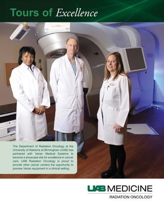
  • 1. The Department of Radiation Oncology at the University of Alabama at Birmingham (UAB) has partnered with Varian Medical Systems to become a showcase site for excellence in cancer care. UAB Radiation Oncology is proud to provide other cancer centers the opportunity to preview Varian equipment in a clinical setting. Tours of Excellence
  • 2. EXTENSIVE EXPERIENCE with delivering the most advanced radiation therapies available goes hand in hand with compassionate patient care at the UAB Department of Radiation Oncology. About UAB The University of Alabama at Birmingham (UAB) Health System is a network of services that provides a complete continuum of care, including comprehensive cancer care, for patients from Alabama and all over the world. As one of the original National Cancer Institute-designated comprehensive centers created, the UAB Comprehensive Cancer Center has maintained the designation for 40 years. As a part of the UAB Comprehensive Cancer Center, the UAB Department of Radiation Oncology is a world leader in the delivery of cancer care with highly experienced clinical faculty, utilizing the most advanced treatment modalities and technologies available in radiation therapy. Depth of Expertise The UAB Department of Radiation Oncology has achieved an international reputation for leadership through: • A multidisciplinary approach to diagnosis and treatment • An extensive and highly experienced clinical faculty that sub-specializes in the breadth of tumor types • A clinical care team of dosimetrists, nurses, and radiation therapists who are trained and certified to the highest standards. Twelve faculty physicians are board-certified in radiation oncol- ogy. Each clinical faculty physician sub-specializes by tumor type. Many hold PhD degrees in addition to their MD. Physicians stay abreast of the latest breakthroughs and technologies through their research into novel treatment modalities and methods to improve radiation delivery. Because UAB is a teaching institution, its physi- cians are training the radiation oncologists of tomorrow. The department physics team is the largest in Alabama. It includes seven PhD, board-certified, medical physicists. Faculty members support quality patient care, perform research to enhance cancer care via the use of radiation physics, and teach in the UAB physics and resident physician program. Leadership and Innovation The UAB Department of Radiation Oncology is proud of its contributions to advancing the use of radiation to treat cancer. Notable achievements include: • First in the world to use Intrafraction Motion Review tech- nology from Varian to continually monitor tumor location during radiosurgery for lung cancer (2011) James A. Bonner, MD Specialty: Lung, Head & Neck Residency: University of Michigan MD: Wayne State University James A Bonner M John Fiveash, MD Specialty: CNS, GU, Ocular Melanoma, Sarcoma Residency: Medical College of Georgia MD: Medical College of Georgia John Fi eash MDJennifer De Los Santos, MD Specialty: Breast, GYN Residency: University of Texas-MD Anderson Cancer Center MD: University of Florida Jennifer De Los Rojymon Jacob, MD Specialty: GI, GU, Sarcoma, Benign Disease Fellowship: Royal College of Radiologists, London MD: Kerala University, India Roj mon Jacob MDMichael Dobelbower, MD, PhD Specialty: CNS, GI, GU, Head & Neck, Lung, Benign Disease Residency: University of Alabama at Birmingham MD: Medical College of Ohio PhD: Ohio State University Michael Dobelbo e UAB Department of Radiation Oncology: Clinical Faculty Advanced treatment. Compassionate care. The Kirklin Clinic at Acton Road O. L. Burnett III, MD Specialty: GU, Lymphoma, Pediatrics, Breast, Sarcoma, GI MD: Emory University O L B rnett III MD
  • 3. • Among the first to use the TrueBeam™ system to treat lung, liver, pancreas, head & neck, brain, and spine cancers (2010) • First in the United States to treat cancer patients with RapidArc® radiotherapy (2008) • First in the region to perform intensity-modulated radiation therapy (IMRT) (1999) • Among the first to perform linear accelerator-based radiosurgery • First to clinically use ultrasound-based image guidance (1974). • First department in the region to separate therapeutic radiology from diagnostic radiology (1969). Varian Technology at UAB The UAB Department of Radiation Oncology delivers the following advanced modalities with Varian equipment: IMRT; image-guided radiation therapy (IGRT); and stereotactic body radiation therapy (SBRT). Professionals visiting UAB will be able to view Varian products in action in a high volume department that handles 30,000 treatment visits annually. In addition, department staff are available to answer questions about security, integration and interfaces, support, and many other issues common to large academic radiation oncology departments. During the tour, visitors can see the following Varian technology in use: • TrueBeam™ STx system • Clinac® iX linear accelerators • On-Board Imager® (OBI) kV imaging system for treatment localization - OBI radiographic: kV-kV anatomy matching & OBI CBCT • RapidArc® radiotherapy technology • Varian Real-time Position Management™ (RPM) system for respiratory gating • VariSource™ high dose rate afterloader • Varian Acuity™ treatment planning, simulation, and verifica- tion system for fluoroscopic simulation • ARIA® oncology information system • Eclipse™ treatment planning system for RapidArc, IMRT, IGRT, and SBRT Hazelrig-Salter Radiation Oncology Center Robert Kim, MD Specialty: GU, GYN, Ocular Melanoma, Orbital tumors Residency: University of Alabama at Birmingham MD: Yonsei University, Korea Robert Kim MD Christopher Willey, MD, PhD Specialty: CNS, Head & Neck, Lung, Pancreas Residency: Vanderbilt University Medical Center MD: Medical University of South Carolina PhD: Medical University of South Carolina Christopher Wille Eddy Yang, MD, PhD Specialty: Prostate, Breast, Head & Neck, Lung Residency: Vanderbilt University School of Medicine MD: University of Miami School of Medicine PhD: University of Miami School of Medicine Edd Yang MD PhRuby Meredith, MD, PhD Specialty: Breast, CNS, Lung, Lymphoma Residency: Medical College of Virginia MD: Ohio State University PhD: Indiana University R b Meredith MDKimberly Keene, MD Specialty: Breast, GI, Head & Neck, Pediatrics, Skin Residency: University of Virginia MD: University of Florida Kimberl Keene MD Sharon Spencer, MD Specialty: Head & Neck, Lung, Lymphoma, Pediatrics, Sarcoma, Skin Residency: The University of Alabama at Birmingham MD: University of Alabama at Birmingham Sharon Spencer M Joint venture clinic
  • 4. 3100 Hansen Way, M/S MGM, Palo Alto, CA 94304 For more information, contact your Varian representative. RAD 5832A © 2010, 2012 Varian Medical Systems, Inc. Printed in USA 5/12 (350) UAB Department of Radiation Oncology: Physics Faculty Ivan Brezovich, PhD Professor and Director PhD: Physics, University of Alabama at Birmingham MS: Physics, University of Alabama at Birmingham I an Bre o ich Ph Richard Popple, PhD Associate Professor Postdoctoral Fellowship: University of Texas-MD Anderson Cancer Center Postdoctoral Fellowship: Rice University Richard Popple Ph Jun Duan, PhD Associate Professor PhD: Physics, Florida State University MS: Medical Physics, University of Florida J n D an PhD Prem Pareek, PhD Adjunct Professor Post-Doctoral Fellowship: Allegheny General Hospital PhD: University of Nebraska Prem Pareek PhD Rex Cardan, PhD Assistant Professor Medical Physics Residency: James G. Brown Cancer Center, University of Louisville PhD: University of Texas at San Antonio Re Cardan PhD Xingen Wu, PhD Assistant Professor Postdoctoral Fellowship: St. Jude Children’s Research Hospital Postdoctoral Fellowship: Zhejiang University, China Xingen W PhDSui Shen, PhD Associate Professor Postdoctoral Fellowship: University of California at Davis Medical Center PhD: University of California at Davis S i Shen PhD Advanced treatment. Compassionate care. 1700 6th Avenue South, Birmingham, AL 35249 The Tour Experience With approximately 250,000 patient visits over the past 10 years, the UAB Department of Radiation Oncology offers a unique experience for professionals who want to learn about Varian technology. In 2010 and 2011, the UAB Department of Radiation Oncology hosted 32 visiting delegations. They represented community hospitals and university medical centers in 15 states, Canada, Mexico, and Brazil. UAB seeks to customize each tour to the expressed interests of our visitors in technology and types of cases treated. Radiation oncologists, neurosurgeons, physicists, dosimetrists, radiation therapists, and administrators have the opportunity to meet with UAB counterparts to exchange information and perspectives. The tour is often the beginning of continuing collaborations.
  • 5. 2012 UAB Radiosurgery Program Outcomes UAB Radiosurgery Program Hazelrig-Salter Radiation Oncology Center HSROC 2248 • 1700 6th Avenue South 619 19TH ST S BIRMINGHAM AL 35249-6832 Non-Profit Org. U.S. Postage PAID Permit No. 1256 Birmingham, AL The UAB Comprehensive Cancer Center To refer a patient to the UAB Radiosurgery Program or schedule appointments, contact UAB MIST at 1.800.822.6478. For information about the UAB Radiosurgery Program, visit uabmedicine.org/radiosurgery or uab.edu/radonc
  • 6. The 2012 UAB Radiosurgery Program Outcomes booklet continues our effort to communicate the strides our team has made in providing the best possible care to the citizens of Birmingham and beyond. We believe that the innovative techniques being harnessed within our program make UAB a standout in patient care, research, and education. Advancing treatments for optimal patient care and outcomes, as well as contributing to the body of radiosurgery knowledge, is helping us work toward our ultimate goal of developing better cancer therapeutics. Two of our most exciting and cutting-edge treatments are discussed in this booklet: triggered imaging technique for thoracic radiosurgery and Gamma Knife radiosurgery for pituitary tumors. Each of these care tactics builds on the Radiosurgery Program’s culture of collaboration and aim of providing our patients with care that is as individualized as they are. Triggered imaging for thoracic surgery is the newest development in motion management at UAB and allows our care teams to even more accurately deliver treatment to our patients. Thoracic radiosurgery is a technically complex procedure that requires advanced technologies and multidisciplinary care, which in Alabama are available uniquely at UAB. For the treatment of pituitary adenomas that require salvage treatment, Gamma Knife radiosurgery offers a precise, successful treatment modality. This salvage therapy offers a high rate of controlling the tumor while minimizing potential radiation-induced damage to adjacent normal tissue—an advantage that decreases the risk of neurocognitive impairment and secondary malignancy. These strides in patient treatment combined with our comprehensive team approach are a hallmark of our radiosurgery program. We strive to deliver these treatments with a patient-centered approach that allows for compassionate and superior care for each and every patient. We welcome any questions and comments you may have. If you would like to learn more about the progress of our program, you may contact the Department of Radiation Oncology at 205.934.5670. John B. Fiveash, MDJames A. Bonner, MD James M. Markert, Jr, MD, MPH Kirby I. Bland, MD At UAB, a new technique called triggered imaging is being used to monitor tumor position in real-time during thoracic radiosurgery. Triggered imaging is improving the accuracy and precision of radiosurgery. Radiosurgery is becoming an increasingly important tool for managing lung cancer in non or marginally operable patients, with outcomes comparable to surgery [8]. Numerous multi- institutional clinical trials are ongoing, with early results showing that this approach is safe and can result in cancer-free survivals at three years similar to surgery with less morbidity in the short term [8,10-13]. Reported control rates for thoracic tumors treated with radiosurgery have reached more than 90 percent [14]. Thoracic radiosurgery is technically challenging, requiring accurate targeting of the radiation beam so that the tumor receives the full, ablative radiation dose while dose to healthy tissue is minimized. To assure the best possible outcome, radiosurgery at UAB is performed by a multidisciplinary team comprised of thoracic surgeons, radiation oncologists, and medical physicists. Team members work in close collaboration throughout the entire treatment process, from initial consultation to the radiosurgical procedure, to patient follow up. A particularly complex technical challenge facing thoracic radiosurgery is respiratory motion of the tumor. Tumor motion is highly variable; Tumors at the apex of the lung typically remain stationary, while diaphragmatic tumors can move as much as 4 cm (K. M. Langen., And D. T. L. Jones, “Organ motion and its management,” Int. J. Radiation Oncology Biol. Phys., Vol. 50, No. 1, pp. 265–278, 2001). At UAB, management of tumor motion begins with the thoracic surgeon. Using navigational bronchoscopy, the surgeon implants fiducial markers in the tumor. Conventional bronchoscopic techniques cannot reach many tumors, for which the only other option for implanting fiducial markers is trans- thoracically. The trans-thoracic approach has a pneumothorax rate as high as 30% ( Yousefi S, Collins BT, Reichner CA, Anderson ED, Jamis-Dow C, Gagnon G, Malik S, Marshall B, Chang T, Banovac F. Complications of thoracic computed tomography-guided fiducial placement for the purpose of stereotactic body radiation therapy. Clin Lung Cancer. 2007 Jan;8(4):252-6.), compared to less than 6% for navigational bronchoscopy (Schroeder C, Hejal R, Linden PA. Coil spring fiducial markers placed safely using navigation bronchoscopy in inoperable patients allows accurate delivery of CyberKnife stereotactic radio surgery. J Thorac Cardiovasc Surg. Triggered Imaging Technique for Thoracic Radiosurgery A Message From the Chairs Triggered Imaging Technique for Thoracic Radiosurgery............... 2-3 Gamma Knife Radiosurgery for Pituitary Tumors..................... 4-5 Quality and Outcome Measures...6-7 Publications...............................8 Educational Site Visits.................9 Clinical Faculty.........................10 Contents 2 Merle M. Salter Professor and Chair UAB Department of Radiation Oncology Fay Fletcher Kerner Professor and Chair UAB Department of Surgery Robert Y. Kim Endowed Chair, Professor and Vice Chair UAB Department of Radiation Oncology
 Interim Associate Director for Clinical Research UAB Comprehensive Cancer Center James Garber Galbraith Endowed Chair, Professor and Director UAB Division of Neurosurgery Cover photo: Dr. Sharon Spencer, Dr. Barton Gurthrie, and Dr. Kristen Riley Participating Faculty James A. Bonner, MD Kirby I. Bland, MD Michael C. Dobelbower, MD, PhD John B. Fiveash, MD Barton L. Guthrie, MD Douglas J. Minnich, MD Richard A. Popple, PhD Kristen Riley, MD Sharon A. Spencer, MD Editorial Support John Brinkerhoff Valeria Pacheco-Rubi Joey Slatsky Fresia Vega Above: Dr. Richard Popple
  • 7. 2010 Nov;140(5):1137-42. Epub 2010 Sep 20.; Harley DP, Krimsky WS, Sarkar S, Highfield D, Aygun C, Gurses B. Fiducial marker placement using endobronchial ultrasound and navigational bronchoscopy for stereotactic radiosurgery: an alternative strategy. Ann Thorac Surg. 2010 Feb;89(2):368-73; discussion 373-4). At UAB, we have had no pneumothoraces with fiducial placement. The markers are typically implanted during a diagnostic bronchoscopy, so the patient does not need to undergo an additional procedure. After bronchoscopy, the radiation oncologist and the thoracic surgeon consult to determine the best treatment strategy. Once the decision has been made to use radiosurgery, the patient receives a CT scan to identify the tumor and nearby healthy structures that need to be protected from the radiation. The CT scan is the next stage in the management of tumor motion. During the scan, an optical technique is used to measure the chest motion. The scan is a special type, called a 4D CT, composed of 10 complete 3-dimensional CT image sets. Each CT corresponds to a snapshot at a different point in the respiratory cycle, which is correlated with the chest motion. One method to ensure that the tumor remains within the radiation beam is to simply treat the entire volume encompassed by tumor motion. However, this approach results in a relatively large volume of lung receiving a high radiation dose (Wu J, Li H, Shekhar R, Suntharalingam M, D’Souza W., “An evaluation of planning techniques for stereotactic body radiation therapy in lung tumors,” Radiother Oncol. 2008 Apr;87(1):35-43. Epub 2008 Mar 24). This approach is particularly undesirable in the context of the high, ablative radiation dose delivered by radiosurgery. An alternative approach preferred at UAB is to gate the radiation beam, turning it on only at the end of expiration, when the lung is at rest and the tumor is relatively stationary. The scans are evaluated for tumor motion by the medical physicist, who determines the optimal point in breathing cycle to turn the radiation beam on and off. The medical physicist also locates the fiducial markers in the CT images. The radiosurgery team then develops and tests an individualized treatment plan. The dose distribution is sculpted to tightly conform to the tumor and limit radiation dose to the lung, chest wall, and other healthy tissues. The final and most critical step in motion management is treatment, usually one to five treatments over one to two weeks. Prior to starting radiation delivery, x-rays are taken to ensure that the tumor is in the correct position. The fiducial markers are easily seen in the x-rays and are compared with outlines of the expected position, derived from the 4D CT scan and the preparation by the medical physicist. If the outline and the image on the x-ray do not coincide, the patient is shifted until they do. When the patient is in the correct position and the tumor is centered in the radiation beam, the beam is turned on. During treatment, the same optical technique used during the CT scan is used to track the patient’s breathing. The optical system instructs the radiation beam to turn on at the end of expiration and to turn off as inspiration begins. The newest development in motion management at UAB is triggered imaging. Using triggered imaging, we observe the fiducial marker during treatment delivery. At the beginning of each expiratory cycle, immediately before the radiation beam comes on, an x-ray image is taken. The image is displayed along with a circle around the expected position of the fiducial marker. The radiation oncologist and thoracic surgeon are thus able to monitor the position of the tumor in real time as the treatment progresses. If the patient moves or the breathing pattern changes, treatment is suspended, the position corrected, and treatment resumed. Thoracic radiosurgery is a technically complex procedure requiring advanced technologies and multidisciplinary care, which in Alabama are available uniquely at UAB. The experienced team at UAB will continue to remain at the forefront of innovation as the technologies for thoracic radiosurgery continue to evolve. Pituitary adenomas represent one of the most common intracranial neoplasms. Found in 10-15% of the population, these benign tumors often pose complex management situations. While the majority of pituitary tumors can be treated with medication or surgery alone, a significant proportion require salvage treatment. Pituitary tumors that generally require additional treatment include functional tumors not controlled with surgery or medication and nonfunctional tumors that recur following surgery. Gamma Knife radiosurgery offers a precise, successful treatment modality for pituitary adenomas. Tumor growth can be controlled in 90% of patients treated, frequently with reduction in tumor volume. Radiosurgery effects on biochemical cure vary depending on tumor type. (Sheehan et al 2011). Patient selection for radiosurgery depends on endocrine evaluation, tumor size, location, growth pattern, and pathology. Pituitary adenomas are classified according to size and endocrine profile. Microadenomas, defined as smaller than 10mm in size, rarely cause clinical concern due to size, but may require treatment if they are functionally active. Macroadenomas, larger than 10mm, may cause visual difficulty if the optic pathways become compressed by the tumor. Located at the base of the skull, pituitary tumors occur adjacent to many critical structures such as the optic nerves, optic chiasm, cranial nerves within the cavernous sinuses, carotid artery, and brainstem. The location of these tumors requires specialized knowledge and techniques for management. Additionally, pituitary adenomas often have either hormone overproduction or deficiency. All management decisions regarding these tumors require a multidisciplinary approach. While many tumors require only observation, a significant number have endocrine and anatomical implications that must be addressed. The UAB Neurosurgical Pituitary Disorders Clinic offers comprehensive evaluation and care for patients with pituitary tumors. Following diagnosis, whether for an incidentally found tumor or a symptomatic pituitary adenoma, appropriate evaluation includes imaging review, endocrine evaluation, and often ophthalmologic evaluation. Observation, medical therapy, surgery, and radiation therapy comprise the armamentarium of treatment options for pituitary tumors. Gamma Knife Radiosurgery for Pituitary Tumors 43 Above: Dr. Douglas Minnich and Dr. Michael Dobelbower Above: Fiducial marker during treatment delivery
  • 8. Functional tumors, those that result in overproduction of hormones, often require multi-modality treatment. Prolactinomas are the most common functional pituitary tumors. For prolactinomas, medical therapy with dopamine agonists is the standard of care for first line treatment. However, for patients not controlled with medication or who do not tolerate medication, surgery and radiation may be utilized. Pituitary tumors resulting in acromegaly, from excess growth hormone and Cushing’s disease from excess ACTH, require treatment regardless of size. Surgery is the first line of treatment for the majority of these tumors. In cases where a surgical cure is not achieved, additional therapy is paramount due to the significant increase in morbidity and mortality if hormone overproduction is not controlled. For patients with Cushing’s disease, there is no available medical treatment to suppress steroid production. Radiosurgery offers a potential for cure. In acromegaly, controversy exists regarding the timing of radiation therapy related to medical therapy. Medical therapy is often successful in normalizing growth hormone production, but at a significant yearly financial cost. Without controversy, is the use of radiation when patients are not controlled with medical therapy. However, there may be utility in radiation treatment in an attempt to shorten the length of time a patient requires medical therapy. Data suggests radiosurgery offers a greater than 50% rate of cure for growth hormone secreting pituitary tumors. (Sheehan et al 2011) For residual nonfunctional adenomas following surgery, Gamma Knife radiosurgery is considered if there is observed tumor growth over time or if the pathology is atypical pituitary adenoma, indicating a potentially higher chance of tumor recurrence. The recurrence rate of pituitary adenomas following surgery is reported around 20%. Recurrence is influenced by extent of resection and tumor pathology. Patients with pituitary adenomas are followed postoperatively with yearly imaging. The majority of tumor recurrence is seen in the first five to seven years postoperatively, but can occur later. Following any radiation to the sella, patients should have a yearly endocrine evaluation. Secondary hypopituitarism is the most common side effect of radiosurgery for pituitary adenomas. The incidence of secondary hormone deficits increases with time, thus necessitating long-term endocrine surveillance. Gamma Knife radiosurgery may have a decreased rate of endocrine dysfunction over fractionated radiation due to the ability to precisely deliver radiation to the tumor and limit radiation to the normal gland in some patients. (Taussky et al 2011) In addition to minimizing dose to the normal pituitary gland, radiosurgery allows for treatment delivery that minimizes radiation to adjacent normal brain cells. This precision decreases the risk of neurocognitive impairment and secondary malignancy from radiation. Appropriate patient selection and experienced treatment planning help to minimize the risks of radiosurgery. The anatomical location of the pituitary tumor necessitates careful evaluation and planning to limit toxicity to critical structures. Gamma Knife, with frame based head fixation, offers the most precise method of radiation delivery. In this area, millimeters matter. At UAB, we feel strongly that Gamma Knife precision allows us to perform safe, successful radiosurgery for pituitary tumors. For more information or to refer a patient to the Multidisciplinary Pituitary Clinic: Contact Michel Thomas, Office Assistant to Dr. Riley, at 205-996-2461. Gamma Knife Stereotactic Body Radiation Therapy 0 500 1000 1500 2000 2500 3000 3500 4000 4500 5000 1992 1993 1994 1995 1996 1997 1998 1999 2000 2001 2002 2003 2004 2005 2006 2007 2008 2009 2010 2011 Timeline of our Success Quality and Outcome Measure Timeline of our success SELECTED DISEASE SITES 1992 First patient treated with stereotactic radiosurgery (linac) 1995 First CNS case treated with Gamma Knife 1999 First FDA-approved IMRT- delivering device 2001 First in Alabama to offer RPM Gating System 2005 First in Alabama to treat with stereotactic body radiation therapy 2008 First in the U.S. to treat with volumetric arc therapy (RapidArc™) 2010 One of the world’s first facilities to offer TrueBeam system (third in the United States) 2011 First in the world to use “Triggered Imaging” Technology from Varian Medical Systems to continually monitor tumor location during radiosurgery for lung cancer The UAB Radiosurgery Program offers state-of-the-art treatment therapies and technologies for a wide variety of body sites, including central nervous system (CNS), lung, spine, and others. CNS tumors essentially are treated with the Gamma Knife. Tumors or malformations of the liver, lung, spine, and other body sites are treated using Stereotactic Body Radiation Therapy (SBRT). The following charts show the outcome measures of selected body sites treated with cranial radiosurgery and SBRT at UAB. 6 Sheehan JP, Pouratian N, Steiner L, Laws ER, Vance ML. Gamma Knife surgery for pituitary adenomas: factors related to radiological and endocrine outcomes. J Neurosurg. 2011 Feb: 114 (2) 303-9. Taussky P, Kalra R, Coppens J, Mohebali J, Jensa R, Couldwell WT. Endocrinological outcome after pituitary transposition (hypophysopexy) and adjuvant radiotherapy for tumors involving the cavernous sinus. J Neurosurg. 2011 Jul; 115(1): 55-62. Gamma Knife 2114 Benign 432 Malignant 1083 Trigeminal Neuralgia 409 Vascular 188 Seizure 2 Stereotactic Body Radiation Therapy 363 Brain 36 Lung 129 Liver 24 Other 53 Spine 121 5
  • 9. 0 500 1000 1500 2000 2500 3000 3500 4000 4500 5000 1992 1993 1994 1995 1996 1997 1998 1999 2000 2001 2002 2003 2004 2005 2006 2007 2008 2009 2010 2011 0 10 20 30 40 50 60 70 80 90 100 2005 2006 2007 2008 2009 2010 2011 2012 Radiosurgery Noteworthy Publications Clark GM, Popple RA, Prendergast BM, Spencer SA, Thomas EM, Stewart JG, Guthrie BL, Markert JM, Fiveash JB: Plan quality and treatment planning technique for single isocenter cranial radiosurgery with volumetric modulated arc therapy. Practical Radiation Oncology. Published online February 1, 2012. Citation Pending. Clark G, Popple R, Young PE, Fiveash J: Feasibility of single- isocenter volumetric modulated arc radiosurgery for the treatment of multiple brain metastases. Int J Radiat Oncol Biol Phys. 2010 Jan 1;76(1):296-302. Fiveash J, Guthrie BG, Duan J, Markert JM, DeLosSantos JF, Keene KS, Spencer SA, Dobelbower MC, Arafat W, Popple RA. A Phase II Isotoxicity Study of Spinal Radiosurgery/SBRT. Int. J. Radiat. Oncol. Biol. Phys. 2010 November; 78(3) Suppl: S278. Parker JN, Zheng X, Luckett W, Markert JM, Cassady KA. Strategies for the rapid construction of conditionally-replicating HSV-1 vectors expressing foreign genes as anticancer therapeutic agents. Mol Pharm. 2011 Feb 7;8(1):44-9. Epub 2010 Dec 17. Review. PMID: 21142023 Pearson BE, Markert JM, Fisher WS, Guthrie BL, Fiveash JB, Palmer CA, Riley K. Hitting a moving target: evolution of a treatment paradigm for atypical meningiomas amid changing diagnostic criteria. Neurosurg Focus. 2008;24(5):E3. PMID: 18447742 Popple RA, Dieterich S, Duan J, Fiveash JB. Dependence of Dose- volume Values on Calculation Method for Paraspinal Radiosurgery. Int. J. Radiat. Oncol. Biol. Phys. 2010 November; 78(3) Suppl: S783. Popple RA, Fiveash JB, Brezovich IA, Bonner JA: RapidArc radiation therapy: first year experience at the University of Alabama at Birmingham. Int J Radiat Oncol Biol Phys. 2010 Jul 1;77(3):932-41. Prendergast BM, Bonner JA, Popple RA, Spencer SA, Fiveash JB, Keene KS, Cerfolio RJ, Minnich DJ, Dobelbower MC. Dosimetric analysis of imaging changes following pulmonary stereotactic body radiation therapy. J Med Imagina Radiat Oncol 2011 Feb;55(1):90-6. Prendergast Brendan M, Popple Richard A., Clark Grant M., Spencer Sharon A., Guthrie Bart, Markert James, Fiveash John B: Improved clinical efficacy in CNS stereotactic radiosurgery using a flattening filter free linear accelerator. Journal of Radiosurgery and SBRT. Accepted for publication Journal of Radiosurgery and SBRT, August 9, 2011. Citation Pending. Sawrie SM, Fiveash JB. Caudell, JJ: Stereotactic Body Radiation Therapy for Liver Metastases and Primary Hepatocellular Carcinoma: Normal Tissue Tolerances and Toxicity. Cancer Control April 2010, Vol. 17, No. 2:111-119 Spencer S, Swaid N, Barton G, Young P, Wong W, Meredith RF, Markert J, Fisher W, Wu X, Nordal R, Fiveash J. Impact of Dose Rate on Outcomes of Gamma Knife Radiosurgery in Patients with Face Pain. Radiosurgery 2010 7:360-5. Sperduto PW, Kased N, Roberge D, Xu Z, Shanley R, Luo X, Sneed PK, Chao ST, Weil RJ, Suh J, Bhatt A, Jensen AW, Brown PD, Shih HA, Kirkpatrick J, Gaspar LE, Fiveash JB, Chiang V, Knisely JPS, Sperduo CM, Lin N, Mehta M: Summary Report on The Graded Prognostic Assessment: An Accurate and Facile Diagnosis-Specific Tool to Estimate Survival for Patients with Brain Metastases. J Clin Oncol, 29, 2011. Sperduto PW, Kased N, Roberge D, Xu Z, Shanley R, Luo X, Sneed PK, Chao ST, Weil RJ, Suh J, Bhatt A, Jensen AW, Brown PD, Shih HA, Kirkpatrick J, Gasper LE, Fiveash JB, Chiang V, Knisely JP, Sperduto CM, Lin N, Mehta M: Effect of Tumor Subtype on Survival and the Graded Prognostic Assessment for Patients with Breast Cancer and Brain Metastases. Int J Radiat Oncol Biol Phys 2011, April 14. Stewart JG, Sawrie SM, Bag A, Han X, Fiveash JB: Management of Brain Metastases. Current Treatment Options in Neurology. 2010 Jul;12(4):334-46. Vaphiades MS, Spencer SA, Riley K, Francis C, Deitz L, Kline LB. Radiation-induced ocular motor cranial nerve palsies in patients with pituitary tumor. J Neuroophthalmol. 2011 Sep;31(3):210-3. The Leksell Gamma Knife is a highly advanced technology that delivers 201 tightly focused cobalt radiation beams to one point in the brain. The radiation beams and doses are so precise they affect only the targeted tissue and generally spare the surrounding healthy tissue. Stereotactic Body Radiation Therapy (SBRT) uses a high dose of radiation shaped to conform to the patient’s tumor. It delivers radiation to the intended target and avoids healthy tissue. Small tumors are accurately identified and located with precise coordinates. Quality and Outcome Measure CRANIAL RADIOSURGERY PROCEDURES SBRT PROCEDURES 87
  • 10. Visiting Institution Date of Visit Gulfport Memorial Hospital – Gulfport, MS 1/31/2011 Rush University – Chicago, IL 2/17/2011 Torrance Memorial Medical Center – Torrance, CA 2/18/2011 University of Kentucky – Lexington, KY 3/24/2011 Memorial Hospital – Chattanooga, TN 4/29/2011 Hospital Israelita Albert Einstein – São Paulo, Brasil 4/12/2011 West Michigan Cancer Center – Kalamazoo, MI 5/6/2011 Corpus Christi Cancer Center – Corpus Christi, TX 5/20/2011 Medical Center at Bowling Green – Bowling Green, KY 6/3/2011 Memorial Hospital – Gulfport, MS 6/9/2011 Eastern Health-Cancer Care Program Dr. H. Bliss Murphy Cancer Centre – NL, Canada 6/16/2011 Baptist Hospital – Miami, FL 7/1/2011 University of Puerto Rico Cancer Center – San Juan, Puerto Rico 8/4/2011 8/5/2011 Vanderbilt University Medical Center – Nashville, TN 8/26/2011 Jackson-Madison County General Hospital – Jackson, TN 9/8/2011 9/9/2011 University of Tennessee Hospital – Knoxville, TN 9/23/2011 Cancer Treatment Centers of America – Tulsa, OK 10/28/2011 Renown Medical Center – Reno, NV 11/11/2011 Radiological Associates of Sacramento – Sacramento, CA 11/18/2011 Hospital Médica Sur – Mexico City, D.F., Mexico 12/9/2011 Tours of Excellence UAB Site Visits 2011 UAB Radiosurgical Clinical Faculty James A. Bonner, MD Radiation Oncology Specialties: lung, head and neck Ivan Brezovich, PhD Medical Physicist Specialty: physics O.L. Burnett III, MD Radiation Oncology Specialties: GU, gynecological, lymphoma, pediatrics, breast, sarcoma, GI Rex A. Cardan, PhD Medical Physicist Specialty: physics Robert Cerfolio, MD Thoracic Surgery Specialty: thorax Melissa Chambers, MD Neurosurgery Specialties: brain tumors Jennifer De Los Santos, MD Radiation Oncology Specialties: breast, gynecological, lung, lymphoma, sarcoma, skin Michael Dobelbower, MD, PhD Radiation Oncology Specialties: benign disease, CNS, GI, GU, head and neck Juan Duan, PhD Medical Physicist Specialty: physics Winfield S. Fisher, MD Neurosurgery Specialties: brain tumors, face pain, vascular John Fiveash, MD Radiation Oncology Specialties: CNS, GU, gynecological, ocular melanoma, pediatrics, sarcoma Barton L. Guthrie, MD Neurosurgery Specialties: brain tumors, face pain Rojymon Jacob, MD Radiation Oncology Specialties: CNS, GI, GU, sarcoma, benign disease Kimberly Keene, MD Radiation Oncology Specialties: breast, GI, head and neck, pediatrics, skin Robert Kim, MD Radiation Oncology Specialties: GU, gynecololgical, ocular melanoma, orbital tumors James A. Markert, MD Neurosurgery Specialties: brain tumors, spinal radiosurgery, trigeminal neuralgia Ruby Meredith, MD, PhD Radiation Oncology Specialties: benign disease, breast, CNS, GI, head and neck, lung, lymphoma, orbital tumors, skin Douglas J. Minnich, MD Thoracic Oncology Specialty: thorax Richard Popple, PhD Medical Physicist Specialty: physics Prem Pareek, PhD Medical Physicist Specialty: physics Kristen Riley, MD Neurosurgery Specialties: brain tumors, epilepsy, spine Sui Shen, PhD Medical Physicist Specialty: physics Sharon Spencer, MD Radiation Oncology Specialties: breast, CNS, GI, gynecological, head and neck, lung, lymphoma, orbital tumors, ocular melanoma, pediatrics, sarcoma, skin Christopher Willey, MD, PhD Radiation Oncology Specialties: breast, CNS, head and neck, lung, pancreas Xingen Wu, PhD Medical Physicist Specialty: physics Eddy Yang, MD Radiation Oncology Specialties: lung, GU, breast, head and neck 109 Partial listing of programs visiting the University of Alabama at Birmingham to learn about treatment techniques on the TrueBeam linear accelerator
  • 11. 2011 UAB Radiosurgery Program Outcomes UAB Radiosurgery Program Hazelrig-Salter Radiation Oncology Center HSROC 2248 • 1700 6th Avenue South 619 19TH ST S BIRMINGHAM AL 35249-6832 Non-Profit Org. U.S. Postage PAID Permit No. 1256 Birmingham, AL The UAB Comprehensive Cancer Center To refer a patient to the UAB Radiosurgery Program or schedule appointments, contact UAB MIST at 1.800.822.6478. For more information about the UAB Radiosurgery Program, visit uabmedicine.org/radiosurgery or uab.edu/radonc.
  • 12. The 2011 UAB Radiosurgery Program Outcomes booklet continues our effort to provide our friends and colleagues an informative picture of how we are handling our mission to provide care to the citizens of Alabama and the region. In UAB’s culture of collaboration, the Department of Radiation Oncology and the Department of Surgery developed the UAB Radiosurgery Program. This special approach to patient care provides every patient requiring stereotactic radiation surgery with a reasoned and thorough evaluation of their situation, resulting in a recommended treatment plan. Treatment outcomes are completed as patients are treated and followed. The goal is to optimize treatments and add to the body of knowledge of the field. As this interspecialty relationship has flourished, the program has maintained growth and the outstanding score in patient satisfaction you will see in this report. As an update, we are pleased to report that the linear accelerator based radiosurgery program moved into a new building, the Hazelrig-Salter Radiation Oncology Center, in March 2010, providing our patients and their families with a more comfortable, attractive setting. Included in the new space is one of the first TrueBeam radiation devices in the world. TrueBeam is living up to its promise of delivering precise radiosurgical treatments in significantly less time than previously possible with other machines. For our patients, reduced treatment time means more accurate delivery and increased comfort. The improvement in delivery accuracy reduces the potential for collateral damage to nearby healthy tissue. This type of continually updated technology, a faculty with more than 253 total years of experience in radiosurgery, and a clinical team that understands and supports our patients’ individual needs all combine to pursue our goal of eventually curing cancer. We invite your questions and comments. If you wish to learn more about the progress of our program, you may contact the Department of Radiation Oncology at 205.934.5670. Kirby I. Bland, MD Lung cancer is a disease that is too well known by too many people. Only 100 years ago, lung cancer was considered a rare and uncommon entity [1]. Medical literature at that time regarding lung cancer was limited to small studies and individual reports of an uncommon disease [2-5]. Now, scarcely three generations later, it is a leading cause of death and morbidity in the United States, with approximately 196,000 cases diagnosed each year. Of those, 158,000 will die from their disease. Surgical resection of lung cancer has long been considered the standard of care when attempting to cure patients when the disease is diagnosed early and in a well- localized fashion. Unfortunately, many patients present with advanced disease that is not amenable to operative resection. Other patients, who otherwise would have resectable disease, are not candidates for surgery because of comorbidities such as heart disease. For patients who are unable to undergo surgical resection, high-dose radiation that is delivered daily for several weeks has been used in an effort to cure. This approach has produced less than satisfying results [6, 7]. Now, with the advent of thoracic radiosurgery, outcomes that are more comparable to surgery are possible [8]. Radiosurgery is not a new technology. It has been used for many years to treat cancers in the central nervous system [9]; however, its use in the lung has been Thoracic Radiosurgery A Message From the Chairs 1. Witschi H. A Short History of Lung Cancer. Toxicological Sciences. 2001;64:4- 6. 2. Ryn TC, Meyer FW. Bronchogenic Carcinoma. U.S. Naval Bulletin. 1949;49(5):863-867. 3. Hirsch EF. Bronchogenic Carcinoma of the Lung. Illinois Med J. 1949;95(4):241- 243. 4. Corsello JN, O’Brien WB. Primary Bronchogenic Carcinoma, a report of 47 cases. Rhode Island Med J. 1947;30(1):15- 20. 5. Eagan JC. Bronchgenic Carcinoma of the Lung; report of a case. Nebraska State Med J. 1948;31:94-98. 6. Haffty B, et al. Results of radical radiation therapy in clinical stage 1, technically operable non-small cell lung cancer. Int J Radiat Oncol Biol Phys. 1998;69-73. 7. Dosoretz D, et al. Radiation therapy in the management of medically inoperable carcinoma of the lung: Results and implications for future treatment strategies. Int J Radiat Oncol Biol Phys. 1992;24:3. 8. Palma D, Visser O, Lagerwaard F, Slotman B, Belderbos J, Senan S. A Population- Based Matched-Pair Comparison of Stereotactic Radiotherapy vs. Surgery for the Treatment of Stage I NSCLC in Elderly Patients. Chicago Multidiciplinary Symposium in Thoracic Oncology, Chicago, Dec 2010. 9. Leksell L. The Steroetaxic Method and Radiosurgery of the Brain. Acta chir Scand. 1951;102:316. Thoracic Radiosurgery............. 2-3 Truebeam: Image Guided Radiotherapy and Radiosurgery... 4-5 Locations...................................5 Quality and Outcome Measures...6-7 Publications...............................8 Faculty Presentations..................9 Educational Site Visits.................9 Clinical Faculty.........................10 Contents 2 Merle M. Salter Professor and Chair UAB Department of Radiation Oncology Fay Fletcher Kerner Professor and Chair UAB Department of Surgery Cover photo: Depicts a patient on UAB’s new Truebeam with Dr. Douglas Minnich, Dept of Surgery, and Dr. Chris Dobelbower, Dept of Radiation Oncology James A. Bonner, MD Participating Faculty James A. Bonner, M.D. Kirby I. Bland, M.D. Michael C. Dobelbower, M.D., Ph.D. John B. Fiveash, M.D. Barton L. Guthrie, M.D. Douglas J. Minnich, M.D. Richard A. Popple, Ph.D. Christopher D. Willey, M.D., Ph.D. Editorial Team John C. Brinkerhoff Catina M. Diggs Valeria Pacheco-Rubi Fresia Vega-Thompson Data Collection Support Ginna Blaylock Kathy Bowman Joey P. Slatsky
  • 13. a far more challenging problem for numerous reasons. The first among these is that the lung is in motion. Thus, the challenge is to hit a moving target with great precision. Other challenges include visualizing small tumors with great accuracy and dose calculation challenges in the lungs that are unique from other sites in the body. Technological advances in radiation treatment machines, such as the Varian TrueBeam™ STx radiosurgical machine and the superDimension® navigational bronchoscopy system, have solved many of the problems associated with thoracic radiosurgery. In fact, numerous currently ongoing clinical trials are testing the safety and efficacy of expanding the use of thoracic radiosurgery. Early results from several institutions have shown that this approach is not only safe, but also can produce similar cancer-free survivals to surgery at 3 years and have less morbidity in the short term [8,10-13]. In fact, control rates for thoracic tumors treated with radiosurgery now range from 80 percent to more than 90 percent [14]. Thoracic radiosurgery at UAB is performed by a multidisciplinary team including thoracic surgeons, radiation oncologists, dosimetrists, and medical physicists. The process begins with the diagnosis of malignancy. New tools for the diagnosis of cancer with minimally invasive approaches, such as navigational bronchoscopy are used at UAB to diagnose the malignancy and to place markers into the tumor for targeting by the radiation machine. Once a diagnosis is made, patients undergo a specialized planning CT scan to identify the tumor and nearby structures that need to be protected from the radiation. The radiosurgery team then develops and tests an individualized treatment plan. Radiation is subsequently delivered, usually in one to five treatments over the next 1 to 2 weeks, with each treatment generally lasting less than 30 minutes. The treatments are performed on an outpatient basis, are painless, and only require that the patient lie still during treatment. Thoracic radiosurgery is an exciting and promising new therapy for patients with medically inoperable early-stage lung cancer. The ultimate role that thoracic radiosurgery will have in the treatment of lung cancer is yet to be defined. Large clinical trials evaluating its efficacy are exploring new indications for this treatment, and the long-term effects remain unknown. What is clear is that thoracic radiosurgery does offer a chance for cure in patients who previously would have had limited treatment options. 10. Rusthoven KE, Kavanagh BD, Burri SH, Chen C, Cardenes H, Chidel MA, Pugh TJ, Kane M, Gaspar LE, Schefter TE. Multi-Institutional Phase I/II Trial of Stereotactic Body Radiation Therapy for Lung Metastases. Journal of Clin Oncol. 2009;27(10). 11. Timmerman R, Papiez L, McGarry R, Likes L, DesRosiers C, Frost S, Williams M. Extracranial Stereotactic Radioablation Results of a Phase I Study in Medically Inoperable Stage I Non-small Cell Lung. Chest. 2003;124:1946-1955. 12. Fakiris AJ, McGarry RC, Yiannoutsos CT, Papiez L, Williams M, Henderson MA, Timmerman R. Steroetactic Body Radiation Therapy for Early-Stage Non-Small-Cell Lung Carcinoma: Four-Year Results of a Prospective Phase II Study. Int J Radiat Oncol Biol Phys. 2009;75(3):677-682. 13. Louie AV, Rodrigues G, Hannouf M, Palma DA, Cao JQ, Yaremko BP, Malthaner R, Mocanu JD, Zaric GS. Is Stereotactic Body Radiotherapy Warranted in Medically Operable Stage I NSCLC? A Markov Model Based Decision Analysis. Chicago Multidisciplinary Symposium in Thoracic Oncology, Chicago, Dec 2010. 14. Timmerman RD, Park C, Kavanagh BD. The North American Experience with Stereotactic Body Radiation Therapy in Non-small Cell Lung Cancer. J Thorac Oncol. 2007;2(7) Supplement 3. Last year, the UAB Department of Radiation Oncology was among the first institutions in the world to deploy a TrueBeam™ system for image-guided radiotherapy and radiosurgery. Designed to treat a moving target with unprecedented speed and accuracy, TrueBeam incorporates numerous technical innovations that dynamically synchronize imaging, patient positioning, motion management, and treatment delivery during a radiotherapy or radiosurgery procedure. One important feature of the TrueBeam system is its high-intensity mode, which makes it possible to deliver doses up to four times faster than can be accomplished with other radiosurgery machines, significantly shortening treatment times. Cutting down treatment time by a factor of two to four makes a big difference to patients and can enhance treatment accuracy by leaving less time for tumor motion during dose delivery. Using the TrueBeam system, a standard intensity-modulated treatment that would typically take 10 minutes can be completed in less than two minutes. Simple RapidArc treatments, which used to be done in 2 minutes, can now be completed in 1 minute. UAB clinicians have used the TrueBeam system to deliver fast, highly precise treatment for tumors of the brain, spine, lung, liver, prostate, head and neck, and pancreas. The system is extremely flexible, allowing for selection of an optimal treatment approach in each case, from intensity-modulated radiotherapy (IMRT) to stereotactic radiosurgery (SRS), from stereotactic body radiotherapy (SBRT) to volumetric arc (RapidArc®) therapy. In addition, a new gated RapidArc capability allows it to be used with tumors that are subject to respiratory motion, such as many tumors of the lung or liver. “Intelligent” automation further speeds treatments with an up to fivefold reduction in the number of steps needed for imaging, positioning, and treating patients. A nine-field IMRT treatment that would have required 52 separate steps or mouse- clicks using earlier generations of technology can now be completed in less than ten TrueBeam: State-of-the-Art Image- Guided Radiotherapy and Radiosurgery 43
  • 14. steps. As a result, UAB radiation therapists can focus more of their attention on the patient and on the progress of the treatment. The precision of a TrueBeam system is measured in increments of less than a millimeter. This accuracy is made possible by the system’s sophisticated architecture, which establishes a new level of synchronization between imaging, patient positioning, motion management, beam shaping, and dose delivery technologies. Accuracy checks are performed every 10 milliseconds throughout the treatment. More than 100,000 data points are monitored continually as a treatment progresses, ensuring that the system maintains a true isocenter, or focal point of treatment. The TrueBeam imager, which is used to localize a tumor just prior to treatment, can generate 3-D anatomical images in 60 percent less time, with a 25 percent reduction in X-ray dose to the patient, when compared with earlier generations of technology. We are excited about this powerful and fully integrated high-end system and regard it as a significant step forward in our ongoing commitment to providing patients with access to the best of available contemporary radiosurgical technology. LOCATIONS 0 100 200 300 400 Gamma Knife Sterotactic Body Radiosurgery Therapy Patients Stereotactic Radiosurgery Special Procedures 2005 2006 2007 2008 2009 2010 S p e c i a l P r o c e d u r e s o n S e l e c t e d D i s e a s e S i t e s Gamma Knife Benign Malignant Trigeminal Neuralgia Vascular 1883 393 981 352 157 Stereotactic Body Radiation Therapy Brain Lung Liver Spine Other 265 13 94 19 89 50 UAB Highlands Cranial radiosurgery with the Leksell Gamma Knife® 1201 11th Avenue South Birmingham, AL 35205 The Kirklin Clinic® at Acton Road SBRT with TomoTherapy® and with the Varian EX® linear accelerator 2145 Bonner Way Birmingham, AL 35243 Hazelrig-Salter Radiation Oncology Center SBRT with the Varian iX linear accelerator and TrueBeam accelerator 1700 6th Avenue South Birmingham, AL 35233 0 500 1000 1500 2000 2500 3000 3500 4000 1992 1993 1994 1995 1996 1997 1998 1999 2000 2001 2002 2003 2004 2005 2006 2007 2008 2009 2010 Timeline of Our Success 4500 5000 NumberofPatientsTreated 1999 2001 1995 1992 2005 2008 2010 Quality and Outcome Measure Timeline of our success SELECTED DISEASE SITES 1992 First patient treated with stereotactic radiosurgery (linac) 1995 First CNS case treated with Gamma Knife 1999 First FDA-approved IMRT- delivering device 2001 First in Alabama to offer RPM Gating System 2005 First in Alabama to treat with stereotactic body radiation therapy 2008 First in the U.S. to treat with volumetric arc therapy (RapidArc™) 2010 One of the world’s first facilities to offer TrueBeam system (third in the United States) The UAB Radiosurgery Program offers state-of-the-art treatment therapies and technologies for a wide variety of body sites, including central nervous system (CNS), lung, spine, and others. CNS tumors essentially are treated with the Gamma Knife. Tumors or malformations of the liver, lung, spine, and other body sites are treated using SBRT. The following charts show the outcome measures of selected body sites treated with cranial radiosurgery and SBRT at UAB. 65
  • 15. 0 10 20 30 40 50 60 70 80 90 100 2005 2006 2007 2008 2009 2010 2011 Radiosurgery Noteworthy Publications Brown PD, Kee AY, Eshleman JS, Fiveash JB. Adjuvant whole brain radiotherapy: strong emotions decide but rationale studies are needed: in regard to Brown et al. (Int J Radiat Oncol Biol Phys. 2008;70:1305-1309). In reply to Drs. Larson and Sahgal. Int J Radiat Oncol Biol Phys. 2009 Sep;75(1):316-7. Clark GM, Popple RA, Young PE, Fiveash JB. Feasibility of single-isocenter volumetric modulated arc radiosurgery for treatment of multiple brain metastases. Int J Radiat Oncol Biol Phys. 2010;76(1):296-302. Dobelbower MC, Nabell L, Markert J, Carroll W, Said-Al-Naief N, Meredith RF. Cancer of the Tonsil presenting as Central Nervous System Metastasis: A Case Report. Head & Neck. 2009;31:127-30. Prendergast BM, Bonner JA, Popple RA, Spencer SA, Fiveash JB, Keene KS, Cerfolio RJ, Minnich DJ, Dobelbower MC. Dosimetric analysis of imaging changes following pulmonary stereotactic body radiation therapy. J Med Imaging Radiat Oncol. 2011;55(1):90-6. Prendergast BM, Popple RA, Spencer SA, Minnich DJ, Dobelbower MC. Flattening filter-free mode improves clinical efficiency for pulmonary and hepatic SBRT in American College of Radiation Oncology Annual Meeting. San Diego, Feb 2011. Sawrie SM, Fiveash JB, Caudell JJ. Stereotactic body radiation therapy for liver metastases and primary hepatocellular carcinoma: normal tissue tolerances and toxicity. Cancer Control. 2010;17(2):111-119. Spencer SA, Swaid S, Guthrie B, Young P, Wond W, Meredith RF, Markert J, Fisher W, Wu J, Nordal R, Fiveash JB. Impact of Dose Rate on Outcomes of Gamma Knife Radiosurgery in Patients with Face Pain. McDermott MW (ed): Radiosurgery. Basel, Garger, 2010, 7: 360-365. Stewart JG, Sawrie SM, Bag A, Han X, Fiveash JB. Management of Brain Metastases. Curr Treat Options Neurol. 2010;12(4):334-346. 2010 Radiosurgery publication mistake: The following publication is not from our Dr. Sharon Spencer-UAB. Spencer SS. Gamma knife radiosurgery for refractory medial temporal lobe epilepsy: Too little, too late? Neurology. 2008;70(19):1654-5. No abstract available. PMID: 18458224 [PubMed - indexed for MEDLINE] The Leksell Gamma Knife is a highly advanced technology that delivers 201 tightly fo- cused cobalt radiation beams to one point in the brain. The radiation beams and doses are so precise they affect only the targeted tissue and relatively spare the surround- ing healthy tissue. Stereotactic Body Radiation Therapy (SBRT) uses a high dose of radiation shaped to conform to the patient’s tumor. It delivers radiation to the intended target and avoids healthy tissue. Small tumors are accurately identi- fied and located with precise coordinates. Quality and Outcome Measure CRANIAL RADIOSURGERY PROCEDURES SBRT PROCEDURES 87
  • 16. Michael Dobelbower, MD, PhD Thoracic Radiosurgery, How we got here (and what we think we know) 8th Annual Simon Kramer Institute Oncologic Symposium, Simon Kramer Institute of Therapeutic Oncology, New Philadelphia, PA Audience: Physicians with practices related to oncology May 22, 2010 John B. Fiveash, MD Initial Clinical Experience with TrueBeam ASTRO Convention, UCSD, San Diego, CA October 30, 2010 Eclipse/TrueBeam Clinical Demonstration University of Florida Radiosurgery Course, Orlando, FL December 10, 2010 Advancing Technology for Therapeutic Gain (Clinical Forums CME) Denver, CO January 26, 2011 Christopher D. Willey, MD, PhD SBRT and Clinical Applications in Radiation Therapy Eastern Shore Oncology Conference, Salisbury, MD November 12, 2009 4D IGRT – Certain Phase of Respiration American Association of Medical Dosimetrists Region IV Dosimetry Conference, Burlington, VT October 24, 2009 Adaptive Radiotherapy: New Technologies & New Applications for IG-IMRT, SBRT, and SRS Varian Clinical Solutions Forum, Old Greenwich, CT March 12, 2009 Faculty Presentations UAB Radiosurgical Clinical Faculty James A. Bonner, MD Radiation Oncology Specialties: lung, head and neck Ivan Brezovich, PhD Medical Physicist Specialty: physics O.L. Burnett III, MD Radiation Oncology Specialties: GU, gynecological, lymphoma, pediatrics, breast, sarcoma, GI Robert Cerfolio, MD Thoracic Surgery Specialty: thorax Jennifer De Los Santos, MD Radiation Oncology Specialties: breast, gynecological, lung, lymphoma, sarcoma, skin Michael Dobelbower, MD, PhD Radiation Oncology Specialties: benign disease, CNS, GI, GU, head and neck Juan Duan, PhD Medical Physicist Specialty: physics Winfield S. Fisher, MD Neurosurgery Specialties: brain tumors, face pain, vascular John Fiveash, MD Radiation Oncology Specialties: CNS, GU, gynecological, ocular melanoma, pediatrics, sarcoma Barton L. Guthrie, MD Neurosurgery Specialties: brain tumors, face pain Rojymon Jacob, MD Radiation Oncology Specialties: CNS, GI, GU, sarcoma, benign disease Kimberly Keene, MD Radiation Oncology Specialties: breast, GI, head and neck, pediatrics, skin Robert Kim, MD Radiation Oncology Specialties: GU, gynecololgical, ocular melanoma, orbital tumors James A. Markert, MD Neurosurgery Specialties: brain tumors, spinal radiosurgery, trigeminal neuralgia Ruby Meredith, MD, PhD Radiation Oncology Specialties: benign disease, breast, CNS, GI, head and neck, lung, lymphoma, orbital tumors, skin Douglass J. Minnich, MD Thoracic Oncology Specialty: thorax Richard Popple, PhD Medical Physicist Specialty: physics Prem Pareek, PhD Medical Physicist Specialty: physics Kristen Riley, MD Neurosurgery Specialties: brain tumors, epilepsy, spine Sui Shen, PhD Medical Physicist Specialty: physics Sharon Spencer, MD Radiation Oncology Specialties: breast, CNS, GI, gynecological, head and neck, lung, lymphoma, orbital tumors, ocular melanoma, pediatrics, sarcoma, skin Christopher Willey, MD, PhD Radiation Oncology Specialties: breast, CNS, head and neck, lung, pancreas Xingen Wu, PhD Medical Physicist Specialty: physics Eddy Yang, MD Radiation Oncology Specialties: lung, GU, breast, head and neck Educational Site Visits to UAB • McLeod Medical Center, August 2010 • Renown Medical Center, Reno, NV, September 2010 • Exeter Hospital Manchester, NH, October 2010 • Mayo Clinic, Jacksonville, FL, October 2010 • Baptist Memorial Hospital-DeSoto, Southaven, MS, November 2010 • Landenau Hospital, Wynnewood, PA, November 2010 • Memorial Medical Center, Modesto, CA, November 2010 • University of Arkansas For Medical Sciences, Little Rock, AR, December 2010 • Florida Hospital, Orlando, FL, December 2010 109
  • 17. T h e U A B C o m p r e h e n s i v e C a n c e r C e n t e r To refer a patient to the UAB Radiosurgery Program or schedule appointments, contact UAB MIST at 1.800.822.6478. For more information about the UAB Radiosurgery Program, visit uabmedicine.org/radiosurgery. outcomes 2010 UAB Radiosurgery Program
  • 18. Amessagefrom contents chair let ters 2-3 histor y 4 qualit y and outcome measures 5-6 program over view 7 patient experience/ locations 8 research/publications 9 clinical facult y 10 0 00 00 00 00 Gamma Knife Sterotactic Body Radiosurgery Therapy Stereotactic Radiosurgery Special Procedures 2005 2006 2007 2008 2009 S p e c i a l P r o c e d u r e s o n S e l e c t e d D i s e a s e S i t e s Gamma Knife Benign Malignant Trigeminal Neuralgia Vascular 1642 350 861 301 130 Stereotactic Body Radiation Therapy Brain Lung Liver Spine Other 172 2 57 8 63 42 5-6 7 Participating Faculty Ivan A. Brezovich, Ph.D. Michael C. Dobelbower, M.D., Ph.D. John B. Fiveash, M.D. Barton L. Guthrie, M.D. Richard A. Popple, Ph.D. Sui Shen, Ph.D. Editorial Team John C. Brinkerhoff Linda F. Gunter Valeria M. Pacheco-Rubi Fresia W. Vega Data Collection Support Mark E. Bassett Jordan M. DeMoss Ronnie A. Hathorne Teresa L. Honeycutt Joey P. Slatsky contributingteam The UAB Radiosurgery Program is proud to introduce the first of its Outcomes book series. The Outcomes book contains a thorough description of the program and provides valuable data on patient volume and outcome measures on selected treatment procedures and disease sites. For more information about the UAB Radiosurgery Program, visit uabmedicine.org/radiosurgery. Radiosurgery Outcomes 2010 2 James A. Bonner, M.D. Chair, Department of Radiation Oncology The University of Alabama at Birmingham This is our inaugural UAB Radiosurgery Program Outcomes book. I am hopeful that our 2010 edition provides you with some valuable insights into the clinical progress occurring in the fields of stereotactic radiosurgery (SRS) and stereotactic body radiation therapy (SBRT). Patients who place their trust in our care are our greatest priority. It is our mission to combine excellence in clinical care, research, and education toward the pursuit of curing cancer for our patients. As an institution, we have chosen to develop a multidisciplinary approach to the treatment of patients with complicated tumors requiring stereotactic radiation therapy. This program, as part of the UAB Comprehensive Cancer Center, has successfully integrated sub-specialized faculty and staff from both the Department of Radiation Oncology and the Department of Surgery. This structure will lead to further innovations, revolutionizing the diagnosis and treatment of patients with complicated cancer processes. Tumors that were untreatable just a few years ago now can be treated successfully with SRS or SBRT. Furthermore, our faculty and staff understand that the diagnosis of cancer is a life-altering event for both the patient and their loved ones. Having the most advanced technology available with a highly experienced faculty is not enough. Our team of associates makes a point to understand our patients’ specific needs and subsequently provides compassionate care and social support services to ease these trying times. As you explore this Outcomes book, I hope you find it to be a valuable tool as you learn more about the progress in SRS and SBRT and how it can help you and your patients. For further information, you may contact the Department of Radiation Oncology at (205)934-5670. Sincerely, James A. Bonner, M.D. Merle M. Salter Professor and Chair UAB Department of Radiation Oncology 8
  • 19. I In April 1992 the first patient in Alabama was treated at UAB with stereotactic radiosurgery for a primary brain tumor. Physics team members modified a standard linear accelerator to provide the extra precision required for this exacting procedure. Because radiosurgery was in its early stages and commercial turnkey equipment was not available, many of the instruments and devices were designed and manufactured in the laboratory. The institution-designed equipment provided for submillimeter precision—the most accurate delivery reported at that time.1 The 1992 multidisciplinary team included neurosurgeons, radiation oncologists, and medical physicists. With the expansion of this modality to arteriovenous malformations and brain metastases, the number of patients benefiting from radiosurgery increased rapidly to the point that a system dedicated to central nervous system treatments became necessary. The UAB Radiosurgery Program added a Leksell Gamma Knife® (model B) in 1995. The first Gamma Knife was replaced in 2004 with a more advanced system that included automatic positioning (model C). With more than 4,300 patient treatments performed by the end of 2009, the UAB Radiosurgery program is one of the most experienced programs in the nation. Further progress in linac technology and image guidance made it possible to extend stereotactic radiosurgery to areas beyond the brain. In 1999 UAB placed the Nomos Peacock® system into operation and initiated its stereotactic body radiation therapy (SBRT) program. This device was the first FDA-cleared, intensity- modulated radiation therapy (IMRT) device available. UAB was the first program in Alabama to treat a patient with IMRT and 32nd in the world. In 2001 a system based on a multileaf collimator with sliding window technology replaced the Nomos Peacock system, substantially shortening treatment delivery time. This technology allowed UAB faculty to treat tumors located near critical structures such as the spinal cord, heart, and gastrointestinal tract. Additionally, in 2001 UAB was the first center in Alabama to offer the Real-time Position Management™ (RPM) system, a noninvasive, video-based system that allows for clean imaging and treatment of lung, breast, and upper abdominal sites. RPM works by measuring the patient’s breathing patterns (their gate) and aligning their respiratory cycle to the tumor’s position. Only when alignment is correct is the linear accelerator allowed to emit a beam of radiation. UAB’s installation of the 14th TomoTherapy® unit in the world in 2004 was another first in Alabama. The TomoTherapy unit was the first clinically viable CT-based image guidance platform for radiation therapy. With the ability to image a tumor immediately before the application of the therapy beam, targeting precision was greatly enhanced increasing the physician’s ability to treat complicated tumors with radiation. Building on its longstanding experience with radiosurgery and SBRT, in May 2008 UAB became the first institution in the United States to treat patients with the newly developed volumetric arc therapy (RapidArc). The system provides high-quality CT images with greatly shortened treatment times, reducing the possibility of patient movement between imaging and radiation delivery. UAB physicists were instrumental in the final research stages of development and testing of RapidArc before its FDA approval. In June 2010, UAB added the TrueBeam STx, the most advanced tool in our radiosurgery armamentarium. The TrueBeam STx was designed from the ground up to provide state-of-the-art radiotherapy techniques and to develop the techniques of the future. Flattening filter-free radiosurgical beams deliver the highest dose rates available on any radiation delivery system, up to four times faster than standard linear accelerators. In combination with RapidArc delivery technology, the TrueBeam STx can complete radiosurgery in minutes rather than hours. Currently UAB offers a variety of advanced technologies for frame- based or frameless radiosurgery and SBRT for tumors. UAB brings together a multidisciplinary team of radiation oncologists, neurosurgeons, and physicists with decades of experience in radiosurgery to design and evaluate each treatment plan. The radiosurgery team at UAB continues to evaluate, pursue, and develop the most advanced technology available for cancer treatment in the world. History o ur Kirby I. Bland, M.D. Chair, Department of Surgery The University of Alabama at Birmingham We are delighted to introduce our first UAB Radiosurgery Program Outcomes book. The UAB Radiosurgery Program began in 1992, and since then we have successfully treated thousands of patients. We remain one of the busiest radiosurgical centers in the world. Our goal is to offer every patient compassionate, superior care by maximizing the value of our encounter with each patient. The UAB Radiosurgery Program accomplishes this in a number of ways. First and foremost is the unique collaborative effort among surgeons and radiation oncologists who are members of the UAB Comprehensive Cancer Center. This unique approach provides every patient with a thoughtful and thorough evaluation of their situation and therapeutic options. Second is the broad array of contemporary radiosurgical technology that is available to best carry out the treatment plan. Finally, we follow up with each patient and focus on outcomes such that treatments can be optimized, as we understand more about the value of our approach to the spectrum of disorders you will see in this report. The results of our attention to patient needs and maximizing our value to the patient is evidenced by our growth and consistently high patient satisfaction depicted in this report. We take this as an indication of excellent service to our patients and the community. It is our mission to continue along this path of optimal patient care. Sincerely, Kirby I. Bland, M.D. Fay Fletcher Kerner Professor and Chair UAB Department of Surgery 3 Amessagefrom 1Brezovich, Ivan, Prem Pareek, Eugene Plott, and Richard Jennelle.“Quality Assurance System to Correct for Errors Arising from Couch Rotation in LINAC-Based Stereotactic Radiosurgery.” Int. J. Radiation Oncology Biol. Phys Vol. 38 (1997): 883-890.
  • 20. The Leksell Gamma Knife is a highly advanced technology that delivers 201 tightly focused cobalt radiation beams to one point in the brain. The radiation beams and doses are so precise they affect only the targeted tissue and relatively spare the surrounding healthy tissue. 0 50 100 150 200 250 300 350 400 1992 1993 1994 1995 1996 1997 1998 1999 2000 2001 2002 2003 2004 2005 2006 2007 2008 2009 Cranial Radiosurgery Procedures Cranial Radiosurgery Procedures Stereotactic Body Radiation Therapy (SBRT) uses a high dose of radiation shaped to conform to the patient’s tumor. It delivers radiation to the intended target and avoids healthy tissue. Small tumors are accurately identified and located with precise coordinates. 0 10 20 30 40 50 60 70 2005 2006 2007 2008 2009 SBRT Procedures Selected Disease Sites The UAB Radiosurgery Program offers state-of-the-art treatment therapies and technologies for a wide variety of body sites including central nervous system (CNS), lung, spine, and others. CNS tumors essentially are treated with the Gamma Knife. Tumors or malformations of the liver, lung, spine, and other body sites are treated using SBRT. The following charts show the outcome measures of selected body sites treated with cranial radiosurgery and SBRT at UAB.0 100 200 300 400 Gamma Knife Sterotactic Body Radiosurgery Therapy Patients Stereotactic Radiosurgery Special Procedures 2005 2006 2007 2008 2009 S p e c i a l P r o c e d u r e s o n S e l e c t e d D i s e a s e S i t e s Gamma Knife Benign Malignant Trigeminal Neuralgia Vascular 1642 350 861 301 130 Stereotactic Body Radiation Therapy Brain Lung Liver Spine Other 172 2 57 8 63 42 5 6 0 500 1000 1500 2000 2500 3000 3500 4000 1992 1993 1994 1995 1996 1997 1998 1999 2000 2001 2002 2003 2004 2005 2006 2007 2008 2009 Timeline of Our Success 4500 5000 NumberofPatientsTreated 1999 2001 1995 1992 2005 2008 Timeline of Our Success 1992 First patient treated with stereotactic radiosurgery (linac) 1995 First CNS case treated with Gamma Knife 1999 First FDA-approved IMRT- delivering device 2001 First in Alabama to offer RPM Gating System 2005 First in Alabama to treat with stereotactic body radiation therapy 2008 First in the U.S. to treat with volumetric arc therapy (RapidArc™) Q ua l i t y a n d Ou t c o m e M e a s ur e s SBRT Procedures
  • 21. A experience PATIENT The UAB Radiosurgery Program strives to provide high quality health care with compassion. To track our success and to measure our patient satisfaction we ask our patients about their experience with our clinical services, personnel, and facilities. We attend to every detail, from parking issues to checkout services. Even though we have consistently been highly rated by our patients as an outstanding health care provider, we are dedicated to improving our services. 0.0 25.0 50.0 75.0 100.0 OverallMeanScore Cranial Radiosurgery Patient Satisfaction Overall 4Q06 n=10 1Q07 n=17 2Q07 n=10 3Q07 n=10 4Q07 n=9 1Q08 n=4 2Q08 n=5 3Q08 n=12 4Q08 n=5 1Q09 n=5 2Q09 n=12 3Q09 n=2 94.2 92.3 98.6 93.1 95.2 96.8 93.3 91.6 92.0 86.7 90.5 85.8 Note:The patient satisfaction chart for Hazelrig-Salter Radiation Oncology Center includes overall performance for Stereotactic Body Radiation Therapy (SBRT). At UAB a team of sub-specialists from multiple disciplines—radiation oncology, surgery, medical oncology, GYN oncology, radiology, and pathology— evaluate multiple parameters related to an individual patient’s cancer and derive a treatment plan based on UAB expertise and current protocols. If radiosurgery is indicated, the patient will be referred to the UAB Radiosurgery Program. Relying on their 18 years of experience treating patients with complicated tumors, the radiation oncologists and surgeons will design a patient-specific plan and implement it with the most advanced technology available. The UAB Radiosurgery Program is a recognized national leader in providing quality comprehensive care and using state-of-the-art technology. Starting in 1992 with a linear accelerator, the program added the Leksell Gamma Knife in 1995. Soon thereafter the program expanded its treatment procedures by introducing stereotactic body radiation therapy (SBRT). SBRT enabled physicians to treat spinal and lung tumors with high precision. Technological leadership on treatment therapies contributes to the program’s success, but the UAB Radiosurgery Program also offers an extensive, highly qualified group of neurosurgeons and radiation oncologists with many years of experience in this field that sub- specialize in the full range of tumor types. Cranial radiosurgery at UAB offers patients with certain disorders a safe, effective alternative to conventional neurosurgery. The program offers cranial radiosurgery on the Leksell Gamma Knife at UAB Highlands. The highly advanced technology allows UAB specialists to treat arteriovenous malformations, benign and malignant brain tumors, select vascular malformations, and other functional brain disorders without an incision and without damage to healthy tissue. The UAB Radiosurgery Program offers SBRT on the TomoTherapy unit at The Kirklin Clinic at Acton Road and also on the Varian iX linear accelerator with RapidArc at the Hazelrig-Salter Radiation Oncology Center. In addition, in June 2010, TrueBeam Technology became available at the Hazelrig-Salter Radiation Oncology Center. This highly advanced radiation therapy allows physicians to deliver high-energy X-ray beams precisely to tumor targets throughout the body. Physicians can use higher doses of radiation and reduce toxicity, resulting in fewer side effects and shorter treatment times as compared with other treatment modalities. Lungs are the most common SBRT treatment site, but spine, liver, and other sites also can be treated. UAB offers cranial radiosurgery and SBRT as part of its comprehensive cancer program recognized for its excellent care, innovative research, specialists, and advanced technology. Stereotactic Radiosurgery a t U A B Top left: Leksell Gamma Knife® Bottom left:Varian TrueBeam™ STx Bottom right:TomoTherapy Hi·Art® 7 Locations UAB Highlands Cranial radiosurgery with the Leksell Gamma Knife® 1201 11th Avenue South Birmingham, AL 35205 The Kirklin Clinic® at Acton Road SBRT with TomoTherapy® and with the Varian EX® linear accelerator 2145 Bonner Way Birmingham, AL 35243 Hazelrig-Salter Radiation Oncology Center SBRT with the Varian iX linear accelerator and TrueBeam accelerator 1700 6th Avenue South Birmingham, AL 35233 8
  • 22. Hitting a moving target: Evolution of a treatment paradigm for atypical meningiomas amid changing diagnostic criteria Pearson BE, Markert JM, Fisher WS, Guthrie BL, Fiveash JB, Palmer CA, Riley K. Neurosurgy Focus. 2008; 24(5):E3. PMID: 18447742 [PubMed - indexed for MEDLINE] Predictors of distant brain recurrence for patients with newly diagnosed brain metastases treated with stereotactic radiosurgery alone Sawrie SM, Guthrie BL, Spencer SA, Nordal RA, Meredith RF, Markert JM, Cloud GA, Fiveash JB. Int. J Radiat Oncol Biol Phys. 2008 Jan 1; 70(1):181-6. Epub 2007 Sep 4. PMID: 17768015 [PubMed - indexed for MEDLINE] Gamma knife radiosurgery for refractory medial temporal lobe epilepsy: Too little, too late? Spencer SS. Neurology. 2008 May 6;70(19):1654-5. No abstract available. PMID: 18458224 [PubMed - indexed for MEDLINE] Treatment of adults with recurrent malignant glioma Nabors LB, Fiveash J. Expert Rev Neurother. 2005 Jul;5(4):509-14. Review. PMID: 16026234 [PubMed - indexed for MEDLINE] Brain metastases Shaffrey ME, Mut M, Asher AL, Burri SH, Chahlavi A, Chang SM, Farace E, Fiveash JB, Lang FF, Lopes MB, Markert JM, Schiff D, Siomin V, Tatter SB, Vogelbaum MA. Curr Probl Surg. 2004 Aug;41(8):665-741. Review. No abstract available. PMID: 15354117 [PubMed - indexed for MEDLINE] Radionecrosis of the inferior occipital lobes with altitudinal visual field loss after gamma knife radiosurgery Monheit BE, Fiveash JB, Girkin CA. J Neuroophthalmol. 2004 Sep;24(3):195-9. PMID: 15348983 [PubMed - indexed for MEDLINE] Initial treatment of melanoma brain metastases using gamma knife radiosurgery: An evaluation of efficacy and toxicity Radbill AE, Fiveash JF, Falkenberg ET, Guthrie BL, Young PE, Meleth S, Markert JM. Cancer. 2004 Aug 15;101(4):825-33. PMID: 15305416 [PubMed - indexed for MEDLINE] 10 faculty UA B R a d i o s u r g i c a l C l i n i c a l James A. Bonner, M.D. Radiation Oncology Specialties: lung, head and neck Ivan Brezovich, Ph.D. Medical Physicist Specialties: physics O.L. Burnett III, M.D. Radiation Oncology Specialties: G.U., gynecological, lymphoma, pediatrics, breast, sarcoma, G.I. Jennifer De Los Santos, M.D. Radiation Oncology Specialties: breast, gynecological, lung, lymphoma, sarcoma, skin Michael Dobelbower, M.D., Ph.D. Radiation Oncology Specialties: benign disease, CNS, G.I., G.U., head and neck Juan Duan, Ph.D. Medical Physicist Specialties: physics Winfield S. Fisher, M.D. Neurosurgery Specialties: brain tumors, face pain, vascular advancements r e s e ar c h UAB is one of America’s premier research universities, with a world-renowned academic medical center and 80 interdisciplinary research centers. UAB consistently ranks among the top 20 academic medical centers in funding from the National Institutes of Health. The UAB Radiosurgery Program contributes to this success by exploring new research methods and performing studies and clinical trials in an effort to bring new solutions and hope for our patients and their families. Two prospective clinical trials of radiosurgery have been conducted at UAB. Four others are planned and may be potentially performed. Active or completed studies include: • A phase 2 trial of temozolomide and radiosurgery in patients with 1 to 4 brain metastases. In this trial systemic chemotherapy was utilized in an attempt to decrease the risk of new brain tumors after radiosurgery alone. • A phase 2 trial of spinal radiosurgery. In this study the quality assurance procedures for spinal stereotactic radiation were defined. Patients were treated with a single large dose of focused radiation instead of 2 to 6 weeks of treatment. John Fiveash, M.D. Radiation Oncology Specialties: CNS, G.U., gynecological, ocular melanoma, pediatrics, sarcoma Barton L. Guthrie, M.D. Neurosurgery Specialties: brain tumors, face pain Rojymon Jacob, M.D. Radiation Oncology Specialties: CNS, G.I., GU, sarcoma, benign disease Kimberly Keene, M.D. Radiation Oncology Specialties: breast, G.I., head and neck, pediatrics, skin Robert Kim, M.D. Radiation Oncology Specialties: G.U., gynecololgical, ocular melanoma, orbital tumors James A. Markert, M.D. Neurosurgery Specialties: brain tumors, spinal radiosurgery, trigeminal neuralgia Ruby Meredith, M.D., Ph.D. Radiation Oncology Specialties: benign disease, breast, CNS, G.I., head and neck, lung, lymphoma, orbital tumors, skin Richard Popple, Ph.D. Medical Physicist Specialties: physics Prem Pareek, Ph.D. Medical Physicist Specialties: physics Kristen Riley, M.D. Neurosurgery Specialties: brain tumors, epilepsy, spine Sui Shen, Ph.D. Medical Physicist Specialties: physics Sharon Spencer, M.D. Radiation Oncology Specialties: breast, CNS, G.I., gynecological, head and neck, lung, lymphoma, orbital tumors, ocular melanoma, pediatrics, sarcoma, skin Christopher Willey, M.D., Ph.D. Radiation Oncology Specialties: breast, CNS, head and neck, lung, pancreas Xingen Wu, Ph.D. Medical Physicist Specialties: physics 9 publications n o t e w o r t h y