SlideShare a Scribd company logo
1 of 35
Download to read offline
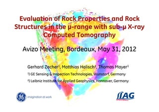
Evaluation of Rock Properties and Rock
Structures in the µ-range with sub-µ X-ray
         Computed Tomography

  Avizo Meeting, Bordeaux, May 31, 2012

    Gerhard Zacher1, Matthias Halisch², Thomas Mayer1
    1)   GE Sensing & Inspection Technologies, Wunstorf, Germany
    ²) Leibniz Institute for Applied Geophysics, Hannover, Germany
Content

1. Introduction & Fundamentals
2. nanotom CT / resolution comparison
3. Scan results for geological samples
4. Conclusion & Outlook


                                          2/
                                         GE /
Introduction & Fundamentals


X-ray tubes   Microfocus - nanofocus


                                                        3/
                                                       GE /
Introduction & Fundamentals

Requirements
Geometry and Resolution

M=FDD/FOD
U=(M-1)F (on the detector)
Vx=P/M

detector pixel P<< U
F predominates resolution

detector pixel P >> U
Pixel- / Voxelsize predominates resolution
                                                                    4/
                                                                   GE /
Introduction & Fundamentals

Resolution and Detail Detectability
Detail detectability of the nanofocus tube

Conclusion:
detail detectability
is no measure                   5 µm                    5 µm
for sharpness


                                500 nm                  500 nm


Focal Spot                  ≈2.5 µm                 ≈ 0.8 µm
                                                                    5/
                                                                   GE /
Introduction & Fundamentals

Resolution and Detail Detectability
Resolution

Focal spot size influence:

    ≈2.5 µm ∅          ≈1.5 µm ∅       ≈0.8 µm ∅


 2 µm bars         2 µm bars        0.6 µm bars

                                                          6/
                                                         GE /
Content

1. Introduction & Fundamentals
2. nanotom CT / resolution comparison
3. Scan results for geological samples
4. Conclusion & Outlook


                                          7/
                                         GE /
Nanotom CT / resolution comparison

nanotom m
ultra-high resolution
nanoCT system

X-ray tube:
nanofocus < 800 nm spot size
180 kV / 15 W, tube cooling

X-ray detector:
Cooled flat panel, 7.4 Mpixel,
11 Mpixel virtual detector
100 µm pixel size

Manipulator:
5 axis stepper motors,
granite-based,
high-precision air bearing


                                                               8/
                                                              GE /
Nanotom CT / resolution comparison


Principle of CT
Acquisition

of projections
during step-by-step
rotation by 360°

Steps < 1°


                 The acquisition of radiographic data is the
                 elementary measuring process in CT
                                                                   9/
                                                                  GE /
nanotom CT / resolution comparison


 Principle of CT: Reconstruction Method
 Example: spark plug


   projection            inversion             log + filter     line    back-projection
                                                                profile
Acquisition of 600 projections       600 back projections               3D visualization


                                                                                            10 /
                                                                                           GE /
Nanotom CT / resolution comparison

microfocus         nanoCT                      microCT
CT
vs.
nanofocus
CT
of a dried
fern


             Image resolution:
             nanoCT: < 1 µm             microCT: ≈4 µm         11 /
                                                              GE /
Nanotom CT / resolution comparison

nanofocus
CT

of a dried
fern


             • Example for resolving smallest features
                ≤ 1µm                                         12 /
                                                             GE /
Content

1. Introduction & Fundamentals
2. nanotom CT / resolution comparison
3. Scan results for geological samples
4. Conclusion & Outlook


                                          13 /
                                         GE /
Scan data of geological samples

Bentheimer   Bentheimer sandstone
sandstone
(Ø 5 mm)


Vx = 1 µm

                                                   A

                        A
                                                   B


                              B


              1 mm

             3D volume of CT scan. Quartz (grey), (A) clay (brown),
             (B) feldspar (blue) and high absorbing minerals (red).    14 /
             Right: pore space is separated (green)                   GE /
Scan data of geological samples

Bentheimer   Bentheimer sandstone
sandstone
(Ø 5 mm)


Vx = 1 µm


             Electron microscope images of clay aggregation (left)
             and highly weathered feldspar (right)                    15 /
                                                                     GE /
Scan data of geological samples

Bentheimer   Bentheimer sandstone
sandstone
(Ø 5 mm)


Vx = 1 µm


                              feldspar


              1 mm
             Comparison of CT result (left) and thin section (right).
             Histogram shows several peaks for different phases          16 /
             (air, clay (Illite), quartz, feldspar, denser minerals.    GE /
Scan data of geological samples

Bentheimer   Bentheimer sandstone
sandstone       Increasing inhomogeneity of samples
(Ø 5 mm)


Vx = 1 µm


                                                  Representative?

                                                    Scale
              1 mm
                                                  problem?

             For different sandstones (Bentheimer, Oberkirchener
             and Flechtinger) porosity has been evaluated by        17 /
                                                                   GE /
             different methods. Range differs a lot.
Scan data of geological samples

Bentheimer   Bentheimer sandstone
sandstone     Bentheimer Sandstone                Flechtingen Sandstone
(Ø 5 mm)


Vx = 1 µm


              1 mm

              Mittlere Porosität: ~ 22.5 %        Mittlere Porosität: ~ 7 %
              Repräsentatives Scan-Volumen:       Repräsentatives Scan-Volumen:
              1000x1000x1000 Voxel                > 1750x1750x1750 Voxel


             Porosity (CT) with respect to volume size for different               18 /
                                                                                  GE /
             sandstones
Scan data of geological samples

Bentheimer   Bentheimer sandstone
sandstone    outlook
(Ø 5 mm)
             • Further linking CT informationen to rock physik:
                 • inner surface
Vx = 1 µm        • pore size distribution / NMR
Avizo        • Preparation of CT data for modelling (pore scale)
fluid flow
simulation


                                                                   19 /
                                                                  GE /
Scan data of geological samples

Bentheimer   Bentheimer sandstone
sandstone    video
(Ø 5 mm)


Vx = 1 µm

Avizo
fluid flow
simulation


                                                     20 /
                                                    GE /
Scan data of geological samples

pyroclastic
rock
(Ø 1 mm)
                                           zoomed
Vx = 1 µm                                   area

yz-slice


               1 mm
      3mm


              yz-slice with different grains with high porosity or
                  fractures and bigger pores
                                                                       21 /
                                                                      GE /
Scan data of geological samples

pyroclastic
rock
(Ø 1 mm)

Vx = 1 µm

yz-slice


               1 mm
      3mm


              Zoom into yz-slice with measurement of thin wall: 1.8 µm

                                                                     22 /
                                                                    GE /
Scan data of geological samples

Etna
pyroclastic
rock
(fresh’11)
(Ø 10 mm)

Vx = 5 µm

xy-slice


               1 mm


     3mm
              xy-slice through 5x5x5mm cube used later for flow
                  simulation
                                                                     23 /
                                                                    GE /
Scan data of geological samples

Etna          Etna pyroclastic rock
pyroclastic
rock
(fresh’11)
(Ø 10 mm)

Vx = 5 µm

3D volume


               1 mm


    3mm
              The surface is composed of 18 Mio. faces and
              represents the stone matrix. Shadows enhance the     24 /
              spatial impression.                                 GE /
Scan data of geological samples

Etna          Etna pyroclastic rock
pyroclastic
rock
(fresh’11)
(Ø 10 mm)
Vx = 5 µm
Avizo
fluid flow
simulation


               1 mm


    3mm
              The colored volume rendering shows the velocity’s
              magnitude within the pore space. The particle plot    25 /
                                                                   GE /
              shows the actual vector field using cones.
Scan data of geological samples

Etna          Etna pyroclastic rock
pyroclastic
rock
(fresh’11)
(Ø 10 mm)
Vx = 5 µm
Avizo
wall
thickness


               1 mm


    3mm
              The pore space is visualized with volume rendering.
              The matrix’ thickness has been calculated and is       26 /
                                                                    GE /
              visualized on the surface.
Scan data of geological samples

Etna          Etna pyroclastic rock
pyroclastic
rock
(fresh’11)
(Ø 10 mm)
Vx = 5 µm
Avizo
fluid flow
simulation


               1 mm


    3mm
              The color slice intersects the velocity field calculated
              with XLab Hydro and visualizes the vector field.            27 /
                                                                         GE /
              Colors give the velocity’s magnitude.
Scan data of geological samples

3D view of a
Nummulite
Lower Eocene
53 million
years old
(Ø 2 mm)

Courtesy of
R. Speijer, K.U.
Leuven, Belgium

Vx = 1 µm


                    1 mm
       3mm


                   Transparent 3D view

                                                                    28 /
                                                                   GE /
Scan data of geological samples

3D view of a
Nummulite
Lower Eocene
53 million
years old
(Ø 2 mm)

Courtesy of
R. Speijer, K.U.
Leuven, Belgium

Vx = 1 µm


                    1 mm


                   Sliced 3D view to show the delicate internal structures

                                                                              29 /
                                                                             GE /
Scan data of geological samples

Slice view of a                             zoomed
Nummulite                                    area
Lower Eocene
53 million
years old
(Ø 2 mm)

Courtesy of
R. Speijer, K.U.
Leuven, Belgium

Vx = 1 µm


                    1 mm


                   Xy slice through center plain

                                                                              30 /
                                                                             GE /
Scan data of geological samples

Slice view of a
Nummulite
Lower Eocene
53 million
years old
(Ø 2 mm)

Courtesy of
R. Speijer, K.U.
Leuven, Belgium

Vx = 1 µm


                    1 mm
       3mm


                   Zoomed xy slice through center plain with measured pore
                      2.3 µm
                                                                         31 /
                                                                        GE /
Content

1. Introduction & Fundamentals
2. nanotom CT / resolution comparison
3. Scan results for geological samples
4. Conclusion & Outlook


                                          32 /
                                         GE /
Conclusions
• State of the art high resolution tube based X-ray CT with
   the phoenix nanotom offers

    • Comparable (or higher) spatial resolution to SRµCT
        setups due to nanofocus tube (ease of use, lower cost
        and faster analysis)

    • Wide variety of geological samples can be analysed

    • Data of a whole 3D volume offers numerous qualitative
        AND quantitative interpretations

    • New insights in rock materials for geo science
                                                                 33 /
                                                                GE /
Outlook

• More quantitative data analysis (like permeability, particle
   size distribution, density distribution, …)
• More input from geoscientists to better generate the
   potential of nanofocus X-ray CT


                                                                  34 /
                                                                 GE /
Contact and further information:

       www.phoenix-xray.com
               or
      www.ge-mcs.com/phoenix


                                    35 /
                                   GE /

More Related Content

Similar to Evaluation of rock properties and rock structures in the μ-range with sub-μ X-ray computed tomography

Time-resolved mirrorless scintillation detector @ KSMPRS2018
Time-resolved mirrorless scintillation detector @ KSMPRS2018Time-resolved mirrorless scintillation detector @ KSMPRS2018
Time-resolved mirrorless scintillation detector @ KSMPRS2018WonjoongCheon
 
Imaging for Radiotherapy delivery and verification
Imaging for Radiotherapy delivery and verificationImaging for Radiotherapy delivery and verification
Imaging for Radiotherapy delivery and verificationMiami Cancer Institute
 
Time-resolved mirrorless scintillation detector @ AAPM2018
Time-resolved mirrorless scintillation detector @ AAPM2018Time-resolved mirrorless scintillation detector @ AAPM2018
Time-resolved mirrorless scintillation detector @ AAPM2018WonjoongCheon
 
Wu - Surface topography investigation for niobium cavities and its implicatio...
Wu - Surface topography investigation for niobium cavities and its implicatio...Wu - Surface topography investigation for niobium cavities and its implicatio...
Wu - Surface topography investigation for niobium cavities and its implicatio...thinfilmsworkshop
 
SimPET a Preclinical PET Insert for Simultaneous PET/MR Imaging
SimPET a Preclinical PET Insert for Simultaneous PET/MR ImagingSimPET a Preclinical PET Insert for Simultaneous PET/MR Imaging
SimPET a Preclinical PET Insert for Simultaneous PET/MR ImagingScintica Instrumentation
 
Design of ultrasound transducer
Design of ultrasound transducer Design of ultrasound transducer
Design of ultrasound transducer SudarshanKanse1
 
Art and medicine_columbia_2013
Art and medicine_columbia_2013Art and medicine_columbia_2013
Art and medicine_columbia_2013oriolespinal
 
S S S A2009 Simulation Study Of Segmentation
S S S A2009 Simulation Study Of SegmentationS S S A2009 Simulation Study Of Segmentation
S S S A2009 Simulation Study Of SegmentationWei Wang
 
CBCT IN IMPLANTOLOGY.pptx
CBCT IN IMPLANTOLOGY.pptxCBCT IN IMPLANTOLOGY.pptx
CBCT IN IMPLANTOLOGY.pptxDr K. Abhilasha
 
10h10 carlos roberto 21 08 botafogo
10h10 carlos roberto 21 08 botafogo10h10 carlos roberto 21 08 botafogo
10h10 carlos roberto 21 08 botafogoslides-mci
 
UMKA NANOTECHNOLOGICAL SYSTEM (www.cmcons.com)
UMKA NANOTECHNOLOGICAL SYSTEM (www.cmcons.com)UMKA NANOTECHNOLOGICAL SYSTEM (www.cmcons.com)
UMKA NANOTECHNOLOGICAL SYSTEM (www.cmcons.com)Alexander Novichkov
 
Image-guided management of uncertainties in scanned particle therapy
Image-guided management of uncertainties in scanned particle therapyImage-guided management of uncertainties in scanned particle therapy
Image-guided management of uncertainties in scanned particle therapyGiovanni Fattori
 
Pet And Radiotherapy For Head And Neck
Pet And Radiotherapy For Head And NeckPet And Radiotherapy For Head And Neck
Pet And Radiotherapy For Head And Neckfondas vakalis
 
PERFORMANCE EVALUATION OF COMPUTED TOMOGRAPHY (CT) SCANNERS
PERFORMANCE EVALUATION OF COMPUTED TOMOGRAPHY (CT) SCANNERSPERFORMANCE EVALUATION OF COMPUTED TOMOGRAPHY (CT) SCANNERS
PERFORMANCE EVALUATION OF COMPUTED TOMOGRAPHY (CT) SCANNERSBhuvi palaniswamy
 

Similar to Evaluation of rock properties and rock structures in the μ-range with sub-μ X-ray computed tomography (20)

Porous rock evaluation with micro ct zacher_ge_copyright
Porous rock evaluation with micro ct zacher_ge_copyrightPorous rock evaluation with micro ct zacher_ge_copyright
Porous rock evaluation with micro ct zacher_ge_copyright
 
Time-resolved mirrorless scintillation detector @ KSMPRS2018
Time-resolved mirrorless scintillation detector @ KSMPRS2018Time-resolved mirrorless scintillation detector @ KSMPRS2018
Time-resolved mirrorless scintillation detector @ KSMPRS2018
 
Cone beam ct
Cone beam ctCone beam ct
Cone beam ct
 
Imaging for Radiotherapy delivery and verification
Imaging for Radiotherapy delivery and verificationImaging for Radiotherapy delivery and verification
Imaging for Radiotherapy delivery and verification
 
Time-resolved mirrorless scintillation detector @ AAPM2018
Time-resolved mirrorless scintillation detector @ AAPM2018Time-resolved mirrorless scintillation detector @ AAPM2018
Time-resolved mirrorless scintillation detector @ AAPM2018
 
Wu - Surface topography investigation for niobium cavities and its implicatio...
Wu - Surface topography investigation for niobium cavities and its implicatio...Wu - Surface topography investigation for niobium cavities and its implicatio...
Wu - Surface topography investigation for niobium cavities and its implicatio...
 
Webinar Geosciences 2012
Webinar Geosciences 2012Webinar Geosciences 2012
Webinar Geosciences 2012
 
Ia3414771479
Ia3414771479Ia3414771479
Ia3414771479
 
SimPET a Preclinical PET Insert for Simultaneous PET/MR Imaging
SimPET a Preclinical PET Insert for Simultaneous PET/MR ImagingSimPET a Preclinical PET Insert for Simultaneous PET/MR Imaging
SimPET a Preclinical PET Insert for Simultaneous PET/MR Imaging
 
Design of ultrasound transducer
Design of ultrasound transducer Design of ultrasound transducer
Design of ultrasound transducer
 
Art and medicine_columbia_2013
Art and medicine_columbia_2013Art and medicine_columbia_2013
Art and medicine_columbia_2013
 
S S S A2009 Simulation Study Of Segmentation
S S S A2009 Simulation Study Of SegmentationS S S A2009 Simulation Study Of Segmentation
S S S A2009 Simulation Study Of Segmentation
 
CBCT IN IMPLANTOLOGY.pptx
CBCT IN IMPLANTOLOGY.pptxCBCT IN IMPLANTOLOGY.pptx
CBCT IN IMPLANTOLOGY.pptx
 
10h10 carlos roberto 21 08 botafogo
10h10 carlos roberto 21 08 botafogo10h10 carlos roberto 21 08 botafogo
10h10 carlos roberto 21 08 botafogo
 
UMKA NANOTECHNOLOGICAL SYSTEM (www.cmcons.com)
UMKA NANOTECHNOLOGICAL SYSTEM (www.cmcons.com)UMKA NANOTECHNOLOGICAL SYSTEM (www.cmcons.com)
UMKA NANOTECHNOLOGICAL SYSTEM (www.cmcons.com)
 
Image-guided management of uncertainties in scanned particle therapy
Image-guided management of uncertainties in scanned particle therapyImage-guided management of uncertainties in scanned particle therapy
Image-guided management of uncertainties in scanned particle therapy
 
Pet And Radiotherapy For Head And Neck
Pet And Radiotherapy For Head And NeckPet And Radiotherapy For Head And Neck
Pet And Radiotherapy For Head And Neck
 
31075
3107531075
31075
 
PERFORMANCE EVALUATION OF COMPUTED TOMOGRAPHY (CT) SCANNERS
PERFORMANCE EVALUATION OF COMPUTED TOMOGRAPHY (CT) SCANNERSPERFORMANCE EVALUATION OF COMPUTED TOMOGRAPHY (CT) SCANNERS
PERFORMANCE EVALUATION OF COMPUTED TOMOGRAPHY (CT) SCANNERS
 
Tomography
TomographyTomography
Tomography
 

More from VSG - Visualization Sciences Group

Autocorrelation and the rose diagram for analyzing structure and anisotropy i...
Autocorrelation and the rose diagram for analyzing structure and anisotropy i...Autocorrelation and the rose diagram for analyzing structure and anisotropy i...
Autocorrelation and the rose diagram for analyzing structure and anisotropy i...VSG - Visualization Sciences Group
 
Studying thermomechanical properties of thermostructural composites using X-r...
Studying thermomechanical properties of thermostructural composites using X-r...Studying thermomechanical properties of thermostructural composites using X-r...
Studying thermomechanical properties of thermostructural composites using X-r...VSG - Visualization Sciences Group
 
3D characterization of microstructure evolution of cast AlMgSi alloys by sync...
3D characterization of microstructure evolution of cast AlMgSi alloys by sync...3D characterization of microstructure evolution of cast AlMgSi alloys by sync...
3D characterization of microstructure evolution of cast AlMgSi alloys by sync...VSG - Visualization Sciences Group
 
Determination of crack-initiating defects in cast aluminium alloys by Non-des...
Determination of crack-initiating defects in cast aluminium alloys by Non-des...Determination of crack-initiating defects in cast aluminium alloys by Non-des...
Determination of crack-initiating defects in cast aluminium alloys by Non-des...VSG - Visualization Sciences Group
 
Building a pore network model from a pore space 3D image to precisely predict...
Building a pore network model from a pore space 3D image to precisely predict...Building a pore network model from a pore space 3D image to precisely predict...
Building a pore network model from a pore space 3D image to precisely predict...VSG - Visualization Sciences Group
 
3D cell lineage reconstruction of early embryogenesis in plant
3D cell lineage reconstruction of early embryogenesis in plant3D cell lineage reconstruction of early embryogenesis in plant
3D cell lineage reconstruction of early embryogenesis in plantVSG - Visualization Sciences Group
 
Visualization of a new generation of climate simulations with Avizo Green
Visualization of a new generation of climate simulations with Avizo GreenVisualization of a new generation of climate simulations with Avizo Green
Visualization of a new generation of climate simulations with Avizo GreenVSG - Visualization Sciences Group
 
Lessons learned and challenges for lab-wide and campus-wide Avizo utilization...
Lessons learned and challenges for lab-wide and campus-wide Avizo utilization...Lessons learned and challenges for lab-wide and campus-wide Avizo utilization...
Lessons learned and challenges for lab-wide and campus-wide Avizo utilization...VSG - Visualization Sciences Group
 
Application of Avizo Fire image processing for substances investigation insid...
Application of Avizo Fire image processing for substances investigation insid...Application of Avizo Fire image processing for substances investigation insid...
Application of Avizo Fire image processing for substances investigation insid...VSG - Visualization Sciences Group
 
Identification of the local mechanical properties of an inorganic fiber-based...
Identification of the local mechanical properties of an inorganic fiber-based...Identification of the local mechanical properties of an inorganic fiber-based...
Identification of the local mechanical properties of an inorganic fiber-based...VSG - Visualization Sciences Group
 
Characterization of porous materials by Focused Ion Beam Nano-Tomography
Characterization of porous materials by Focused Ion Beam Nano-TomographyCharacterization of porous materials by Focused Ion Beam Nano-Tomography
Characterization of porous materials by Focused Ion Beam Nano-TomographyVSG - Visualization Sciences Group
 
Asphalt internal structure characterization with X-Ray computed tomography
Asphalt internal structure characterization with X-Ray computed tomographyAsphalt internal structure characterization with X-Ray computed tomography
Asphalt internal structure characterization with X-Ray computed tomographyVSG - Visualization Sciences Group
 

More from VSG - Visualization Sciences Group (14)

Autocorrelation and the rose diagram for analyzing structure and anisotropy i...
Autocorrelation and the rose diagram for analyzing structure and anisotropy i...Autocorrelation and the rose diagram for analyzing structure and anisotropy i...
Autocorrelation and the rose diagram for analyzing structure and anisotropy i...
 
Studying thermomechanical properties of thermostructural composites using X-r...
Studying thermomechanical properties of thermostructural composites using X-r...Studying thermomechanical properties of thermostructural composites using X-r...
Studying thermomechanical properties of thermostructural composites using X-r...
 
3D characterization of microstructure evolution of cast AlMgSi alloys by sync...
3D characterization of microstructure evolution of cast AlMgSi alloys by sync...3D characterization of microstructure evolution of cast AlMgSi alloys by sync...
3D characterization of microstructure evolution of cast AlMgSi alloys by sync...
 
Determination of crack-initiating defects in cast aluminium alloys by Non-des...
Determination of crack-initiating defects in cast aluminium alloys by Non-des...Determination of crack-initiating defects in cast aluminium alloys by Non-des...
Determination of crack-initiating defects in cast aluminium alloys by Non-des...
 
Building a pore network model from a pore space 3D image to precisely predict...
Building a pore network model from a pore space 3D image to precisely predict...Building a pore network model from a pore space 3D image to precisely predict...
Building a pore network model from a pore space 3D image to precisely predict...
 
3D cell lineage reconstruction of early embryogenesis in plant
3D cell lineage reconstruction of early embryogenesis in plant3D cell lineage reconstruction of early embryogenesis in plant
3D cell lineage reconstruction of early embryogenesis in plant
 
Visualization of a new generation of climate simulations with Avizo Green
Visualization of a new generation of climate simulations with Avizo GreenVisualization of a new generation of climate simulations with Avizo Green
Visualization of a new generation of climate simulations with Avizo Green
 
Lessons learned and challenges for lab-wide and campus-wide Avizo utilization...
Lessons learned and challenges for lab-wide and campus-wide Avizo utilization...Lessons learned and challenges for lab-wide and campus-wide Avizo utilization...
Lessons learned and challenges for lab-wide and campus-wide Avizo utilization...
 
A new 3D pore shape classification using Avizo Fire
A new 3D pore shape classification using Avizo FireA new 3D pore shape classification using Avizo Fire
A new 3D pore shape classification using Avizo Fire
 
Application of Avizo Fire image processing for substances investigation insid...
Application of Avizo Fire image processing for substances investigation insid...Application of Avizo Fire image processing for substances investigation insid...
Application of Avizo Fire image processing for substances investigation insid...
 
3D characterization of asymmetric microfiltration membranes
3D characterization of asymmetric microfiltration membranes3D characterization of asymmetric microfiltration membranes
3D characterization of asymmetric microfiltration membranes
 
Identification of the local mechanical properties of an inorganic fiber-based...
Identification of the local mechanical properties of an inorganic fiber-based...Identification of the local mechanical properties of an inorganic fiber-based...
Identification of the local mechanical properties of an inorganic fiber-based...
 
Characterization of porous materials by Focused Ion Beam Nano-Tomography
Characterization of porous materials by Focused Ion Beam Nano-TomographyCharacterization of porous materials by Focused Ion Beam Nano-Tomography
Characterization of porous materials by Focused Ion Beam Nano-Tomography
 
Asphalt internal structure characterization with X-Ray computed tomography
Asphalt internal structure characterization with X-Ray computed tomographyAsphalt internal structure characterization with X-Ray computed tomography
Asphalt internal structure characterization with X-Ray computed tomography
 

Evaluation of rock properties and rock structures in the μ-range with sub-μ X-ray computed tomography

  • 1. Evaluation of Rock Properties and Rock Structures in the µ-range with sub-µ X-ray Computed Tomography Avizo Meeting, Bordeaux, May 31, 2012 Gerhard Zacher1, Matthias Halisch², Thomas Mayer1 1) GE Sensing & Inspection Technologies, Wunstorf, Germany ²) Leibniz Institute for Applied Geophysics, Hannover, Germany
  • 2. Content 1. Introduction & Fundamentals 2. nanotom CT / resolution comparison 3. Scan results for geological samples 4. Conclusion & Outlook 2/ GE /
  • 3. Introduction & Fundamentals X-ray tubes Microfocus - nanofocus 3/ GE /
  • 4. Introduction & Fundamentals Requirements Geometry and Resolution M=FDD/FOD U=(M-1)F (on the detector) Vx=P/M detector pixel P<< U F predominates resolution detector pixel P >> U Pixel- / Voxelsize predominates resolution 4/ GE /
  • 5. Introduction & Fundamentals Resolution and Detail Detectability Detail detectability of the nanofocus tube Conclusion: detail detectability is no measure 5 µm 5 µm for sharpness 500 nm 500 nm Focal Spot ≈2.5 µm ≈ 0.8 µm 5/ GE /
  • 6. Introduction & Fundamentals Resolution and Detail Detectability Resolution Focal spot size influence: ≈2.5 µm ∅ ≈1.5 µm ∅ ≈0.8 µm ∅ 2 µm bars 2 µm bars 0.6 µm bars 6/ GE /
  • 7. Content 1. Introduction & Fundamentals 2. nanotom CT / resolution comparison 3. Scan results for geological samples 4. Conclusion & Outlook 7/ GE /
  • 8. Nanotom CT / resolution comparison nanotom m ultra-high resolution nanoCT system X-ray tube: nanofocus < 800 nm spot size 180 kV / 15 W, tube cooling X-ray detector: Cooled flat panel, 7.4 Mpixel, 11 Mpixel virtual detector 100 µm pixel size Manipulator: 5 axis stepper motors, granite-based, high-precision air bearing 8/ GE /
  • 9. Nanotom CT / resolution comparison Principle of CT Acquisition of projections during step-by-step rotation by 360° Steps < 1° The acquisition of radiographic data is the elementary measuring process in CT 9/ GE /
  • 10. nanotom CT / resolution comparison Principle of CT: Reconstruction Method Example: spark plug projection inversion log + filter line back-projection profile Acquisition of 600 projections 600 back projections 3D visualization 10 / GE /
  • 11. Nanotom CT / resolution comparison microfocus nanoCT microCT CT vs. nanofocus CT of a dried fern Image resolution: nanoCT: < 1 µm microCT: ≈4 µm 11 / GE /
  • 12. Nanotom CT / resolution comparison nanofocus CT of a dried fern • Example for resolving smallest features ≤ 1µm 12 / GE /
  • 13. Content 1. Introduction & Fundamentals 2. nanotom CT / resolution comparison 3. Scan results for geological samples 4. Conclusion & Outlook 13 / GE /
  • 14. Scan data of geological samples Bentheimer Bentheimer sandstone sandstone (Ø 5 mm) Vx = 1 µm A A B B 1 mm 3D volume of CT scan. Quartz (grey), (A) clay (brown), (B) feldspar (blue) and high absorbing minerals (red). 14 / Right: pore space is separated (green) GE /
  • 15. Scan data of geological samples Bentheimer Bentheimer sandstone sandstone (Ø 5 mm) Vx = 1 µm Electron microscope images of clay aggregation (left) and highly weathered feldspar (right) 15 / GE /
  • 16. Scan data of geological samples Bentheimer Bentheimer sandstone sandstone (Ø 5 mm) Vx = 1 µm feldspar 1 mm Comparison of CT result (left) and thin section (right). Histogram shows several peaks for different phases 16 / (air, clay (Illite), quartz, feldspar, denser minerals. GE /
  • 17. Scan data of geological samples Bentheimer Bentheimer sandstone sandstone Increasing inhomogeneity of samples (Ø 5 mm) Vx = 1 µm Representative? Scale 1 mm problem? For different sandstones (Bentheimer, Oberkirchener and Flechtinger) porosity has been evaluated by 17 / GE / different methods. Range differs a lot.
  • 18. Scan data of geological samples Bentheimer Bentheimer sandstone sandstone Bentheimer Sandstone Flechtingen Sandstone (Ø 5 mm) Vx = 1 µm 1 mm Mittlere Porosität: ~ 22.5 % Mittlere Porosität: ~ 7 % Repräsentatives Scan-Volumen: Repräsentatives Scan-Volumen: 1000x1000x1000 Voxel > 1750x1750x1750 Voxel Porosity (CT) with respect to volume size for different 18 / GE / sandstones
  • 19. Scan data of geological samples Bentheimer Bentheimer sandstone sandstone outlook (Ø 5 mm) • Further linking CT informationen to rock physik: • inner surface Vx = 1 µm • pore size distribution / NMR Avizo • Preparation of CT data for modelling (pore scale) fluid flow simulation 19 / GE /
  • 20. Scan data of geological samples Bentheimer Bentheimer sandstone sandstone video (Ø 5 mm) Vx = 1 µm Avizo fluid flow simulation 20 / GE /
  • 21. Scan data of geological samples pyroclastic rock (Ø 1 mm) zoomed Vx = 1 µm area yz-slice 1 mm 3mm yz-slice with different grains with high porosity or fractures and bigger pores 21 / GE /
  • 22. Scan data of geological samples pyroclastic rock (Ø 1 mm) Vx = 1 µm yz-slice 1 mm 3mm Zoom into yz-slice with measurement of thin wall: 1.8 µm 22 / GE /
  • 23. Scan data of geological samples Etna pyroclastic rock (fresh’11) (Ø 10 mm) Vx = 5 µm xy-slice 1 mm 3mm xy-slice through 5x5x5mm cube used later for flow simulation 23 / GE /
  • 24. Scan data of geological samples Etna Etna pyroclastic rock pyroclastic rock (fresh’11) (Ø 10 mm) Vx = 5 µm 3D volume 1 mm 3mm The surface is composed of 18 Mio. faces and represents the stone matrix. Shadows enhance the 24 / spatial impression. GE /
  • 25. Scan data of geological samples Etna Etna pyroclastic rock pyroclastic rock (fresh’11) (Ø 10 mm) Vx = 5 µm Avizo fluid flow simulation 1 mm 3mm The colored volume rendering shows the velocity’s magnitude within the pore space. The particle plot 25 / GE / shows the actual vector field using cones.
  • 26. Scan data of geological samples Etna Etna pyroclastic rock pyroclastic rock (fresh’11) (Ø 10 mm) Vx = 5 µm Avizo wall thickness 1 mm 3mm The pore space is visualized with volume rendering. The matrix’ thickness has been calculated and is 26 / GE / visualized on the surface.
  • 27. Scan data of geological samples Etna Etna pyroclastic rock pyroclastic rock (fresh’11) (Ø 10 mm) Vx = 5 µm Avizo fluid flow simulation 1 mm 3mm The color slice intersects the velocity field calculated with XLab Hydro and visualizes the vector field. 27 / GE / Colors give the velocity’s magnitude.
  • 28. Scan data of geological samples 3D view of a Nummulite Lower Eocene 53 million years old (Ø 2 mm) Courtesy of R. Speijer, K.U. Leuven, Belgium Vx = 1 µm 1 mm 3mm Transparent 3D view 28 / GE /
  • 29. Scan data of geological samples 3D view of a Nummulite Lower Eocene 53 million years old (Ø 2 mm) Courtesy of R. Speijer, K.U. Leuven, Belgium Vx = 1 µm 1 mm Sliced 3D view to show the delicate internal structures 29 / GE /
  • 30. Scan data of geological samples Slice view of a zoomed Nummulite area Lower Eocene 53 million years old (Ø 2 mm) Courtesy of R. Speijer, K.U. Leuven, Belgium Vx = 1 µm 1 mm Xy slice through center plain 30 / GE /
  • 31. Scan data of geological samples Slice view of a Nummulite Lower Eocene 53 million years old (Ø 2 mm) Courtesy of R. Speijer, K.U. Leuven, Belgium Vx = 1 µm 1 mm 3mm Zoomed xy slice through center plain with measured pore 2.3 µm 31 / GE /
  • 32. Content 1. Introduction & Fundamentals 2. nanotom CT / resolution comparison 3. Scan results for geological samples 4. Conclusion & Outlook 32 / GE /
  • 33. Conclusions • State of the art high resolution tube based X-ray CT with the phoenix nanotom offers • Comparable (or higher) spatial resolution to SRµCT setups due to nanofocus tube (ease of use, lower cost and faster analysis) • Wide variety of geological samples can be analysed • Data of a whole 3D volume offers numerous qualitative AND quantitative interpretations • New insights in rock materials for geo science 33 / GE /
  • 34. Outlook • More quantitative data analysis (like permeability, particle size distribution, density distribution, …) • More input from geoscientists to better generate the potential of nanofocus X-ray CT 34 / GE /
  • 35. Contact and further information: www.phoenix-xray.com or www.ge-mcs.com/phoenix 35 / GE /