
Empfohlen
Weitere ähnliche Inhalte
Was ist angesagt?
Was ist angesagt? (20)
Ähnlich wie Measurement of Radiation (Thimble Ionization Chamber, Free air Ionization Chamber , PM Tube )
Ähnlich wie Measurement of Radiation (Thimble Ionization Chamber, Free air Ionization Chamber , PM Tube ) (20)
Mehr von Upakar Paudel
Mehr von Upakar Paudel (13)
Kürzlich hochgeladen
Kürzlich hochgeladen (20)
Measurement of Radiation (Thimble Ionization Chamber, Free air Ionization Chamber , PM Tube )
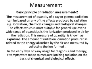
- 1. Measurement Basic principle of radiation measurement-2 The measurement of quantity of x-ray or gamma radiation can be based on any of the effects produced by radiation e.g. ionization, chemical changes and biological changes. The effects which is most suitable for general use over a wide range of quantities is the ionization produced in air by the radiation. This measure of quantity is known as exposure. The amount of radiation ionization produced is related to the energy absorbed by the air and measured by collecting the ion formed . In the early days of x-ray usage for diagnosis and therapy, attempts were made to measure ionizing radiation on the basis of chemical and biological effects
- 2. For instance, radiation effects on the photographic emulsions, changes in the color of some chemical compounds, and reddening of the human skin could be related to the amount of radiation absorbed. For example, in radiotherapy, a unit called skin erythema dose (SED) was defined as that amount of x or gamma radiation that just produced reddening of the human skin. • However, the unit has many drawbacks. Skin erythema depends on many conditions, such as the type of skin, the quality of radiation, the extent of skin exposed, dose fractionation (dose per fraction and interval between fractions), and differences between early and delayed skin reactions.
- 3. • Although the SED was later discarded in favor of a more precisely measurable unit such as the roentgen, the skin erythema was used by physicians as an approximate index of response to the radiation treatments. • In 1928, the International Commission on Radiation Units and Measurements (ICRU) adopted the roentgen as the unit of measuring X and gamma radiation exposure. The unit is denoted by R.
- 4. Exposure • Exposure : The roentgen is a unit of exposure. The quantity exposure is a measure of ionization produced in air by photons. The ICRU defines exposure (X) as the quotient of dQ / dm where dQ is the absolute value of the total charge of the ions of one sign produced in air when all the electrons (negatrons and positrons) liberated by photons in air of mass dm are completely stopped in air. • X= dQ / dm • The SI unit for exposure is coulomb per kilogram (C/kg) but the special unit is roentgen (R).1 • 1R = 2.58 x10⁻⁴ C/kg air
- 6. • According to the definition of roentgen, the electrons produced by photons in a specified volume must spend all their energies by ionization in air enclosed by the plates (region of ion collection) and the total ionic charge of either sign should be measured. However, some electrons produced in the specified volume deposit their energy outside the region of ion collection and thus are not measured. On the other hand, electrons produced outside the specified volume may enter the ion-collecting region and produce ionization there. If the ionization loss is compensated by the ionization gained, a condition of electronic equilibrium exists. Under this condition, the definition of roentgen is effectively satisfied. This is the principle of free-air ionization chamber.
- 7. Free air ionization chamber The free-air, or standard, ionization chamber is an instrument used in the measurement of the roentgen. Generally, such a primary standard is used only for the calibration of secondary instruments designed for field use. The free-air chamber installations are thus confined principally to some of the national standards laboratories.
- 8. A free-air chamber is represented schematically in Fig. An x-ray beam, originating from a focal spot S, is defined by the diaphragm D, and passes centrally between a pair of parallel plates. A high- voltage (field strength of the order of 100 V/cm) is applied between the plates to collect ions produced in the air between the plates. The ionization is measured for a length L defined by the limiting lines of force to the edges of the collection plate C. The lines of force are made straight and perpendicular to the collector by a guard ring G.
- 10. Electrons produced by the photon beam in the specified volume (shaded in Fig. ) must spend all their energy by ionization of air between the plates. Such a condition can exist only if the range of the electrons liberated by the incident photons is less than the distance between each plate and the specified volume. In addition, for electronic equilibrium to exist, the beam intensity (photon fluence per unit time) must remain constant across the length of the specified volume, and the separation between the diaphragm and the ion- collecting region must exceed the electron range in air.
- 11. • If ΔQ is the charge collected in Coulombs and ρ is the density (kg/m3) of air, then the exposure Xp at the center of the specified volume (point P) is: • Xp = ΔQ 1 roentgen ρ. AP L 2.58X10⁻⁴ where Ap is the cross-sectional area (in meters squared) of the beam at point P and L (in meters) is the length of the collecting volume.
- 12. • In practice, it is more convenient to state the exposure (X) at the position of the diaphragm. Suppose f1 and f2 are the distances of the x-ray source to the diaphragm and point P, respectively. Because the intensity at point P and at the diaphragm are related by an inverse square law factor (f1/f2)2, which also relates the area of the beams at the diaphragm and at point P, exposure XD at the diaphragm is given by: • XD = ΔQ 1 roentgen ρ. AD L 2.58 X10⁻⁴ where AD is the diaphragm aperture area
- 13. • Accurate measurements with a free-air ionization chamber require considerable care. A few corrections that are usually applied include: (a) correction for air attenuation; (b) correction for recombination of ions; (c) correction for the effects of temperature, pressure, and humidity on the density of air; and (d) correction for ionization produced by scattered photons
- 14. Drawbacks of a free air ionization chamber. -2 • There are drawbacks on the design of a free-air chamber for the measurement of roentgens for high-energy x-ray beams. As the photon energy increases, the range of the electrons liberated in air increases rapidly. This necessitates an increase in the separation of the plates to maintain electronic equilibrium. Too large a separation, however, creates problems of non-uniform electric field and greater ion recombination. Although the plate separation can be reduced by using air at high pressures, the problems still remain in regard to air attenuation, photon scatter, and reduction in the efficiency of ion collection. Because of these problems, there is an upper limit on the photon energy above which the roentgen cannot be accurately measured. This limit occurs at about 3 MeV.
- 15. Thimble chamber Free-air ionization chambers are too delicate and bulky for routine use. Their main function is in the standardizing laboratories where they can be used to calibrate field instruments such as a thimble chamber. A thimble chamber is basically a ionization chamber. The wall is shaped like a sewing thimble-hence the name. The inner surface of the thimble wall is coated by a special material to make it electrically conducting. This forms one electrode. The other electrode is a rod of low atomic number material such as graphite or aluminum held in the center of the thimble but electrically insulated from it. A suitable voltage is applied between the two electrodes to collect the ions produced in the air cavity
- 16. • For the thimble chamber to be air equivalent, the effective atomic number of the wall material and the central electrode must be such that the system as a whole behaves like a free-air chamber. • Most commonly used wall materials are made either of graphite (carbon), Bakelite, or a plastic coated on the inside by a conducting layer of graphite or of a conducting mixture of Bakelite and graphite
- 18. • The principle of the thimble chamber is illustrated in Fig. In Fig. A, a spherical volume of air is shown with an air cavity at the center. Suppose this sphere of air is irradiated uniformly with a photon beam. Also, suppose that the distance between the outer sphere and the inner cavity is equal to the maximum range of electrons generated in air. If the number of electrons entering the cavity is the same as that leaving the cavity, electronic equilibrium exists. Suppose also that we are able to measure the ionization charge produced in the cavity by the electrons liberated in the air surrounding the cavity. Then by knowing the volume or mass of air inside the cavity, we can calculate the charge per unit mass or the beam exposure at the center of the cavity
- 19. Then the exposure (X)is given by X =Q/(ρx v) Where ρ is the density of air .v is the volume Now if the air wall in Fig. A is compressed into a solid shell as in Fig.B, we get a thimble chamber. Although the thimble wall is solid, it is air equivalent, i.e., its effective atomic number is the same as that of air. In addition, the thickness of the thimble wall is such that the electronic equilibrium occurs inside the cavity, just as it did in Fig.A. As before, it follows that the wall thickness must be equal to or greater than the maximum range of the electrons liberated in the thimble wall.
- 20. Since the density of the solid air-equivalent wall is much greater than that of free air, the thicknesses required for electronic equilibrium in the thimble chamber are considerably reduced. For example, in the 100 to 250 kVp x-ray range, the wall thickness of the thimble (assuming unit density) is about 1 mm and in the case of 60Co gamma rays (average E=1.25 MeV), it is approximately 5 mm. In practice, however, a thimble chamber is constructed with wall thicknesses of 1 mm or less and this is supplemented with close-fitting caps of Plexiglas or other plastic to bring the total wall thickness up to that needed for electronic equilibrium .
- 21. • Care must be taken to ensure that the wall is of the proper thickness for the energy of the radiation being measured. If the wall is too thin, insufficient absorption takes place in the wall and too few secondary electrons are liberated. Also, electrons from outside the chamber can pass through the wall. If the wall is too thick, there will be appreciable attenuation of the X and gamma radiation in the outer layers of the wall resulting in reduced ionization in the air in the wall. If a chamber with a thin wall is to be used for measurement at energies higher than that for which it was designed, the wall can be increased to the required thickness by surrounding the chamber with a build up cap such as a Perspex sheath a few millimeters thick. A thimble chamber is small in size (0.6cc) and very much suitable for routine measurement in hospital, for calibrating x-ray and tele-cobalt units.
- 22. Desirable Chamber Characteristics A practical ion chamber for exposure measurement should have the following characteristics. 1. There should be minimal variation in sensitivity or exposure calibration factor over a wide range of photon energies. 2. There should be suitable volume to allow measurements for the expected range of exposures. The sensitivity (charge measured per roentgen) is directly proportional to the chamber sensitive volume. For example, the reading obtained for a given exposure with a 30-cm3 chamber will be approximately 50 times higher than that obtained with a 0.6-cm3 chamber. However, the ratio may not be exactly 50, because a chamber response also depends on the chamber design.
- 23. 3. There should be minimal variation in sensitivity with the direction of incident radiation. Although this kind of variation can be minimized in the design of the chamber, care is taken to use the chamber in the same configuration with respect to the beam as specified under chamber calibration conditions. 4. There should be minimal stem œleakage. A chamber is known to have stem leakage if it records ionization produced anywhere other than its sensitive volume. 5. The chamber should have been calibrated for exposure against a standard instrument for all radiation qualities of interest. 6. There should be minimal ion recombination losses. If the chamber voltage is not high enough or regions of low electric field strength occur inside the chamber, such as in the vicinity of sharply concave surfaces or corners, ions may recombine before contributing to the measured charge.
- 24. ELECTROMETER The electrometer is basically a charge measuring device. A. String Electrometer B. Other Electrometer Operational amplifiers a. Integrate mode b. Rate mode c. Direct exposure reading mode
- 25. String electrometer :It is a type of electrometer that operates on the principle of a gold-leaf electroscope. Such electrometers are commonly used for the measurement of charge on a condenser chamber. Figure shows the mechanism of a string electrometer used in a Victoreen R meter. The device consists of a string (platinum wire) stretching along a support rod and maintained under tension by a quartz loop attached at one end of the rod. A deflection electrode is mounted near the middle of the string. When the support rod and the platinum wire are positively charged, a negative charge is induced on the deflection electrode.
- 26. • The deflection electrode attracts the wire that moves a distance that depends on the amount of charge on the wire. The deflection of the wire is viewed through a small microscope that shows a shadow of the wire projected on an illuminated scale. • To operate the instrument, the condenser chamber is inserted into the electrometer. This connects the central electrode of the chamber with the support rod and the wire. The chamber and the electrometer are then charged until the shadow of the wire coincides with the zero end of the scale (fully charged position).
- 27. • The voltage across the chamber electrodes at this instant is about 400 V. The chamber is then removed from the electrometer and exposed to radiation. The ionization produced in the thimble air of the chamber reduces the charge on the central electrode. As the chamber is reconnected to the electrometer, the reduction in charge is shared by the chamber and the electrometer. The attraction between the wire and the deflection electrode is decreased and the string shadow moves upscale. The reading on the scale reflects the amount of radiation received by the chamber. The exposure in roentgens can be calculated by multiplying the reading with several correction factors such as temperature and pressure correction, chamber calibration factor for the given quality of radiation, and stem correction
- 29. Other Electrometers The condenser chambers described earlier are detached from the electrometer during exposure and then reattached to measure the charge. Other exposure-measuring devices are available in which the chamber remains connected to the electrometer during exposure
- 30. The cable is long enough so that the electrometer is placed outside the room at the control console of the radiation generator. This arrangement is more convenient than that of the detachable condenser chamber in which the operator must carry the chamber to the room, return to the control console, operate the machine, and then go back in the room to retrieve the chamber for charge measurement.
- 31. Operational Amplifiers • Since the ionization current or charge to be measured is very small, special electrometer circuits have been designed to measure it accurately. The most commonly used electrometers use negative-feedback operational amplifiers. Figure schematically shows three simplified circuits that are used to measure ionization in the integrate mode, rate mode, and direct-reading dosimeter mode. The operational amplifier is designated as a triangle with two input points. The negative terminal is called the inverting terminal and the positive one as the noninverting position. A negative-feedback connection is provided, which contains either a capacitor or a resistor.
- 33. • The operational amplifier has a high open-loop gain (> 104) and a high input impedence (> 1012 ohm). Because of this, the output voltage is dictated by the feedback element, independent of the open-loop gain, and the potential between the positive and negative inputs of the amplifier (called the error voltage) is maintained very low (< 100 mV). For example, if the ionization current is 10-8 A and the resistor in the feedback circuit of Fig. B is 109 ohm, the output voltage will be current times the resistance or 10 V. Assuming open-loop gain of 104, the error voltage between the input terminals of the amplifier will be 10-3 V or 1 mV. This leads to a very stable operation, and the voltage across the feedback element can be accurately measured with the closed-loop gain of almost unity.
- 34. a. In the integrate mode (Fig. A), the charge Q collected by the ion chamber is deposited on the feedback capacitor C. The voltage V across C is read by a voltmeter and is given by Q/C, where C is the capacity. Measurement of this voltage is essentially the measurement of ionization charge. Farmer 0.6 cu cm ion chamber. b. In the rate mode (Fig. B), the capacitor is replaced by a resistance R. Irradiation of the chamber causes an ionization current I to flow through the resistor, generating a voltage V = IR across the resistance. The measurement of this voltage reflects the magnitude of the ionization current
- 35. c. Direct exposure-reading mode: For total capacitative or resistive feedback circuits, the closed-loop gain of the operational amplifier is unity, i.e., the output voltage is given by the voltage across the feedback element. If a variable fraction of the output voltage is fed back to the input as by a voltage divider (Fig.C), the electrometer can be converted into a direct exposure-reading (R or R/min) instrument for a given chamber and a given quality of radiation.
- 36. Condenser Chambers • A condenser chamber is a thimble ionization chamber connected to a condenser. Figure shows a Victoreen condenser chamber, manufactured by Victoreen Instrument Company. The thimble at the right-hand end consists of an approximately air equivalent wall (Bakelite, nylon, or other composition) with a layer of carbon coated on the inside to make it electrically conducting. The conducting layer makes contact with the metal stem. The central electrode (aluminum rod) is connected to a conducting layer of carbon coated on the inside of a hollow polystyrene insulator.
- 37. • This arrangement of an outer metal shield and an inner conducting layer with an insulator in between constitutes an electrical condenser capable of storing charge. The central wire and the thimble's inner conducting surface together also act as a condenser. Thus the chamber has a total capacitance (C) between the central electrode and the outer metal sheath which is given by C= CC + Ct
- 38. Where Cc and Ct are the capacitance of the condenser and thimble, respectively. Usually Cc is much greater than Ct. • The device for charging the condenser chamber and measuring its charge is an electrometer . When fully charged, the potential difference between the carbon layer of the thimble wall and the central electrode is of the order of 400 V. When the chamber is exposed to radiation, electrons are generated in the thimble wall and produce ionization of the air in the thimble cavity. The negative ions are attracted to the positive central electrode and the positive ions are attracted to the negative inner wall. As ions are collected, the charge on the electrodes is reduced. The reduction in charge is proportional to the exposure.
- 39. • In general, all the ionization measured is produced in the air volume within the thimble. Although the ionization is also produced in the air within the hollow portion of the stem, these ions recombine since they are in a field-free region. • Figure shows several Victoreen condenser chambers designed with different sensitivities. The chambers are designated by the maximum exposure that can be measured.
- 40. • . For example, a 100-R chamber is capable of measuring exposures up to 100 R. This chamber has a sensitive volume of about 0.45 cm3. A 25-R chamber has a volume of about 1.8 cm3, which is four times that of 100-R chamber and, therefore, about four times as sensitive, since chamber sensitivity is directly proportional to sensitive volume
- 41. Chamber Sensitivity • Suppose a chamber with volume v is given an exposure X. The charge Q collected is given by • Q= X. ρair .v • where ρair is the density of air and ρair v is the mass of the air volume. The Equation above is in accordance with the definition of exposure, given by exposure Equation. If A is assumed equal to 1.Using appropriate units.
- 42. Q(coulombs) = X(R)x(2.58 x10⁻⁴C/kgair)•ρair (kg/m3)v(m3) If air is assumed to be at standard temperature and pressure (0 degree C and 760 mm Hg), ρair = 1.29 kg/m3. Then, Q=3.33X 10⁻⁴ X.v If C is the total capacitance of the chamber in farads, then the voltage drop V across the chamber is: V= Q/C =3.33X 10⁻⁴ X.v C The voltage drop per roentgen is: V =3.33X 10⁻⁴ .v X C
- 43. The voltage drop per roentgen is known as the sensitivity of the chamber. Thus the chamber sensitivity is directly proportional to the chamber volume and inversely to the chamber capacitance. If the chamber is connected to an electrometer of capacitance Ce used to measure the charge, then the sensitivity of the chamber is modified: V =3.33X 10⁻⁴ .v X C+ Ce
- 44. Stem effect • Figure shows an arrangement used for measuring exposure with a condenser chamber. A chamber is oriented with the axis of the chamber at a right angle to the direction of the beam. Different lengths of the chamber stem are included in the field, depending on the field size. However, the calibration of the thimble chamber against a standard chamber is performed with a fixed field that may cover the entire stem or only a small portion of the stem. If the irradiation of the stem gives rise to ionization that can be measured by the chamber, the chamber reading depends on the amount of the stem in the beam. Thus a correction will be necessary whenever the length of the stem irradiated differs from that irradiated at the time of the chamber calibration
- 45. • Stem effect can be caused by two problems: (a) measurable ionization in the body of the stem and (b) ionization of the air between the end of the chamber and the metal cap. As discussed earlier, the ionization produced in air in the central hollow portion of the stem is normally not measured since this is a field- free region and the ions produced there recombine. However, electrons ejected from the metal stem and the insulator could reach the central electrode and reduce its charge.
- 46. • However, electrons ejected from the metal stem and the insulator could reach the central electrode and reduce its charge. This kind of stem leakage is usually small and occurs only with high-energy radiation (~2 MeV or higher). The stem leakage caused by ionization of the air surrounding the stem end is eliminated or minimized by a metal cap which fits over the end of the chamber and covers up the central electrode. In addition, the end cap attenuates the radiation and reduces the stem ionization. This cap must be in place during irradiation (Fig. 6.6). If the cap does not fit properly over the chamber end, some charge can be collected in the air adjacent to the end of the electrode, thus causing stem leakage. Correction for the stem effect may be as large as 10%.
- 47. • The stem correction may be determined as illustrated in Fig. 6.8. Measurements are made with the chamber oriented in each of the two positions shown. A number of points in the field are selected for such measurements and correction factors are obtained as a function of the stem length exposed relative to the amount of the stem exposed during calibration. Figure 6.9 presents data for a particular chamber which had been calibrated for 60Co exposure with the center of the chamber sensitive volume at the center of a 10x 10 cm field. It appears that in this case the major stem effect is occurring at the stem end where the protective cap seals the chamber
- 48. Solid state detector • There are several solid state systems available for the dosimetry of ionizing radiation. However, none of the systems is absolute each needs calibration in a known radiation field before it can be used for the determination of absorbed dose. • There are two types of solid state dosimeters: (a) integrating type dosimeters (thermo luminescent crystals, radiophoto luminescent glasses, optical density type dosimeters such as glass and film), and .
- 49. (b) electrical conductivity dosimeters (semiconductor junction detectors, induced conductivity in insulating materials). Of these, the most widely used systems for the measurement of absorbed dose are the thermo luminescent dosimeter (TLD), diodes, and film
- 50. Thermoluminescence Dosimetry(TLD) • Many crystalline materials exhibit the phenomenon of thermo luminescence. When such a crystal is irradiated, a very minute fraction of the absorbed energy is stored in the crystal lattice. Some of this energy can be recovered later as visible light if the material is heated. This phenomenon of the release of visible photons by thermal means is known as thermo luminescence (TL).
- 52. • The arrangement for measuring the TL output is shown schematically in Fig. above . The irradiated material is placed in a heater cup or planchet, where it is heated for a reproducible heating cycle. The emitted light is measured by a photomultiplier tube (PMT) which converts light into an electrical current. The current is then amplified and measured by a recorder or a counter.
- 53. • There are several TL phosphors available but the most noteworthy are lithium fluoride (LiF), lithium borate (Li2B4O7), and calcium fluoride (CaF2). Of these phosphors, LiF is most extensively studied and most frequently used for clinical dosimetry. LiF in its purest form exhibits relatively little thermoluminescence. But the presence of a trace amount of impurities (e.g., magnesium) provides the radiation-induced TL. These impurities give rise to imperfections in the lattice structure of LiF and appear to be necessary for the appearance of the TL phenomenon.
- 54. THEORY . Figure below shows an energy-level diagram of an inorganic crystal exhibiting TL by ionizing radiation. In an individual atom, electrons occupy discrete energy levels. In a crystal lattice, on the other hand, electronic energy levels are perturbed by mutual interactions between atoms and give rise to energy bands: the allowed energy bands and the forbidden energy bands.
- 56. • In addition, the presence of impurities in the crystal creates energy traps in the forbidden region, providing metastable states for the electrons. When the material is irradiated, some of the electrons in the valence band (ground state) receive sufficient energy to be raised to the conduction band. The vacancy thus created in the valence band is called a positive hole.
- 57. • The electron and the hole move independently through their respective bands until they recombine (electron returning to the ground state) or until they fall into a trap (metastable state). If there is instantaneous emission of light owing to these transitions, the phenomenon is called fluorescence.
- 58. • If an electron in the trap requires energy to get out of the trap and fall to the valence band, the emission of light in this case is called phosphorescence (delayed fluorescence). If phosphorescence at room temperature is very slow, but can be speeded up significantly with a moderate amount of heating (~300°C), the phenomenon is called thermo luminescence.
- 59. Although not as precise as the ion chamber, TLD's main advantage is in measuring doses in regions where ion chamber cannot be used. For example, TLD is extremely useful for patient dosimetry by direct insertion into tissues or body cavities. Since TLD material is available in many forms and sizes, it can be used for special dosimetry situations such as for measuring dose distribution in the build- up region, around brachytherapy sources, and for personnel dose monitoring.
- 60. Silicon Diodes • Silicon p-n junction diodes are often used for relative dosimetry. Their higher sensitivity, instantaneous response, small size and ruggedness offer special advantages over ionization chambers. They are particularly well suited for relative measurements in electron beams, output constancy checks and in vivo patient dose monitoring. Their major limitations as dosimeters include energy dependence in photon beams, directional dependence, thermal effects, and radiation-induced damage. Modern diodes for dosimetry have been designed to minimize these effects
- 61. • A dosimetry diode consists of a silicon crystal which is mixed or doped with impurities to make p- and n-type silicon. The p-type silicon is made by introducing a small amount of an element from group III of the periodic table (e.g., boron), making it into an electron receptor. When silicon is mixed with a material from group V (e.g., phosphorus) it receives atoms that are carriers of negative charge, thus making it into an electron donor or n-type silicon. • A p-n junction diode is designed with one part of a p- silicon disc doped with an n-type material (Fig. 8.15). The p- region of the diode is deficient in electrons (or contains “holes―) while the n-region has an excess of electrons
- 63. • At the interface between p- and n-type materials, a small region called the depletion zone is created because of initial diffusion of electrons from the n-region and holes from the p-region across the junction, until equilibrium is established. The depletion zone develops an electric field which opposes further diffusion of majority carriers once equilibrium has been achieved
- 64. • When a diode is irradiated electron-hole pairs are produced within the depletion zone. They are immediately separated and swept out by the existing electric field in the depletion zone. This gives rise to a radiation-induced current. The current is further augmented by the diffusion of electrons and holes produced outside the depletion zone within a diffusion length. The direction of electronic current flow is from the n- to the p-region (which is opposite to the direction of conventional current).
- 65. Operation • Figure 8.16A shows schematically a radiation diode detector, which essentially consists of a silicon p-n junction diode connected to a coaxial cable and encased in epoxy potting material. This design is intended for the radiation beam to be incident perpendicularly at the long axis of the detector. Although the collecting or sensitive volume (depletion zone) is not known precisely, it is on the order of 0.2 to 0.3 mm3. It is located within a depth of 0.5 mm from the front surface of the detector, unless electronic buildup is provided by encasing the diode in a buildup material
- 67. • Figure 8.16B shows the diode connected to an operational amplifier with a feedback loop to measure radiation induced current. There is no bias voltage applied. The circuit acts as a current-to-voltage transducer, whereby the voltage readout at point B is directly proportional to the radiation induced current. • Diodes are far more sensitive than ion chambers. Since the energy required to produce an electron-hole pair in Si is 3.5 eV compared to 34eV required to produce an ion-pair in air and because the density of Si is 1,800 times that of air, the current produced per unit volume is about 18,000 times larger in a diode than in an ion chamber. Thus, a diode, even with a small collecting volume, can provide an adequate signal
- 68. Energy Dependence • Because of the relatively high atomic number of silicon (Z = 14) compared to that of water or air, diodes exhibit severe energy dependence in photon beams of non-uniform quality. Although some diodes are designed to provide energy compensation through filtration (59), the issue of energy dependence never goes away and therefore, their use in x-ray beams is limited to relative dosimetry in situations where spectral quality of the beam is not changed significantly, for example, profile measurements in small fields, dose constancy checks. In electron beams, however, the diodes do not show energy dependence as the stopping power ratio of silicon to water does not vary significantly with electron energy or depth. Thus diodes are qualitatively similar to films so far as their energy dependence is concerned. • Some diodes exhibit greater stability and less energy dependence than others. It is therefore incumbent upon the user to establish dosimetric accuracy of a diode by comparative measurements with an ion chamber
- 69. Angular Dependence: Diodes exhibit angular dependence, which must be taken into account if the angle of beam incidence is changed significantly. Again these effects should be ascertained in comparative measurements with a detector which does not show angular dependence Temperature Dependence Diodes show a small temperature dependence that may be ignored unless the change in temperature during measurements or since the last calibration is drastic. The temperature dependence of diodes is smaller than that of an ion chamber. Moreover, their response is independent of pressure and humidity.
- 70. • Radiation Damage A diode can suffer permanent damage when irradiated by ultrahigh doses of ionizing radiation. The damage is most probably caused by displacement of silicon atoms from their lattice positions. The extent of damage will depend upon the type of radiation, energy and total dose. Because of the possibility of radiation damage, especially after prolonged use, diode sensitivity should be checked routinely to assure stability and accuracy of calibration
- 71. Clinical Applications • As previously mentioned, diodes are useful in electron beam dosimetry and in limited situations in photon beam measurements. Most often their use is dictated by the requirements on the detector size. For example, dose profiles or output factors in a small field may pose difficulties in the use of an ion chamber. So a film or a diode response is checked against an ion chamber under suitable benchmark conditions. • Diodes are becoming increasingly popular with regard to their use in patient dose monitoring. Since diodes do not require high voltage bias, they can be taped directly onto the patient at suitable points to measure dose. The diodes are carefully calibrated to provide a check of patient dose at a reference point (e.g., dose at dmax). Different amounts of buildup material can be incorporated to make the diode sample the dose close to the peak dose for a given energy beam. Calibration factors are applied to convert the diode reading into expected dose at the reference point, taking into account source-to-detector distance, field size, and other parameters used in the calculation of monitor units.
- 72. Geiger Muller counter The GM counter consists of a cylindrical cathode with a fine wire anode along its axis. These electrodes are mounted inside a usually thin envelope- often made of glass. Common dimensions are a cathode 2 cm in diameter and 5 cm long and a central wire of 0.2 mm diameter. The device is filled with a special mixture of gases(9 parts of inert such as argon with 1 part alcohol vapour) gas at a pressure of about 10 cm of Hg.
- 73. • GM counter operated is usually battery operated at a voltage of 500-900 V. Lower voltage is used for smaller tubes, some especial tubes operated even at 1300V. • The meter connected to the GM probe gives reading in mR/hr or counts per minute. • Since most GM counters are commonly calibrated for 662 kev photon of cs-137 ,
- 74. • The GM survey meters are more sensitive than ionization chambers. Because voltage pulses generated in GM tubes are independent of the energy deposited, they cannot discriminate between energies and types of radiation. These counters are almost 100% efficient for alpha and beta particles but have only 1 to 2 % efficiency for counting gamma and x-rays. These survey meters must be calibrated annually .
- 76. Scintillation counter A scintillation counter consists of the following : a. Scintillation detector b. Photo multiplier (PM) tube c.Preamplifier d. Pulse Height Analyzer (PHA) e. Recording device
- 78. NaI(TI) detector Pure sodium iodide do not produce any scintillation after interaction with gamma ray at room temp. However if it is doped with a trace amount (0.1-.4%) of thallium as an activator. The most commonly used scintillation detector is NaI(TI). The choice of NaI(TI) crystals for gamma ray detection is primarily due to the high density (3.67 g/cu. cm)of the crystal and high atomic no. of Iodine. The NaI(TI) crystals are hermetically sealed in aluminum container. Also the entrance and side of the crystal are coated with a reflecting substance (magnesium oxide) so that light photons are reflected towards the photocathode of the PM tube.
- 79. The crystal are fragile and must be handled with care. The NaI(TI) detector are made of various sizes for different types of instrument. The thyroid probes and well counters , smaller and thicker crystals are used and where as larger and thinner crystal are employed in scintillation cameras
- 80. Photomultiplier tube • A PM tube consists of a light –sensitive photocathode at one end , a series (usually 10) of metallic electrodes known as dynodes in the middle and an anode at the other end – all enclosed in a vacuum glass tube. • The photocathode is usually an alloy of cesium and antimony that releases electron after absorption of light photon. The PM tube is fixed on to the NaI(TI) crystal with the photocathode facing the crystal by a special optical grease or connected to the crystal using light pipes.
- 81. • A high voltage of 1000 V is applied between the photocathode and the anode of the PM tube in steps of 50-150 V between dynodes .when light photon from the NaI(TI) crystal strike the photocathode , photoelectrons are emitted, which are accelerated toward the next (i.e. first) closest dynode by the voltage difference between electrodes. Approximately one to three photoelectron are produced from the photocathode per 7 to 10 light photons.
- 82. • Each of these photoelectrons is accelerated to the second dynode and emits two to four electrons upon impingement. The accelerated electrons strike the successive dynodes and more electrons are emitted. The process of multiplication continues until the last dynode is reached, where a pulse of 100000 to 100000000 electron is produced. The pulse is then attracted to the anode and finally delivered to the preamplifier.
- 83. • The amplitude of the pulse is proportional to the no. of light photons received by the photocathode and in turn to the energy of the gamma ray photon absorbed in the detector. Thus , the size of pulse, that emerges from the anode is proportional to the energy of the incident photon in the crystal. Hence this detector can be used to distinguish between photon of different energy.
- 84. The applied voltage must be very stable, because slight changes in dynode voltage cause a great variation in electron multiplication factor. Preamplifier: The pulse from the PM tube is small in amplitude and is initially amplified by a preamplifier. Linear amplifier: A linear amplifier amplifies further the signal from the preamplifier and delivered it to the pulse height analyzer for analysis of its amplitude.
- 85. PULSE HEIGHT ANALYZER(PHA): A PHA is a device that selects for counting only those pulse falling within preselected voltage intervals and rejected all others . DISPLAY or STORAGE: Pulses processed by PHA can be displayed on a cathode ray tube or can be counted for a preset count or time by a scaler- timer device. A rate meter can be used to display the pulse in terms of counts per minute (cpm)or count per second(cps).
- 86. • Scintillation crystal are commonly used in gamma cameras and CT scanners.