(Rocky) Jaipur Call Girl - 09521753030 Escorts Service 50% Off with Cash ON D...
Proteinuria as a clinical tool
1. SPOTLIGHT
Proteinuria tests as useful tools in
clinical practice
Complete this course
and earn
1 CME POINT
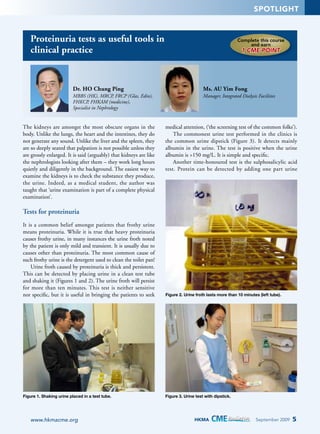
Dr. HO Chung Ping
Ms. AU Yim Fong
MBBS (HK), MRCP, FRCP (Glas, Edin),
FHKCP, FHKAM (medicine),
Specialist in Nephrology
Manager, Integrated Dialysis Facilities
The kidneys are amongst the most obscure organs in the
body. Unlike the lungs, the heart and the intestines, they do
not generate any sound. Unlike the liver and the spleen, they
are so deeply seated that palpation is not possible unless they
are grossly enlarged. It is said (arguably) that kidneys are like
the nephrologists looking after them – they work long hours
quietly and diligently in the background. The easiest way to
examine the kidneys is to check the substance they produce,
the urine. Indeed, as a medical student, the author was
taught that ‘urine examination is part of a complete physical
examination’.
medical attention, (‘the screening test of the common folks’).
The commonest urine test performed in the clinics is
the common urine dipstick (Figure 3). It detects mainly
albumin in the urine. The test is positive when the urine
albumin is >150 mg/L. It is simple and specific.
Another time-honoured test is the sulphosalicylic acid
test. Protein can be detected by adding one part urine
Tests for proteinuria
It is a common belief amongst patients that frothy urine
means proteinuria. While it is true that heavy proteinuria
causes frothy urine, in many instances the urine froth noted
by the patient is only mild and transient. It is usually due to
causes other than proteinuria. The most common cause of
such frothy urine is the detergent used to clean the toilet pan!
Urine froth caused by proteinuria is thick and persistent.
This can be detected by placing urine in a clean test tube
and shaking it (Figures 1 and 2). The urine froth will persist
for more than ten minutes. This test is neither sensitive
nor specific, but it is useful in bringing the patients to seek
Figure 2. Urine froth lasts more than 10 minutes (left tube).
Figure 1. Shaking urine placed in a test tube.
Figure 3. Urine test with dipstick.
www.hkmacme.org
HKMA
September 2009
5
2. SPOTLIGHT
to three parts 3% sulphosalicylic acid and observing the
turbidity (Figures 4, 5 and 6). The test can be made semiquantitative by comparing the intensity of the turbidity
(Figures 7 and 8). This test is not commonly available
in most clinics. It tests both albumin and other heavier
proteins in the urine. Thus the test will be positive in cases
with ‘tubular proteinuria’ or ‘myeloma kidney urine’. Since
the proteins in such conditions are heavy proteins, they are
not detected by the common urine dipstick.
The traditional use of proteinuria tests
Figure 4. Sulphosalicylic acid test.
The tests mentioned above are semi-quantitative only.
They are useful in the management of glomerulonephropathies and nephrotic syndrome (Figure 9). Relapses
can be detected by the proteinuria tests carried out at home
by the patients.
These tests only give a measure of the urine albumin
(or protein) concentration; therefore, they will be affected
by the hydration state of the patient. If a patient gives early
morning urine, the urine albumin may be 100 mg/dL. After
he drinks plenty of water and the same test is repeated on the
new urine sample, the concentration may drop to 30 mg/dL.
For this reason, a quantitative estimation of urine protein
per day is more reliable and a 24-hour urine collection is
useful in this regard.
Advances made in the past two decades provided answers to
the following questions:
1. Is the current dipstick urine test sensitive enough?
2. Does the quantitative urine albumin excretion have any
bearing on the clinical management of renal patients?
3. Is there a less cumbersome method to measure daily
urine protein excretion?
4. Are there any therapies for proteinuria?
Figure 5. Adding urine to sulphosalicylic acid.
Figure 7. Performing multiple tests.
Figure 6. Observing the turbidity.
Figure 8. Different concentrations reflected by turbidity.
6
HKMA
September 2009
www.hkmacme.org
3. SPOTLIGHT
Figure 9. A patient with nephrotic syndrome.
The discovery of microalbuminuria
One of the most significant recent discoveries in nephrology
is microalbuminuria in diabetic patients. It was known that
in patients with incipient diabetic nephropathy, there was
increased urine albumin excretion at a concentration not
yet detectable by the common urine dipstick. It was called
‘microalbuminuria’. The term ‘micro’ referred to the low
urine albumin concentration and not the molecular size, as
the albumin detected are normal albumin molecules [1].
In quantitative terms, normal urine contains around
20 mg/L of albumin and dipsticks can only detect around
150 mg/L of albumin. A patient is said to have macroalbuminuria if urine albumin reaches the concentration
which can be detected by the dipstick. Albumin excretion
between 20–150 mg/L is thus called microalbuminuria.
It is an early indictor of diabetic nephropathy as the
Figure 10. Urine microalbuminuria
screening with Clinitek 50.
www.hkmacme.org
risk of diabetic nephropathy increases 10–20 times
with microalbuminuria. It is amenable to treatment at this
stage.
The detection of microalbuminuria required radioimmunoassay when it was first discovered and was only available
in reference laboratories. With the development of immunoturbometric methods, the test is now within the reach
of most laboratories. To avoid interference caused by the
urine hydration, it is common to test for both the urine
(micro) albumin and urine creatinine concentration and
the result is expressed as a ratio. Under such circumstances,
urine albumin:creatinine ratio >2.5 mg/mmol in men,
>3.5 mg/mmol (30 mg/g) for women is regarded as
microalbuminuria.
With improvements in medical technology and
automation, the test can now be performed in renal clinics
as a valuable point-of-care test. In the author’s clinic, the
early morning urine sample is first tested for albumin using
the common urine dipstick. If the test is positive, there is
macro-albuminuria and there is no need to do the expensive
microalbuminuria test. If the test is negative, a screening
test using the Clinitek 50 (Bayer diagnostics) machine is
performed (Figure 10). It is a reagent strip test which tests
both the albumin and creatinine. The colour changes are
read by the machine and the urine albumin:creatinine ratio
is calculated (Figure 11). The test takes one minute and is
relatively cheap, but the accuracy is not too high.
If the test is positive, it is immediately confirmed with
the DCA 2000 test (Figure 12), which takes 7 minutes and
is considerably more expensive, but the accuracy is much
higher.
Apart from incipient diabetic nephropathy, the presence
of microalbuminuria also signals the increased risk of
cardiovascular disease. Microalbuminuria is an inflammation
marker like the C-reactive protein. It is thus a predicator of
a cardiovascular event in a hypertensive patient.
Figure 11. Reagent strip with Clinitek 50
machine.
HKMA
Figure 12. Urine microalbuminuria
confirmation with DCA2000.
September 2009
7
4. SPOTLIGHT
Urine albumin excretion as a clinical tool
For renal patients with macro-albuminuria, the daily urine
albumin excretion correlates with the renal outcome.
Patients with proteinuria <1 g/day have a much better renal
prognosis than a patient with urine protein >3 g/day. Thus
urine protein is of paramount importance in the prevention,
diagnosis and risk stratification of renal patients.
In his excellent review article [2], Biff Palmer
summarized the clinical significance of urine albumin
excretion:
1. The magnitude of baseline proteinuria is an accurate
predictor of the renal outcome (the renal prognosis).
2. Reduction in urinary protein excretion correlates with
long-term preservation of renal function.
3. Apart from renal protection, reduction in urine albumin
excretion was associated with reduction in cardiovascular
risk.
From the above, it is clear that increased urine albumin
excretion is a marker of poor renal prognosis. It is an
indication for active interventional measures. Alternatively,
reduction of proteinuria is an indication of the response to
treatment and signifies improved renal prognosis.
Measurement of daily urine protein excretion
Though urine protein excretion is a useful tool, the
collection of 24-hour urine is cumbersome. Recently it was
suggested that we send early morning urine for estimation
of the urine albumin and creatinine concentration. If both
can be expressed in the same unit, (e.g. mg/L), then the
urine albumin:creatinine ratio will be a ‘pure’ number. This
number correlates closely with daily protein excretion in
g/1.73 m2 of body surface area. Thus, a ratio of 3.0 (as with
respective urinary albumin and creatinine concentrations
of 300 and 100 mg/dL) represents daily protein excretion
of approximately 3 g/1.73 m2. The accuracy of the total
protein-to-creatinine ratio is related to the fortuitous
occurrence that daily creatinine excretion is only slightly
greater than 1000 mg (8.8 mmol)/day per 1.73 m2.
Such methods avoided the timed collection of urine. It is
useful for the follow-up of the same patient.
like captopril could reduce proteinuria in type 1 diabetes
mellitus. Later studies showed the renoprotective effects
of angiotensin receptor blockers (ARB) in type 2 diabetes
mellitus patients. They were successful in increasing the
time for doubling of the serum creatinine and other renal
events. Apart from lowering the systemic blood pressure,
the drugs dilate the efferent glomerular arterioles, thus
reducing the glomerular hypertension caused by the chronic
hyperglycaemia. Their anti-proteinuria effect is independent
of blood pressure reduction.
ACEI and ARB were subsequently shown to
reduce proteinuria in non-diabetic renal patients in many
studies. Studies also showed that when proteinuria is
reduced, the progression to end-stage renal failure (ESRD) is
reduced.
ACEI or ARB can delay the onset of ESRD but it can be
prevented only in a minority of cases by either drug alone.
In other words, there is a limit to their reno-protective
effect. A multi-tier approach is therefore needed.
For many years, there were studies researching the use
of a combination of ACEI and ARB, aimed at a complete
blockage of the renin-angiotensin system to achieve better
renal protection. The beneficial effect was confirmed by a
recent meta-analysis. It is recommended that if proteinuria
is greater than 500 mg/day, despite monotherapy with an
ACEI, an ARB can be added (or vice versa). Under such
circumstances, monitoring of the serum creatinine and
potassium is mandatory. Another approach is aldosterone
blockade with spironolactone or eplerenone. There were a
number of small clinical trials which suggested the benefits
of add-on aldosterone antagonists, but the danger of severe
hyperkalaemia is a serious concern.
Apart from lifestyle changes like stopping smoking and
weight reduction, the following measures are useful:
1. Dietary protein restriction – Moderate dietary protein
restriction would reduce the progression of renal disease.
Severe dietary protein restriction (e.g. the GiordanoGiovanetti die’) tends to cause protein malnutrition
and muscle wasting and should be prescribed with great
care.
2. Statin therapy – Some trials have shown the antiproteinuric effect of statin therapy in early renal disease.
Therapeutic intervention for proteinuria
Conclusion
Proteinuria is a marker of renal progression. There is also
evidence that proteinuria is responsible for the interstitial
inflammation and fibrosis, contributing to renal progression
[3]. Reduction of proteinuria is therefore of paramount
importance in patient management.
From the work in diabetic nephropathy, it was shown
that angiotensin converting enzyme inhibitors (ACEI)
Proteinuria exists as a spectrum from microalbuminuria
(>300 mg/day) to severe nephrotic range proteinuria (>3.5 g/
day). Early detection of microalbuminuria in diabetic
patients is the best way to prevent diabetic nephropathy.
For patients with macro-albumunuria, quantitation of
the proteinuria is useful in disease stratification and
monitoring.
8
HKMA
September 2009
www.hkmacme.org
5. SPOTLIGHT
The tests for proteinuria have emerged as cost-effective
and reliable ways to study the status of the kidneys and the
prognosis. Three decades have passed by, and the teaching
of our mentors ‘urine test is part of a complete physical
examination’ still remain a golden principle.
References
1. Ho CP. Chronic kidney disease – old diseases with new global challenges.
HKMA CME Bulletin April, 2008; p 4–7.
2. Palmer BF. Proteinuria as a therapeutic target in patients with chronic
renal failure. Am J Nephrol 2007;27:287–93.
3. Ruggenenti P, et al. Kidney prevention recipes for your office practice.
Kidney International 2005;67(Suppl 94):S136–S141.
Answer these on page 20 or
make an online submission at:
www.hkmacme.org
Please indicate whether the following questions are true or false
1. The easiest way to examine the kidneys is to check the
substance they produce, the urine.
2. Urine froth can be detected by placing urine in a clean
test tube and leaving it to sit for one hour.
3. The common urine dipstick detects mainly albumin in the
urine, and is positive when the urine albumin is >150 mg/L.
4. A quantitative estimation of urine protein per day is more
reliable and a 36-hour urine collection is useful in this
regard.
5. A patient is said to have macro-albuminuria if urine
albumin reaches the 150 mg/L. Albumin excretion
between 20–150 mg/L is thus called microalbuminuria.
6. A urine albumin:creatinine ratio >2.5 mg/mmol in men,
and >3.5 mg/mmol (30 mg/g) for women is regarded as
microalbuminuria.
7. A screening test for microalbuminuria uses the radioimmunoassay.
HalfPage-PI-out.pdf
11/27/08
www.hkmacme.org
8. Apart from incipient diabetic nephropathy, the presence
of microalbuminuria also signals the increased risk of
cardiovascular disease.
9. A ratio of 3.0 (as with respective urinary albumin and
creatinine concentrations of 300 and 100 mg/dL) represents daily protein excretion of approximately 8 g/1.73 m2.
10. Statin therapy may be a useful intervention tool – some
trials have shown the anti-proteinuric effect of statin
therapy in early renal disease.
ANSWERS TO AUGUST 2009
Classification of venous disease
1. True
6. False
11. False
2. True 3. False 4. True
7. True . True
8
9. False
5. True
10. False
6:42:13 PM
HKMA
September 2009
9