1. REVIEW
Dynamic, yet structured: The cell membrane three
decades after the Singer–Nicolson model
G. Vereb*†, J. Szollosi*†‡, J. Matko§, P. Nagy*, T. Farkas¶, L. Vıgh¶, L. Matyus*, T. A. Waldmann ,
¨ ˝ ´ ´ ´
and S. Damjanovich*‡**
*Department of Biophysics and Cell Biology and ‡Cell Biophysical Research Group of the Hungarian Academy of Sciences, Research Center for Molecular
Medicine, Medical and Health Science Center, University of Debrecen, H-4012, Debrecen, Hungary; §Department of Immunology, Lorand Eotvos
´ ¨ ¨
University, H-1117, Budapest, Hungary; ¶Institute of Biochemistry, Biological Research Center, Hungarian Academy of Sciences, H-6701,
Szeged, Hungary; and Metabolism Branch, National Cancer Institute, National Institutes of Health, Bethesda, MD 20892-1374
Contributed by T. Farkas, April 29, 2003
The fluid mosaic membrane model proved to be a very useful metric distribution in the membrane bilayer all indicate a
hypothesis in explaining many, but certainly not all, phenomena molecular heterogeneity and the possible formation of mem-
taking place in biological membranes. New experimental data brane microdomains (12).
show that the compartmentalization of membrane components
can be as important for effective signal transduction as is the Advanced Cell Biophysical and Molecular Biological
fluidity of the membrane. In this work, we pay tribute to the Methodology Provides Quantitative Data on the Static and
Singer–Nicolson model, which is near its 30th anniversary, honor- Dynamic Organization of Membranes
ing its basic features, ‘‘mosaicism’’ and ‘‘diffusion,’’ which predict Membrane dynamics, i.e., the ever changing mobility and prox-
the interspersion of proteins and lipids and their ability to undergo imity relationships of lipid and protein molecules in the plasma
dynamic rearrangement via Brownian motion. At the same time, membrane, have a significant impact on essential cellular pro-
modifications based on quantitative data are proposed, highlight- cesses, such as activation, ligand-receptor recognition, antigen
ing the often genetically predestined, yet flexible, multilevel struc- presentation, intercellular interactions (e.g., between target and
ture implementing a vast complexity of cellular functions. This killer cells), etc. Quantitative measurements of membrane dy-
new ‘‘dynamically structured mosaic model’’ bears the following namics are possible with fluorescence recovery after photo-
characteristics: emphasis is shifted from fluidity to mosaicism, bleaching (13, 14), single-particle tracking techniques (8, 15, 16),
which, in our interpretation, means nonrandom codistribution and optical trapping by laser tweezers (13, 17, 18). Fluorescence
patterns of specific kinds of membrane proteins forming small- correlation spectroscopy, a method with tradition in the study of
scale clusters at the molecular level and large-scale clusters (groups reaction kinetics and molecular interactions in solution (19, 20),
of clusters, islands) at the submicrometer level. The cohesive also has been applied to the study of cellular systems recently (21,
forces, which maintain these assemblies as principal elements of 22). The method allows the determination of absolute molecular
the membranes, originate from within a microdomain structure, concentration, mobility, and comobility in small, confocal vol-
where lipid–lipid, protein–protein, and protein–lipid interactions, ume elements of living cells (23). Confocal laser-scanning mi-
as well as sub- and supramembrane (cytoskeletal, extracellular croscopy (24) at the verge of its resolution limits proved to be
matrix, other cell) effectors, many of them genetically predestined, successful in determining the uneven cell-surface distribution of
play equally important roles. The concept of fluidity in the original various antigens. Scanning near-field optical microscopy
model now is interpreted as permissiveness of the architecture to (NSOM), a method ideal for assessing localization of membrane
continuous, dynamic restructuring of the molecular- and higher- proteins at the resolution of several tens of nanometers, also has
level clusters according to the needs of the cell and as evoked by been gaining space in investigating the cytoplasm membrane
the environment. (25–28), although, as Edidin (29) points out, ‘‘while NSOM
promises much, its application to biology is about where electron
cientific dogmas, let alone models, rarely survive more
S
microscopy was 40 or 50 years ago.’’ These methodologies
than a quarter of a century without significant modifica- supported by digital image processing add valuable information
tions. Around its 30th anniversary, the time seems to be to the dynamic data about the spatial distribution and compart-
ripe for at least a modest modification of an old paradigm. mentation of membrane constituents. In general, these ap-
The Singer–Nicolson f luid mosaic membrane model (S-N proaches provide evidence for the domain-like distribution of
model) (1) predicts lateral and rotational freedom and random lipids and proteins in biological membranes (17, 30, 31).
distribution of molecular components in the membrane. Mem- Restrictions in the lateral mobility of both lipid and protein
branes had been considered by the S-N model as ‘‘a two- components were studied extensively by using the fluorescence-
dimensional oriented solution of integral proteins . . . in the recovery-after-photobleaching technique, measuring the diffu-
viscous phospholipid bilayer’’ (1–3). Now it is known, however, sion of fluorescently labeled membrane components from non-
that this freedom of protein (and lipid) mobility is far from being bleached areas into a small, bleached spot. Lateral diffusion
unrestricted. One of the earliest indications of a nonrandom parameters of MHC molecules (9) were highly dependent on the
distribution of proteins was provided by the discovery of cocap- bleached-spot size. Because diffusion rate in a lipid bilayer is
ping (4). The emerging evidence on hierarchically built supramo- expected to be independent of this size, one plausible explana-
lecular protein complexes (5–7) hindered diffusion of proteins in tion is the mosaic-like domain structure of the biological mem-
the membrane (8–10), and the existence of distinct membrane branes that restrict the barrier-free path of proteins and can be
domains termed ‘‘rafts’’ (11) also contradicts the S-N model. partly responsible for the clustered arrangements of membrane
Therefore, Jacobson et al. (2) have correctly stated, “Most proteins.
membrane proteins do not enjoy the continuous unrestricted
lateral diffusion. . . . Instead, proteins diffuse in a more compli-
cated way that indicates considerable lateral heterogeneity in Abbreviations: S-N model, Singer–Nicolson fluid mosaic membrane model; FRET, fluores-
membrane structure, at least on a nanometer scale.’’ The great cence resonance energy transfer; TCR, T cell antigen receptor.
variety of phospholipid molecular species, the differences in †G.V. and J.S. contributed equally to this work.
their molecular shapes and physical properties, and their asym- **To whom correspondence should be sent at the * address. E-mail: dami@jaguar.dote.hu.
www.pnas.org cgi doi 10.1073 pnas.1332550100 PNAS July 8, 2003 vol. 100 no. 14 8053– 8058
2. cells and do not require cytokine-induced aggregation. This
colocalization was modulated significantly by binding of relevant
ILs. In addition, there is evidence that the IL-15 receptor
-subunit, which shares the - and -subunits with IL-2R , also
can form preassembled supramolecular structures with IL-2R
and the ‘‘common’’ -chain (53–55). These data (37) have
challenged the frequently applied paradigm that multisubunit
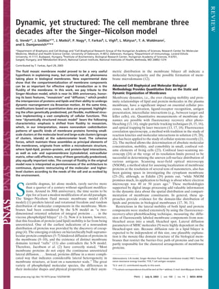
Fig. 1. Proteins experience different types of restrictions to translational
diffusion in the plasma membrane. The view of the membrane is shown from
receptors are assembled under the influence of their specific
beneath. A, Proteins showing preferential accumulation in a lipid microdo- ligands and support an alternative model in which preassembled
main may be confined to the area of the microdomain if the activation energy receptor clusters facilitate faster and stronger biological re-
of passing the domain barrier is larger than the kinetic energy of the protein. sponses, because there is no need for lateral diffusion of
The extent to which passing a domain barrier is prohibited is determined by receptors to associate.
the preference of the protein for the lipid environment: if the protein interacts
preferentially and avidly with lipids of the microdomain, it may be reluctant Electron Microscopy and Scanning-Force Microscopy Reveal
to leave. B and C, The cytoskeleton is also important in restricting free, lateral Submicrometer Clusters of Membrane Receptors
diffusion of membrane proteins. Proteins whose intracellular domain is long
FRET measurements detect molecular associations in the 1- to
are unable to pass through a fence composed of a filament of the cytoskeleton
(B), whereas proteins with a short intracellular domain are free to move across
10-nm range. Combined application of electron and scanning-
such a fence (C). D, Associations of proteins experience more viscous force; force microscopy made possible the discovery of a new, higher
therefore, their translational diffusion rate is usually smaller than that of hierarchical level of receptor clustering in lymphoid cells (36).
monomeric proteins. The distribution of ImmunoGold labels attached to receptors
showed a nonrandom pattern, differing from the Poisson distri-
bution assumed for randomly scattered molecules. The method
Single-particle tracking follows the ‘‘random walk’’ of a gold also was used under near-physiological conditions, where the
particle fixed to a cell-surface protein. For the transferrin gold particles were detected with scanning-force microscopy by
receptor and E-cadherin, a barrier-free path of 400 nm was using tapping mode on hydrated samples (36). The observation
determined (8), whereas for wild-type and cytoplasmatically that nanometer-scale islets of MHC class I molecules in the cell
truncated mutant class I MHC, 600 and 1,700 nm were membrane are organized into micrometer-sized ‘‘island groups’’
measured, respectively (13), indicating that, for membrane- was extended to MHC class II (56) and to the IL-2 receptor
spanning proteins, the barrier-free path is in the same range and -subunit and the transferrin receptor (24). Sequential applica-
that barriers to the ‘‘free’’ diffusion are present 2–3 nm below the tion of different-sized gold labels targeted to MHC classes I and
membrane bilayer. As shown in Fig. 1, cytoskeletal elements and II also revealed successively that positive FRET data did not
intracytosolic molecules with functional connections to cell- necessarily mean that each receptor was homo- or heteroasso-
surface receptors have the capacity either to slow down or to ciated in a large population. The degree of association for MHC
completely stop the lateral motion of transmembrane proteins. class II to MHC class I was 66%, but only 25% of MHC class I
molecules were in the molecular vicinity of MHC class II (56).
Fluorescence Resonance Energy Transfer (FRET) Highlights Besides establishing the nonrandom distribution of receptors,
Nanometer-Scale Associations in the Cell Membrane ImmunoGold labeling also provided for estimating the average
It has been suggested that membrane proteins can be codistrib- size of higher-order molecular clusters (or island groups) (24).
uted nonrandomly, and this nonrandom yet dynamic codistribu- These were in the range of 400–800 nm for various receptors, in
tion pattern even may be controlled genetically (32–35). This good correlation with mean barrier-free paths reported earlier
assumption has been supported by a significant amount of (8, 13). Confocal laser-scanning microscopy of fluorescently
experimental data (36, 37). A major asset in studying these labeled, live, and fixed cells followed by surface reconstruction
codistributions was the application of FRET to cellular systems. and spatial autocorrelation analysis confirmed the existence of
In FRET, an excited fluorescent dye (donor) donates energy to such receptor clusters with sizes essentially equal to those
an acceptor dye if the separation distance between them is 1–10 deduced from electron microscopy (24).
nm. In the 1970s, cell-surface lectins were the first to be
investigated by FRET (38, 39). Since the early 1980s, more Lipid Rafts: The Functional Equivalent of Receptor Islands?
systematic and better established data acquisition and evaluation Numerous studies directed at the plasma membrane have pro-
methods of FRET were introduced in flow cytometry (35, 40). vided evidence for the existence of distinct domains in the
A quantitative method was introduced in digital fluorescence submicron range (24–26, 36, 56–60). Paralleling these observa-
microscopy that exploited the differences in photobleaching tions, the term ‘‘lipid rafts’’ was coined based on studies of
kinetics of donor dyes in the presence and absence of acceptors epithelial cell polarity and gained widespread popularity in the
(36, 41). Recently, a new method, based on systematic and past years (11). Biochemical analysis suggested that rafts consist
normalized FRET measurements between individual receptor of cholesterol and sphingolipids in the exoplasmic leaflet of the
pairs yielding a receptor map by triangulation, has been intro- lipid bilayer and cholesterol and phospholipids with saturated
duced to describe the exact two-dimensional topology of recep- fatty acids in the endoplasmic leaflet (61). They are surrounded
tor clusters (7, 37). The homo- and heteroassociations of MHC by more fluid membrane domains, which are abundant in
class I and II (42), the IL-2 receptor -subunit and intercellular unsaturated fatty acids (62). Polyunsaturated phosphatidylcho-
adhesion molecule 1 (43–45), MHC molecules and transferrin lines and phosphatidylethanolamines (63) including ethanol-
receptors (46), CD4 and CD8 antigens (47, 48), the T cell antigen amine plasmalogens were detected in rafts by quantitative
receptor (TCR) CD3 complex (49), tetraspan molecules (CD53, electrospray ionization and mass spectrometry (64), probably
CD81, CD82) and CD20 with MHC class I and class II (50), the reflecting a regulatory mechanism that offsets the rigidifying
three subunits of the multisubunit IL-2 receptor (37), the tumor effect of cholesterol (63). In addition, 18:1 and 22:6 phosphati-
necrosis factor receptor (51), Fas (CD95; ref. 52), and many dylethanolamines and ethanolamine plasmalogens are prone to
others have been analyzed in detail. Many receptors were found form nonbilayer phases (65, 66), and the association of soluble
to be oligomers preassembled without ligand binding, including proteins (such as G proteins) to membranes depends on the
tumor necrosis factor receptor, Fas, and TCR. Results for the propensity of reverse hexagonal-phase areas (67). This obser-
IL-2 receptor indicated that its subunits are preassembled in T vation highlights the possible role of lipid rafts in signal-
8054 www.pnas.org cgi doi 10.1073 pnas.1332550100 Vereb et al.
3. transduction processes. Isolating lipid rafts as detergent-resistant tigen 1 (LFA-1) ligands (86). The surface components of anti-
membrane domains, followed by Western blotting, indicated that gen-presenting cells are also integral to these clusters; MHC–
rafts indeed are efficient concentrators of various proteins, many peptide complexes are found in the central supramolecular
of them active in cell signaling. Among these were glycosylphos- activating complex, whereas intercellular adhesion molecule 1,
phatidylinositol-anchored proteins (68), cholesterol-linked and the LFA-1 counterreceptor, is concentrated in the peripheral
palmitoylated proteins such as Hedgehog (69), Src-family ki- supramolecular activating complex. Initially, the TCRs are not
nases and -subunits of heterotrimeric G proteins (70), cytokine necessarily engaged in the center, but as cell–cell interaction
receptors (71), and integrins (72). develops, they translocate from the periphery into the center of
The question is how these biochemically identified, detergent- the synapse (87, 88). According to the serial triggering model, in
resistant membrane domains correspond to the observed con- this manner, very few peptide-presenting MHC molecules can
finement of membrane proteins. As Jacobson and Dietrich (73) activate a high number of TCRs (89). It appears important in
pointed out, ‘‘. . . the nature of the in vivo correlate of such terms of the interaction that palmitoylation of the membrane-
detergent-resistant membranes remains enigmatic. In principle, proximal cysteines of CD4 and the association of CD4 with Lck
microscopy should be able to determine whether the postulated contribute to the enrichment of CD4 in lipid rafts (90) and that,
rafts exist.’’ Recently, it has been shown that indeed microscopic furthermore, K channels also reside in rafts in the molecular
equivalents of rafts, or, rather, aggregates of rafts, can be vicinity of TCR (91). The structure of the CTL–target cell
detected by using high-resolution confocal microscopy and that contact is similar to that observed between T cell and antigen-
their disassembly by depletion or in situ complexation of cho- presenting cell, a ring of adhesion proteins surrounding the inner
lesterol not only destroys their morphology (24) but also impairs signal molecule domain. Lytic granule secretion occurs in a
their signaling capabilities; for example, in T cells, it hinders separate domain within the adhesion ring (92). As for the
Stat5 and Stat3 phosphorylation via the IL-2 receptor (71). spatiotemporal coordination of signaling by the high-affinity IgE
Based on the notion that rafts essentially are membrane units receptor Fc RI, lipid rafts first concentrate Lyn protein kinases
formed from transport vesicles fusing to the membrane, one while excluding the Fc RIs from these domains. Upon crosslink-
would expect their size to be very small (59). In photonic force ing Fc RIs by the antigen, the receptors rapidly translocate into
microscopic experiments, it was determined that raft size is 50 the lipid rafts followed by their phosphorylation and the subse-
nm in diameter (74), representing 3,500 sphingolipid mole- quent recruitment of Syk and PLC into these domains (93). The
cules. This indicates that membrane patches observed in fluo- latter process also involves accumulation of actin cytoskeleton to
rescence microscopy, bearing raft marker proteins and or lipids, the active domains (82).
probably are aggregates of these basic building blocks. In some A recent attempt to repeat the classic experiment of Frye and
instances, these larger aggregates are not observed in resting Edidin (94) with currently available technologies provided in-
cells (75, 76) and can be seen only upon crosslinking the ‘‘unit teresting data underlining the dynamism of membrane domains.
rafts’’ (75). In other cases, cells in their native state present Cells labeled with different fluorescent antibodies were fused
surface patches of submicrometer size, identifiable as rafts based with each other. Near-field scanning optical microscopic and
on their composition (24, 77). parallel FRET studies revealed that intermixing of micrometer-
scale protein clusters started right after cell fusion, but there was
Rafts Are Dynamic Structures Reshaped as Function Requires a delay of about 20 min in the intermixing of nanometer-scale
A few years ago, little was known about the stability and lifespan protein associations (28), which clearly indicates the hierarchy of
of lipid rafts in living cells. It appears now that, compared with protein associations (Fig. 2). Although these experiments cor-
the relatively stable nature of phases in artificial bilayers, lipid roborate the existence of protein clusters, they emphasize their
rafts in cells are relatively short-lived. As Edidin points out in a dynamism, which may be important for rapidly reshuffling
recent review (78), ‘‘. . . domains are now thought to be smaller protein interactions.
and less stable then they were in 1992.’’ Lipid probes with
saturated chains on average spend 13 ms in one domain (79); the Static and Dynamic Factors Organizing Membrane Domains
average lifetime of stable domains is found to be on the scale of The physical and chemical forces giving rise to membrane
tens of seconds (10). Pulsed EPR measurements indicate a very domains are under intensive investigation (2, 57). One presumes
fast exchange rate between protein-rich and bulk domains in the that several intracellular and extracellular constraints and forces
membrane, reaching residency times as low as 15 s (80). This influence the size and distribution of these clusters, one of them
possibility of rapidly changing composition and location in the being the cholesterol content of the membrane area in question
membrane, as well as the ability to form aggregates of various (11, 95), and changing the cholesterol composition of the cell
sizes, easily can account for the dynamic regulatory role lipid membrane alters the association pattern and signaling properties
rafts play in various signaling processes. Such a function has been of various molecules (71, 95).
established for receptor tyrosine kinases (27) and for immune Recently, it was proposed that lipids tend to adopt a super-
receptors such as the TCR (81) and the IgE receptor (82, 83). lattice distribution in fluid-mixed bilayers and distribution of
The first step in immunoreceptor signaling is represented by phospholipids in these structures is determined by the molecular
ligand-dependent receptor aggregation, followed by receptor shape and the charge of the head group (96, 97). These super-
phosphorylation by tyrosine kinases of the Src family. Lipid rafts lattice structures do not cover the whole membrane area; rather,
have been identified as platforms wherein signal transduction they are in dynamic equilibrium with areas in which lipids are
molecules may interact with the aggregated immunoreceptors. distributed randomly. The presence of proteins can modify these
Multichain immune recognition receptors such as TCR (84) and structures by depleting or attracting certain lipid species because
Fc RI (83) use common mechanisms by which lipid rafts assist of similarities or differences in molecular shapes. However, fatty
in the initiation of signaling. acid and polar head group composition of phospholipids (96), as
The onset of T cell activation is associated with the formation well as the thermotropic and lamellar- to nonlamellar-phase
of the so-called ‘‘immunological synapse’’ (85) between T cells transitions, are controlled precisely in a way that overall fluidity
and the cells that they are recognizing. This synapse starts with is reached below body temperature. Molecular architecture of
the initial clustering of receptors and promotes the centralized certain phospholipids (98) and proportion of nonbilayer-forming
accumulation of TCRs that has been termed the ‘‘central lipids (66) may contribute to this phenomenon. Remarkably, the
supramolecular activating complex’’ with a corresponding pe- liquid-ordered- to liquid-disordered-phase transition tempera-
ripheral ring, consisting of lymphocyte function-associated an- ture of rafts proper is 13–15°C above (99) the main transition
Vereb et al. PNAS July 8, 2003 vol. 100 no. 14 8055
4. Fig. 3. Association of proteins can be induced by selective accumulation of
proteins in distinct lipid microdomains (a) or by specific protein–protein
interactions (b). (a) The membrane contains lipid microdomains with distinct
lipid compositions. These membrane areas harbor different sets of proteins.
Green lipid molecules preferentially accumulate proteins whose transmem-
brane domain is displayed in black and also proteins that are attached to the
extracellular leaflet of the membrane (glycosylphosphatidylinositol-anchored
proteins). The mechanism for the selective accumulation of proteins in a given
lipid environment can be explained by a preference of proteins for the
chemical (hydrophobicity) or physical (membrane thickness, microviscosity)
properties of the lipid microdomain. Nanometer-sized protein associations
Fig. 2. Dynamics in the hierarchical association of membrane proteins. can be considered a lipid-mediated interaction in this case. (b) Specific pro-
Imaging of nanometer- and micrometer-sized protein clusters give an over- tein–protein interactions mediated by transmembrane proteins or ligands
view of the hierarchical association of membrane proteins. Two cell samples binding to them also may be responsible for the generation of protein
previously labeled with different fluorescent antibodies (green and red sym- associations.
bols) were fused. Lipid rafts (blue circles) are known to accumulate a specific
set of proteins. Micrometer-sized protein clusters exchanged components
with each other, but this process respected lipid microdomain barriers: pro- molecules from the two cells did not intermix even after 80 min
teins known to be in different membrane microdomains never intermixed and maintained their monomeric behavior. It was proposed that
with each other. After a lag period of 20 min, intermixing of nanometer- the small size of the microdomain allowed such a strong inter-
sized protein clusters also took place. However, this process was not as action with the membrane-spanning parts of the protein mole-
widespread as the intermixing of micrometer-sized clusters, because some
proteins (e.g., MHC class II) did not show a significant ability to move from one
cule that the cohesive forces prevented the fusion of these
nanometer-sized cluster to another. microdomains. Interestingly, class II molecules did intermix with
class I molecules of the other cell. MHC class I probably was
accommodated in larger rafts, which could incorporate the small
temperature of membranes (100) because of the high cholesterol rafts of class II molecules in their entirety.
and sphingomyelin content. Under pathological conditions such As for the assembly and maintenance of protein clusters, the
as neurodegenerative diseases, this balance is upset. internalization-recycling machinery is one important candidate
Lipid rafts often are looked at as structures originating solely ‘‘force’’: proteins are recruited to sites of endosome formation,
from lipid–lipid interactions. One should not, however, overlook which gives rise to protein associations (107, 108). SOS bonds
the fact that proteins and protein–lipid interactions could be between integral membrane proteins were implicated in the case
equally important in the formation, maintenance, and dynamics of human killer cell-activating receptors expressed in the plasma
of these domains. In fact, one can imagine the basic unit of a raft membrane of natural killer cells as multimeric complexes (109).
as a single protein molecule surrounded by lipids that are Partitioning of covalently linked, saturated acyl chains into
especially suitable as its environment regarding polarity and liquid-ordered phase domains is likely to be an important
steric complementarity. In addition to the classic investigations mechanism for targeting proteins to rafts, whereas prenylated
(101), where very precise predictions were given about the proteins tend to be excluded from there (110). Thus, even
membrane-spanning -helices, recent data also suggest that the without direct protein–protein interactions, preferential accu-
exact composition and nature of -helical structures will influ- mulation of certain proteins in a lipid domain may induce homo-
ence heavily how and in what phospholipid environment this or heteroassociation as well as formation of micrometer-sized
polypeptide chain can be hosted, if at all (102). This alone would protein clusters (Fig. 3).
be enough to account for protein patterns in cell membranes that
are predestined genetically, as we have predicted decades ago Do We Need a New Paradigm?
(32). The primary and secondary structure of proteins newly Recent data that do not fit the S-N model can be summarized as
synthesized, together with the sorting capabilities in vesicular follows: (i) nonrandom codistribution patterns of receptors in
transport, can easily determine which proteins will be embedded the plasma membrane at different hierarchical levels; (ii) qua-
into a certain lipid environment and which other proteins will be sipermanent molecular contacts to cytoskeletal elements and
their immediate neighbors. For example, it has been demon- signal-transducing molecules; (iii) much shorter barrier-free
strated that the transmembrane domain of CD40 (103) and path than expected for unrestricted diffusion; (iv) domain struc-
influenza hemagglutinin (104) determines their partitioning into ture of the lipid components of membranes has the capacity to
lipid rafts. segregate or colocalize membrane proteins; (v) participation of
On a similar note but looking at the dynamic side, changes in integral membrane proteins in the maintenance of membrane
the structure of proteins [a most evident example being those domains suggests that proteins are as important as structural
occurring after their interaction with other proteins or those elements as lipids; and (vi) dynamic reorganization of protein
generated by changes in membrane potential (105)], as well as elements in membrane domains allows for streamlined cellular
changes in the constitution or thickness of the various lipid responses and is restricted by protein–lipid and protein–protein
regions, can easily cause reshuffling of the components of interactions.
lipid rafts so that the best steric energetic stability is achieved Thus, the straightforward application of the S-N model as a
again (106). frame of events is impossible without introducing a new concept.
The strength of interaction between proteins and their imme- This new concept has the following attributes emphasizing the
diate lipid environment is well characterized when lymphoid colocalization, comobility, and nonrandom codistribution of a
cells with labeled MHC molecules are fused (28). MHC class II significant number of cell-surface molecules: (i) the mobility of
8056 www.pnas.org cgi doi 10.1073 pnas.1332550100 Vereb et al.
5. the cell-surface (transmembrane, glycosylphosphatidylinositol- regulated nature of vesicular transport processes; and (xii)
anchored, or any other type of) proteins is restricted by lipid- identification of the origin and characteristics of microdomains
domain segregation and the length of the free diffusion pathway and receptor assemblies therein may help us understand the
covered without bumping into boundaries; (ii) membrane pro- immediate past and future of cells, their activation state, and
teins may colocalize with each other on the 1- to 10-nm scale in reactivity. Such signals may carry diagnostic, prognostic, or even
a homologous or heterologous fashion, making the mosaicism of therapeutic values if these nonrandom receptor patterns can be
the S-N model prevalent; (iii) a second hierarchical level of linked to diseases affecting the different states and or altered
protein clustering ranging to several hundred nanometers can be genetic material of the cell.
observed for many membrane proteins; (iv) proteins or protein In light of the above attributes, we must understand that
clusters frequently are accommodated by lipid rafts organized by biological details are far more complicated than the resolving
weak or strong interactions above, inside, or below the cell power of a simple model, which describes generalized, uniform
membrane; (v) some receptor types (e.g., tumor necrosis factor behavior of molecules in the membrane. The S-N model is valid,
and IL receptors) are in a preassembled supramolecular forma-
and free diffusion can occur within domain borders, where
tion even in the absence of their physiological ligands and may
molecular interactions do not interfere. This means that the
form a tighter formation upon ligand binding; (vi) ligand-evoked
receptor aggregations (e.g., epidermal growth factor receptor emphasis must be shifted from the fluidity to the mosaicism of
family) are distinctly different from preformed oligomers, yet the S-N model. Mosaicism can restrict free diffusion through one
may serve as equally important amplifying factors of transmem- of the following ways: (i) lipid domain structure, (ii) cytoskeletal
brane signaling; (vii) the smallest microdomains can be consid- or other cytosolic interactions, or (iii) homo- and heteroasso-
ered modules that accommodate membrane proteins either ciations with other integral proteins. These interactions have the
alone or in functional oligomers preassembled from subunits, capacity to increase the lifetime of an intermolecular encounter,
and these can be the building units of larger signaling domains thereby increasing the possibilities for bi- or multilateral inter-
(such as those formed in the immunological synapse); (viii) the actions, which sometimes simply are called receptor crosstalk.
-helical membrane-spanning parts of transmembrane proteins Hence, the overall mobility of molecular elements of the mem-
are matched in length and shape by the aliphatic side chains of brane can be accepted with the above restrictions, making the
lipids constituting the membrane domains that preferentially membrane a heavily compartmentalized, quasi-two-dimensional
accommodate them; (ix) the localization of proteins in different structure, which is more mosaic-like than fluid. In this two-
lipid regions of the plasma membrane can be determined dimensional plane, diffusion, intermolecular forces, the ever
genetically because the amino acid sequence of the transmem- changing membrane potential, and extracellular influences can
brane domain and sequence-dependent covalent modifications dynamically generate and destroy supramolecular structures. We
define the possible, specific lipid–protein interactions; (x) pro- propose that this new model of the cell membrane be called the
teins are likely to have an equally important role in determining dynamically structured mosaic model.
the constituents, structure, and dynamics of membrane domains;
(xi) whereas artificial lipid bilayers tend to spontaneously form This work was supported in part by Hungarian Academy of Sciences
segregated structures, the dynamics and specificity in the living Grants OTKA T037831, T034393, TS 040773, TS 044836, T042618,
cell membrane are provided by specific protein–protein and T043061, T043087, and T043509. G.V. is a Bekesy Fellow of the
´ ´
protein–lipid interactions, as well as the targeted and sensitively Hungarian Ministry of Education.
1. Singer, S. J. & Nicolson, G. L. (1972) Science 175, 720–731. 25. Vereb, G., Meyer, C. K. & Jovin, T. M. (1997) in Interacting Protein Domains:
2. Jacobson, K., Sheets, E. D. & Simson, R. (1995) Science 268, 1441–1442. Their Role in Signal and Energy Transduction, NATO ASI series, ed.
3. Sheets, E. D., Simson, R. & Jacobson, K. (1995) Curr. Opin. Cell Biol. 7, 707–714. Heilmeyer, L. M. G., Jr. (Springer, New York), Vol. H102, pp. 49–52.
4. Goding, J. W. & Layton, J. E. (1976) J. Exp. Med. 144, 857. 26. Hwang, J., Gheber, L. A., Margolis, L. & Edidin, M. (1998) Biophys. J. 74,
5. Damjanovich, S., Gaspar, R., Jr., & Pieri, C. (1997) Q. Rev. Biophys. 30, 67–106.
´ ´ 2184–2190.
6. Damjanovich, S., Matko, J., Matyus, L., Szabo, G. J., Szollosi, J., Pieri, C.,
´ ´ ´ ¨ ˝ 27. Nagy, P., Jenei, A., Kirsch, A. K., Szollosi, J., Damjanovich, S. & Jovin, T. M.
¨ ˝
Farkas, T. & Gaspar, R. J. (1998) Cytometry 33, 225–234.
´ ´ (1999) J. Cell Sci. 112, 1733–1741.
7. Damjanovich, S., Bene, L., Matko, J., Matyus, L., Krasznai, Z., Szabo, G.,
´ ´ ´ 28. Nagy, P., Matyus, L., Jenei, A., Panyi, G., Varga, S., Matko, J., Szollosi, J.,
´ ´ ¨ ˝
Pieri, C., Gaspar, R. & Szollosi, J. (1999) Biophys. Chem. 82, 99–108.
´ ´ ¨ ˝ Gaspar, R., Jovin, T. M. & Damjanovich, S. (2001) J. Cell Sci. 114, 4063–4071.
´ ´
8. Kusumi, A., Sako, Y. & Yamamoto, M. (1993) Biophys. J. 65, 2021–2040. 29. Edidin, M. (2001) Traffic 2, 797–803.
9. Edidin, M. (1993) J. Cell Sci. Suppl. 17, 165–169. 30. Edidin, M. J. (1988) Immunol. Today 9, 218–219.
10. Dietrich, C., Yang, B., Fujiwara, T., Kusumi, A. & Jacobson, K. (2002) 31. Cherry, R. J., Smith, P. R., Morrison, I. E. & Fernandez, N. (1998) FEBS Lett.
Biophys. J. 82, 274–284. 430, 88–91.
11. Simons, K. & Ikonen, E. (1997) Nature 387, 569–572. 32. Damjanovich, S., Somogyi, B. & Tron, L. (1981) in Advances in Physiological
´
12. Shaikh, S. R., Dumaual, A. C., Jenski, L. J. & Stillwell, W. (2001) Biochim.
Sciences, Neural Communication and Control, eds. Szekely, G., Labos, E. &
´ ´
Biophys. Acta 1512, 317–328.
Damjanovich, S. (Pergamon, Oxford), Vol. 30, pp. 9–21.
13. Edidin, M., Zuniga, M. C. & Sheetz, M. P. (1994) Proc. Natl. Acad. Sci. USA
33. Damjanovich, S., Tron, L., Szollosi, J., Zidovetzki, R., Vaz, W. L., Regateiro,
¨ ˝
91, 3378–3382.
F., Arndt-Jovin, D. J. & Jovin, T. M. (1983) Proc. Natl. Acad. Sci. USA 80,
14. Jacobson, K., Elson, E., Koppel, D. & Webb, W. (1982) Nature 295, 283–284.
5985–5989.
15. Anderson, C. M., Georgiou, G. N., Morrison, I. E., Stevenson, G. V. & Cherry,
34. Damjanovich, S., Szollosi, J. & Tron, L. (1992) Immunol. Today 13, A12–A15.
¨ ˝ ´
R. J. (1992) J. Cell Sci. 101, 415–425.
16. Simson, R., Sheets, E. D. & Jacobson, K. (1995) Biophys. J. 69, 989–993. 35. Szollosi, J. & Damjanovich, S. (1994) in Mobility and Proximity in Biological
¨ ˝
17. Edidin, M., Kuo, S. C. & Sheetz, M. P. (1991) Science 254, 1379–1382. Membranes, eds. Damjanovich, S. & Edidin, M. (CRC, Boca Raton, FL), pp.
18. Sako, Y. & Kusumi, A. (1995) J. Cell Biol. 129, 1559–1574. 49–108.
19. Magde, D., Elson, E. L. & Webb, W. W. (1974) Biopolymers 13, 29–61. 36. Damjanovich, S., Vereb, G., Schaper, A., Jenei, A., Matko, J., Starink, J. P.,
´
20. Ehrenberg, M. & Rigler, R. (1976) Q. Rev. Biophys. 9, 69–81. Fox, G. Q., Arndt-Jovin, D. J. & Jovin, T. M. (1995) Proc. Natl. Acad. Sci. USA
21. Brock, R., Vamosi, G., Vereb, G. & Jovin, T. M. (1999) Proc. Natl. Acad. Sci.
´ 92, 1122–1126.
USA 96, 10123–10128. 37. Damjanovich, S., Bene, L., Matko, J., Alileche, A., Goldman, C. K., Sharrow,
´
22. Schwille, P., Haupts, U., Maiti, S. & Webb, W. W. (1999) Biophys. J. 77, S. & Waldmann, T. A. (1997) Proc. Natl. Acad. Sci. USA 94, 13134–13139.
2251–2265. 38. Fernandez, S. M. & Berlin, R. D. (1976) Nature 264, 411–415.
23. Elson, E. L. (2001) Traffic 2, 789–796. 39. Chan, S. S., Arndt-Jovin, D. J. & Jovin, T. M. (1979) J. Histochem. Cytochem.
24. Vereb, G., Matko, J., Vamosi, G., Ibrahim, S. M., Magyar, E., Varga, S.,
´ ´ 27, 56–64.
Szollosi, J., Jenei, A., Gaspar, R., Jr., Waldmann, T. A., et al. (2000) Proc. Natl.
¨ ˝ ´ ´ 40. Szollosi, J., Damjanovich, S., Mulhern, S. A. & Tron, L. (1987) Prog. Biophys.
¨ ˝
Acad. Sci. USA 97, 6013–6018. Mol. Biol. 49, 65–87.
Vereb et al. PNAS July 8, 2003 vol. 100 no. 14 8057
6. 41. Jovin, T. M. & Arndt-Jovin, D. J. (1989) in Cell Structure and Function by 75. Harder, T., Scheiffele, P., Verkade, P. & Simons, K. (1998) J. Cell Biol. 141,
Microspectrofluorimetry, eds. Kohen, E. & Hirschberg, J. G. (Academic, San 929–942.
Diego), pp. 99–117. 76. Kenworthy, A. K., Petranova, N. & Edidin, M. (2000) Mol. Biol. Cell 11,
42. Szollosi, J., Damjanovich, S., Balazs, M., Nagy, P., Tron, L., Fulwyler, M. J.
¨ ˝ ´ 1645–1655.
& Brodsky, F. M. (1989) J. Immunol. 143, 208–213. 77. Pyenta, P. S., Holowka, D. & Baird, B. (2001) Biophys. J. 80, 2120–2132.
43. Szollosi, J., Damjanovich, S., Goldman, C. K., Fulwyler, M. J., Aszalos, A. A.,
¨ ˝ ´ 78. Edidin, M. (2001) Trends Cell Biol. 11, 492–496.
Goldstein, G., Rao, P., Talle, M. A. & Waldmann, T. A. (1987) Proc. Natl. 79. Schutz, G. J., Kada, G., Pastushenko, V. P. & Schindler, H. (2000) EMBO J.
Acad. Sci. USA 84, 7246–7250. 19, 892–901.
44. Edidin, M., Aszalos, A., Damjanovich, S. & Waldmann, T. A. (1988)
´ 80. Kawasaki, K., Yin, J. J., Subczynski, W. K., Hyde, J. S. & Kusumi, A. (2001)
J. Immunol. 141, 1206–1210. Biophys. J. 80, 738–748.
45. Burton, J., Goldman, C. K., Rao, P., Moos, M. & Waldmann, T. A. (1990) 81. Langlet, C., Bernard, A. M., Drevot, P. & He, H. T. (2000) Curr. Opin.
Proc. Natl. Acad. Sci. USA 87, 7329–7333. Immunol. 12, 250–255.
46. Matyus, L., Bene, L., Heyligen, H., Raus, J. & Damjanovich, S. (1995)
´ 82. Holowka, D., Sheets, E. D. & Baird, B. (2000) J. Cell Sci. 113, 1009–1019.
Immunol. Lett. 44, 203–208. 83. Wofsy, C., Vonakis, B. M., Metzger, H. & Goldstein, B. (1999) Proc. Natl.
47. Mittler, R. S., Goldman, S. J., Spitalny, G. L. & Burakoff, S. J. (1989) Proc. Acad. Sci. USA 96, 8615–8620.
Natl. Acad. Sci. USA 86, 8531–8535. 84. Bini, L., Pacini, S., Liberatori, S., Valensin, S., Pellegrini, M., Raggiaschi, R.,
48. Lee, P. U. & Kranz, D. M. (2003) Mol. Immunol. 39, 687–695. Pallini, V. & Baldari, C. T. (2003) Biochem. J. 369, 301–309.
49. De la Hera, A., Muller, U., Olsson, C., Isaaz, S. & Tunnacliffe, A. J. (1991) 85. Paul, W. E. & Seder, R. A. (1994) Cell 76, 241–251.
J. Exp. Med. 173, 7–17. 86. Krummel, M. F. & Davis, M. M. (2002) Curr. Opin. Immunol. 14, 66–74.
50. Szollosi, J., Horejsi, V., Bene, L., Angelisova, P. & Damjanovich, S. (1996)
¨ ˝ 87. Dustin, M. L. (2002) J. Clin. Invest. 109, 155–160.
J. Immunol. 157, 2939–2946. 88. Hiltbold, E. M., Poloso, N. J. & Roche, P. A. (2003) J. Immunol. 170,
51. Chan, F. K., Chun, H. J., Zheng, L., Siegel, R. M., Bui, K. L. & Lenardo, M. J. 1329–1338.
(2000) Science 288, 2351–2354. 89. Valitutti, S., Muller, S., Cella, M., Padovan, E. & Lanzavecchia, A. (1995)
52. Siegel, R. M., Frederiksen, J. K., Zacharias, D. A., Chan, F. K., Johnson, M., Nature 375, 148–151.
Lynch, D., Tsien, R. Y. & Lenardo, M. J. (2000) Science 288, 2354–2357. 90. Fragoso, R., Ren, D., Zhang, X., Su, M. W., Burakoff, S. J. & Jin, Y. J. (2003)
53. Tagaya, Y., Burton, J. D., Miyamoto, Y. & Waldmann, T. A. (1996) EMBO J. Immunol. 170, 913–921.
J. 15, 4928–4939. 91. Panyi, G., Bagdany, M., Bodnar, A., Vamosi, G., Szentesi, G., Jenei, A.,
´ ´
54. Waldmann, T. A. & Tagaya, Y. (1999) Annu. Rev. Immunol. 17, 19–49. Matyus, L., Varga, S., Waldmann, T. A., Gaspar, R., et al. (2003) Proc. Natl.
´ ´ ´
55. Waldmann, T. A. (2003) Annu. Rev. Immunol. 21, 1–27. Acad. Sci. USA 100, 2592–2597.
56. Jenei, A., Varga, S., Bene, L., Matyus, L., Bodnar, A., Bacso, Z., Pieri, C.,
´ 92. Stinchcombe, J. C., Bossi, G., Booth, S. & Griffiths, G. M. (2001) Immunity
Gaspar, R., Jr., Farkas, T. & Damjanovich, S. (1997) Proc. Natl. Acad. Sci.
´ ´ 15, 751–761.
USA 94, 7269–7274. 93. Kovarova, M., Tolar, P., Arudchandran, R., Draberova, L., Rivera, J. &
57. Edidin, M. (1997) Curr. Opin. Struct. Biol. 7, 528–532. Draber, P. (2001) Mol. Cell. Biol. 21, 8318–8328.
58. Kenworthy, A. K. & Edidin, M. (1998) J. Cell Biol. 142, 69–84. 94. Frye, L. D. & Edidin, M. (1970) J. Cell Sci. 7, 319–335.
59. Varma, R. & Mayor, S. (1998) Nature 394, 798–801. 95. Rothberg, K. G., Ying, Y. S., Kamen, B. A. & Anderson, R. G. (1990) J. Cell
60. Horejsi, V. (2002) Trends Immunol. 23, 562–564. Biol. 111, 2931–2938.
61. Fridriksson, E. K., Shipkova, P. A., Sheets, E. D., Holowka, D., Baird, B. & 96. Somerharju, P., Virtanen, J. A. & Cheng, K. H. (1999) Biochim. Biophys. Acta
McLafferty, F. W. (1999) Biochemistry 38, 8056–8063. 1440, 32–48.
62. Schroeder, R., London, E. & Brown, D. (1994) Proc. Natl. Acad. Sci. USA 91, 97. Virtanen, J. A., Cheng, K. H. & Somerharju, P. (1998) Proc. Natl. Acad. Sci.
12130–12134. USA 95, 4964–4969.
63. Blom, T. S., Koivusalo, M., Kuismanen, E., Kostiainen, R., Somerharju, P. & 98. Farkas, T., Kitajka, K., Fodor, E., Csengeri, I., Lahdes, E., Yeo, Y. K.,
Ikonen, E. (2001) Biochemistry 40, 14635–14644. Krasznai, Z. & Halver, J. E. (2000) Proc. Natl. Acad. Sci. USA 97, 6362–6366.
64. Pike, L. J., Han, X., Chung, K. N. & Gross, R. W. (2002) Biochemistry 41, 99. Gousset, K., Wolkers, W. F., Tsvetkova, N. M., Oliver, A. E., Field, C. L.,
2075–2088. Walker, N. J., Crowe, J. H. & Tablin, F. (2002) J. Cell. Physiol. 190, 117–128.
65. Giorgione, J., Epand, R. M., Buda, C. & Farkas, T. (1995) Proc. Natl. Acad. 100. Tablin, F., Oliver, A. E., Walker, N. J., Crowe, L. M. & Crowe, J. H. (1996)
Sci. USA 92, 9767–9770. J. Cell. Physiol. 168, 305–313.
66. Lohner, K. (1996) Chem. Phys. Lipids 81, 167–184. 101. Hartmann, E., Rapoport, T. A. & Lodish, H. F. (1989) Proc. Natl. Acad. Sci.
67. Escriba, P. V., Ozaita, A., Ribas, C., Miralles, A., Fodor, E., Farkas, T. & USA 86, 5786–5790.
Garcia-Sevilla, J. A. (1997) Proc. Natl. Acad. Sci. USA 94, 11375–11380. 102. Lewis, R. N., Zhang, Y. P., Hodges, R. S., Subczynski, W. K., Kusumi, A.,
68. Horejsi, V., Cebecauer, M., Cerny, J., Brdicka, T., Angelisova, P. & Drbal, K. Flach, C. R., Mendelsohn, R. & McElhaney, R. N. (2001) Biochemistry 40,
(1998) Immunol. Lett. 63, 63–73. 12103–12111.
69. Rietveld, A., Neutz, S., Simons, K. & Eaton, S. (1999) J. Biol. Chem. 274, 103. Bock, J. & Gulbins, E. (2003) FEBS Lett. 534, 169–174.
12049–12054. 104. Scheiffele, P., Roth, M. G. & Simons, K. (1997) EMBO J. 16, 5501–5508.
70. Resh, M. D. (1999) Biochim. Biophys. Acta 1451, 1–16. 105. Fromherz, P. (1988) Proc. Natl. Acad. Sci. USA 85, 6353–6357.
71. Matko, J., Bodnar, A., Vereb, G., Bene, L., Vamosi, G., Szentesi, G., Szollosi,
´ ´ ¨ ˝ 106. Bene, L., Szollosi, J., Balazs, M., Matyus, L., Gaspar, R., Ameloot, M., Dale,
¨ ˝ ´ ´ ´ ´
J., Gaspar, R., Horejsi, V., Waldmann, T. A., et al. (2002) Eur. J. Biochem. 269,
´ ´ R. E. & Damjanovich, S. (1997) Cytometry 27, 353–357.
1199–1208. 107. Gheber, L. A. & Edidin, M. (1999) Biophys. J. 77, 3163–3175.
72. Baron, W., Decker, L., Colognato, H. & ffrench-Constant, C. (2003) Curr. 108. Tang, Q. & Edidin, M. (2001) Biophys. J. 81, 196–203.
Biol. 13, 151–155. 109. Olcese, L., Cambiaggi, A., Semenzato, G., Bottino, C., Moretta, A. & Vivier,
73. Jacobson, K. & Dietrich, C. (1999) Trends Cell Biol. 9, 87–91. E. (1997) J. Immunol. 158, 5083–5086.
74. Pralle, A., Keller, P., Florin, E. L., Simons, K. & Horber, J. K. (2000) J. Cell 110. Melkonian, K. A., Ostermeyer, A. G., Chen, J. Z., Roth, M. G. & Brown, D. A.
Biol. 148, 997–1008. (1999) J. Biol. Chem. 274, 3910–3917.
8058 www.pnas.org cgi doi 10.1073 pnas.1332550100 Vereb et al.