SlideShare ist ein Scribd-Unternehmen logo
1 von 49
Downloaden Sie, um offline zu lesen
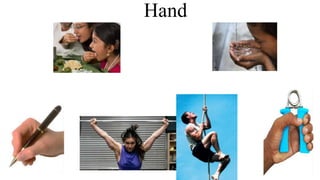
Hand
Hand
• Composed of
• Bones
• Muscles
• Ligaments
• To allow a large amount of
movements
Hand
• Has 2 surfaces
• Palmar
• Dorsal
Palm – Features
• Hollow
• Eminence
• Creases
Palm – Features
• Palmar surface
• Thick skin
• Devoid of
• Hairs & sebaceous glands
• Connected with deep fascia by
• Many fibrous septa
• Helps grip by restricting mobility of skin
• Has fat
• Act as cushion
• To withstand pressure
Palm - features
• Has
• Creases or lines
• 3 types
• Flexure lines
• Papillary ridges
• Langer lines
Creases - Flexure lines
• Visible marking
• Lies close to joints
• On medial 4 fingers
• 2 interphalangeal creases
• Proximal crease only marks joint
• Crease between palm & fingers
• Do not represent the metacarpo-
phalangeal joint
• Joint lies 2 cm proximal to crease
• Distal skin crease of wrist
• Represents proximal border of flexor
retinaculum
• At the midpoint
• Median nerve passes behind
Creases - Flexure lines
• Wrist
• Proximal
• Distal
• Palm
• Longitudinal
• Mid-palmar
• Radial
• Transverse
• Proximal
• Longitudinal
• Applied anatomy – Simian’s crease
• Phalanges
• Proximal
• Middle
• Distal
Creases – Papillary ridges
• Otherwise - Finger prints
• Peculiar over the flexor surface of
distal phalanges
• Corresponds to
• Pattern of underlying dermal papilla
• Ducts of sweat gland opens
• 3 types
• Whorl, loop & arch
• Helps in personal identity
• Dermatoglyphics
• Science of finger prints
Creases – Langer lines
• Produced by
• Bundles of collagen fibres of dermis
• On dorsum
• Transverse
• On palm
• Longitudinal
Hand - Dorsum
• Dorsal surface
• Thin skin
• Presence of
• Hair & sebaceous & sweat glands
• Beneath skin
• Dorsal digital nerves
• Dorsal venous plexus
• Drains not only
• Dorsal surface but also palm
• To avoid the pressure of grip
Superficial fascia
• Subcutaneous tissue
• Contains
• Fat
• Palmaris brevis
• Lies across the base of hypothenar
eminence
• Superficial transverse ligament of palm
• Across roots of medial 4 fingers
Palmaris brevis
• Lies across the base of hypothenar eminence
• Origin
• Flexor retinaculum & central part of palmar
aponeurosis
• Insertion
• Dermis of ulnar border of hand
• Nerve supply
• Superficial branch of ulnar nerve
• Action
• Improves the grip
• Morphology
• Remnant of Panniculus carnosus
•
Superficial transverse ligament of Palm
• Modification of superficial fascia
• Stretches across
• Free margin of the webs of medial four
fingers
• Structures passing deep to it
• Digital vessels & nerves
Cutaneous nerves
• Palmar cutaneous branch of
median– in lower part of
forearm – passes into hand
above flexor retinaculum –
supplying skin over thenar
eminence & middle of palm
• Palmar cutaneous of ulnar –
near lower forearm –supplies
skin of medial 1/3 of palm
Deep fascia
• Thickened at 3 sites
• Flexor retinaculum
• Palmar aponeurosis
• Fibrous flexor sheath
Palmar aponeurosis
• Triangular in shape
• Occupies the central area of the palm
• Has
• Apex
• Base
• Apex
• Blends with distal border of flexor
retinaculum
• Continuous with palmaris longus tendon
• Base
• Lies at bases of medial four fingers
• Divides into four digital slips
• Further each digital slip divided into
• Superficial & deep sets of fibres
Palmar aponeurosis – superficial & deep fibres
• Superficial fibres
• Joins with dermis
• Blend with superficial
transverse ligament of palm
• Deep fibres
• Divides into 2 bands
• Attaches to
• Deep transverse ligament of
palm
• Palmar ligament of
metacarpophalangeal joint
• Bases of proximal phalanges
• Fibrous flexor sheaths
Palmar aponeurosis – space between the slips
• Intervals between the four digital
slips
• Connected by transverse fibres
• Structures passing
• Digital vessels
• Digital nerves
• Lumbricals
Palmar aponeurosis – medial & lateral palmar septa
• Medial septum
• From medial margin
• To palmar surface of shaft of 5th
metacarpal
• Lateral septum
• From lateral margin
• To 1st metacarpal bone
• Septa subdivide the palm into
fascial spaces
Palmar aponeurosis – functions
• Powerful grip
• By firm attachment of overlying skin
• Prevents bow-stringing
• Of flexor tendons
• Protects
• Vessels & nerves
• Provides
• Origin to palmaris brevis
• Potential spaces
• Septa from aponeurosis forms
• Peculiarity from plantar aponeurosis
• Absence of digital slip to thumb
• Allows free movement of thumb
• Plantar aponeurosis
• Has 5 slips
• Morphology
• Degenerated part of palmaris longus tendon
Palmar aponeurosis
• Thickened deep fascia of the hand
• Triangular in shape
• Occupies the central area of the palm
• The apex is attached to the distal border
of flexor retinaculum and receives the
insertion of palmaris longus tendon.
• Base divides at the bases of the fingers
into four slips that pass into the fingers
• Functions
• Gives firm attachment to the overlying skin
and improves the grip.
• Protects the underlying tendons, vessels &
nerves
Palmar aponeurosis (Central part)
• Thickened deep fascia of the hand
• Triangular in shape
• Occupies the central area of the palm
• Apex is attached to the distal border of
flexor retinaculum and receives the
insertion of palmaris longus tendon.
• Base divides at the bases of the fingers
into four slips that pass into the fingers
• Functions:
• Gives firm attachment to the overlying skin
and improves the grip.
• Protects the underlying tendons, vessels &
nerves
Dupuytren’s contracture
• Hypertrophy of palmar
aponeurosis –progressive
fibrosis (ulnar side)
• Shortening & thickening of
fibrous bands from aponeurosis
to little and ring fingers
Intrinsic muscles of hand
Intrinsic muscles
• Thenar
• Hypothenar
• Lumbricals
• Interossei muscles
Intrinsic Muscles
• Situated totally within the hand
• Divided into 4 groups:-
• Lateral group
• Four thenar muscles
• Medial group
• Three hypothenar muscles
• Palmaris brevis
• Central group:
• Four lumbricals
• Four palmar interossei
• Four dorsal interossei
• All muscles are supplied by
• C 8 & T 1 spinal segments
• Through median & ulnar nerves
Lumbricals
• Earth worm
• 4 lumbricals
• Numbered from lateral to medial
• Arise from 4 tendons of FDP
• Inserted into extensor expansion
• Numbered from lateral to medial
• First & second are
• Unipennate
• Supplied by Median nerve
• Third & fourth are
• Bipennate
• Supplied by deep branch of ulnar
• Left Hand
Lumbricals - origin
• I lumbrical
• From radial side of tendon for index
finger
• II lumbrical
• From radial side of tendon of middle
finger
• III lumbrical
• From adjacent sides of the tendons for
middle & ring fingers
• IV lumbrical
• From adjacent sides of the tendons for
ring & little fingers
• Right Hand
Lumbricals
• Insertion
• All lumbricals pass backwards on radial side
of
• 2nd , 3rd , 4th & 5th metacarpo-phalangeal joints
respectively
• Insert on lateral angle of extensor expansion
• Action
• Flex the metacarpo-phalangeal joints
• Extend the interphalangeal joints
Thenar muscles
• Abductor pollicis brevis
• Flexor pollicis brevis
• Opponens pollicis
• Adductor pollicis
Abductor pollicis brevis
• Origin
• Tubercle of Scaphoid
• Crest of Trapezium
• Flexor retinaculum
• Insertion
• Lateral aspect of proximal phalanx of
thumb
• Near base
• Action
• Abduction & medial rotation
• At
• Metacarpophalangeal joint
• Carpometacarpal joint
• Right angle to the thumb
• Nerve supply
• Recurrent branch of Median (C 8 &
T1)
Flexor pollicis brevis
• Origin
• Superficial head
• Crest of Trapezium
• Flexor retinaculum
• Deep head
• Trapezoid
• Capitate
• Insertion
• Lateral aspect of proximal phalanx of
thumb
• Near base
• Action
• Flexion of carpometacarpal &
metacarpophalangeal joints
• Nerve supply
• Recurrent branch of Median (C 8 & T1)
Opponens pollicis
• Origin
• Tubercle of trapezium
• Flexor retinaculum
• Insertion
• I metacarpal
• Lateral margin & lateral
palmar aspect
• Action
• Opposition
• At carpo metacarpal joint
• Rotating & flexing the
metacarpal on trapezium
Thenar eminence
• Raised region between wrist & base of
thumb
• Muscles
• Abductor pollicis brevis(median)
• Opponens pollicis(median)
• Flexor pollicis brevis(median & ulnar)
• Adductor pollicis(ulnar)
Carpal tunnel syndrome
Adductor pollicis
• Origin
• Oblique head – capitate, trapezoid &
bases of 2nd & 3rd metacarpal bones
• Transverse head – distal 2/3rd of shaft
of 3rd metacarpal bone
• Insertion
• Ulnar side of base of proximal phalanx
of thumb
• Action
• Transverse head – adductor &flexor of
thumb
• Nerve supply – Ulnar nerve (deep)
C8 &T1
Interossei
• Deepest structure of hand
• Between the metacarpal bones
• 8 muscles present
• Arranged in 2 groups
• Palmar interossei (4)
• Dorsal interossei (4)
Palmar interossei
• Superficial than dorsal interossei
• Smaller ones
• Unipennate
• Adduct the fingers towards middle finger
• No adductors for middle finger
• Origin
• 1st & 2nd
• Medial side of shafts of 1st & 2nd metacarpals
• 3rd & 4th
• From radial side of shafts of 4th & 5th metacarpals
• Insertion
• Base of proximal phalanx of corresponding digit on
side of their origin
• Dorsal digital expansion
2
3
4
Dorsal interossei
• Deepest structure
• Fill up intermetacarpal spaces
• Bipennate
• Abductors
• Little & thumb have their own
abductors
• Middle finger require abduction on
both sides
Dorsal interossei
• Fills up all (4) intermetacarpal spaces
• Origin
• Contiguous sides of shafts of
metacarpals
• Leaves a gap in between their origin
• Space between first dorsal interossei
transmits
• Radial artery
• Gap between 2nd 3rd & 4th dorsal
interossei transmits
• Proximal perforating arteries
Dorsal interossei
• Insertion
• Proximal phalanges of 2nd, 3rd & 4th
• Middle finger gets insertion on both
sides
• 1st & 2nd
• Radial side of bases of proximal
phalanges of index & middle
• 3rd & 4th
• Ulnar side of bases of proximal
phalanges of middle & ring fingers
• Nerve supply
• Deep branch of ulnar
• Lateral & medial angles of dorsal digital expansion of
• Index, middle & ring fingers receives
• Interossei
Hypothenar muscles
• Abductor digiti minimi
• Flexor digiti minimi
• Opponens digiti minimi
Abductor digiti minimi
• Origin
• Pisiform, tendon of flexor carpi ulnaris
• Insertion
• Ulnar side of base of proximal phalanx
• Action
• Abductor of little finger
• Nerve supply
• Ulnar (deep) C8 & T1
Flexor digiti minimi brevis
• Origin
• Hook of hamate
• Flexor retinaculum
• Insertion
• Ulnar side of base of proximal phalanx
of little finger
• Action
• Flexor of proximal phalanx of little
finger
• Nerve supply
• Ulnar (deep) C8 & T1
Opponens digiti minimi
• Origin
• Hook of hamate
• Flexor retinaculum
• Insertion
• Ulnar border of 5th metacarpal
• Action
• Flexes 5th metacarpal
• Nerve supply
• Ulnar (deep) C8 & T1
Deep branch of ulnar nerve
• Course
• Passes between abductor digiti
minimi & flexor digiti minimi
• Accompanied by deep branch of ulnar
artery
• Perforates opponens digiti minimi
• Follow deep palmar arch
Branches
• Muscular
• Abductor digiti minimi
• Flexor digiti minimi brevis
• Opponens digiti minimi
• 3rd & 4th lumbricals
• Adductor pollicis
• Flexor pollicis brevis
• Interossei
• Articular
• Intercarpal, carpometacarpal joints
• Blood vessels – ulnar & palmar arteries
Median nerve contd…
• Muscular branches to Thenar muscles
– Abductor pollicis brevis
– Flexor pollicis brevis
– Opponens pollicis
• Branches from digital nerves supply
– 1st & 2nd lumbricals

Weitere ähnliche Inhalte

Was ist angesagt?

Was ist angesagt? (20)

Slideshow: Anterior Leg
Slideshow: Anterior LegSlideshow: Anterior Leg
Slideshow: Anterior Leg
 
Hand
Hand Hand
Hand
 
Retinacula of the hand
Retinacula of the handRetinacula of the hand
Retinacula of the hand
 
Extensor retinaculum & dorsal digital expansion Dr.N.Mugunthan
Extensor retinaculum & dorsal digital expansion Dr.N.MugunthanExtensor retinaculum & dorsal digital expansion Dr.N.Mugunthan
Extensor retinaculum & dorsal digital expansion Dr.N.Mugunthan
 
Muscles of the forearm
Muscles of the forearmMuscles of the forearm
Muscles of the forearm
 
7. anatomy of the leg and dorsum of the foot.
7. anatomy of the leg and dorsum of the foot.7. anatomy of the leg and dorsum of the foot.
7. anatomy of the leg and dorsum of the foot.
 
Blood vessel, Innervation and lymph system of lower limb
Blood vessel, Innervation and lymph system of lower limbBlood vessel, Innervation and lymph system of lower limb
Blood vessel, Innervation and lymph system of lower limb
 
Popliteal Fossa.pptx
Popliteal Fossa.pptxPopliteal Fossa.pptx
Popliteal Fossa.pptx
 
The arm
The armThe arm
The arm
 
Popliteal fossa
Popliteal fossaPopliteal fossa
Popliteal fossa
 
Femoral sheath
Femoral sheathFemoral sheath
Femoral sheath
 
Slideshow: Femoral Triangle
Slideshow: Femoral TriangleSlideshow: Femoral Triangle
Slideshow: Femoral Triangle
 
Slideshow: Hand
Slideshow: Hand Slideshow: Hand
Slideshow: Hand
 
Slideshow: The Foot
Slideshow: The FootSlideshow: The Foot
Slideshow: The Foot
 
Popliteal fossa
Popliteal fossa   Popliteal fossa
Popliteal fossa
 
Brachial artery,radial & ulnar artery,plamar arches
Brachial artery,radial & ulnar artery,plamar archesBrachial artery,radial & ulnar artery,plamar arches
Brachial artery,radial & ulnar artery,plamar arches
 
Medial compartment of thigh
Medial compartment of thighMedial compartment of thigh
Medial compartment of thigh
 
9. posterior compartment of the forearm
9. posterior compartment of the forearm9. posterior compartment of the forearm
9. posterior compartment of the forearm
 
The foot
The footThe foot
The foot
 
Pectoral muscles
Pectoral musclesPectoral muscles
Pectoral muscles
 

Ähnlich wie Hand Anatomy Guide

Ähnlich wie Hand Anatomy Guide (20)

Hand-spaces and arterial arches.pptx
Hand-spaces and arterial arches.pptxHand-spaces and arterial arches.pptx
Hand-spaces and arterial arches.pptx
 
10. anatomy of the palm of the hand
10. anatomy of the palm of the hand10. anatomy of the palm of the hand
10. anatomy of the palm of the hand
 
HAND SNUFF BOX (1).pptx
HAND SNUFF BOX (1).pptxHAND SNUFF BOX (1).pptx
HAND SNUFF BOX (1).pptx
 
Palm- 1 and 2 .pptx
Palm- 1 and 2 .pptxPalm- 1 and 2 .pptx
Palm- 1 and 2 .pptx
 
Palmar spaces
Palmar spacesPalmar spaces
Palmar spaces
 
lecture 6aThe Fore am.pdf
lecture 6aThe Fore am.pdflecture 6aThe Fore am.pdf
lecture 6aThe Fore am.pdf
 
Hand anatomy new
Hand anatomy newHand anatomy new
Hand anatomy new
 
Hand
HandHand
Hand
 
5 anterior forearm.ppt
5  anterior forearm.ppt5  anterior forearm.ppt
5 anterior forearm.ppt
 
Forearm muscles.pptx
Forearm muscles.pptxForearm muscles.pptx
Forearm muscles.pptx
 
Muscles of hand.pptx
Muscles of hand.pptxMuscles of hand.pptx
Muscles of hand.pptx
 
The hand(2)
The hand(2)The hand(2)
The hand(2)
 
THE Intrinsic muscle OF THE HAND Prof.Ugo 02-10-23.pptx
THE Intrinsic muscle OF THE HAND Prof.Ugo 02-10-23.pptxTHE Intrinsic muscle OF THE HAND Prof.Ugo 02-10-23.pptx
THE Intrinsic muscle OF THE HAND Prof.Ugo 02-10-23.pptx
 
Wrist Joint & Hand
Wrist Joint & HandWrist Joint & Hand
Wrist Joint & Hand
 
Forearm muscles.pptx
Forearm muscles.pptxForearm muscles.pptx
Forearm muscles.pptx
 
Forearm muscles
Forearm musclesForearm muscles
Forearm muscles
 
Hand anatomy
Hand anatomyHand anatomy
Hand anatomy
 
Hand anatom yw
Hand anatom ywHand anatom yw
Hand anatom yw
 
Muscles of Anterior compartment of forearm .pptx
Muscles of Anterior compartment of forearm .pptxMuscles of Anterior compartment of forearm .pptx
Muscles of Anterior compartment of forearm .pptx
 
10-Palm of hand.ppt
10-Palm of hand.ppt10-Palm of hand.ppt
10-Palm of hand.ppt
 

Kürzlich hochgeladen

6 ways Samsung’s Interactive Display powered by Android changes the classroom
6 ways Samsung’s Interactive Display powered by Android changes the classroom6 ways Samsung’s Interactive Display powered by Android changes the classroom
6 ways Samsung’s Interactive Display powered by Android changes the classroomSamsung Business USA
 
How to Make a Duplicate of Your Odoo 17 Database
How to Make a Duplicate of Your Odoo 17 DatabaseHow to Make a Duplicate of Your Odoo 17 Database
How to Make a Duplicate of Your Odoo 17 DatabaseCeline George
 
Congestive Cardiac Failure..presentation
Congestive Cardiac Failure..presentationCongestive Cardiac Failure..presentation
Congestive Cardiac Failure..presentationdeepaannamalai16
 
Narcotic and Non Narcotic Analgesic..pdf
Narcotic and Non Narcotic Analgesic..pdfNarcotic and Non Narcotic Analgesic..pdf
Narcotic and Non Narcotic Analgesic..pdfPrerana Jadhav
 
Tree View Decoration Attribute in the Odoo 17
Tree View Decoration Attribute in the Odoo 17Tree View Decoration Attribute in the Odoo 17
Tree View Decoration Attribute in the Odoo 17Celine George
 
Mythology Quiz-4th April 2024, Quiz Club NITW
Mythology Quiz-4th April 2024, Quiz Club NITWMythology Quiz-4th April 2024, Quiz Club NITW
Mythology Quiz-4th April 2024, Quiz Club NITWQuiz Club NITW
 
Oppenheimer Film Discussion for Philosophy and Film
Oppenheimer Film Discussion for Philosophy and FilmOppenheimer Film Discussion for Philosophy and Film
Oppenheimer Film Discussion for Philosophy and FilmStan Meyer
 
ESP 4-EDITED.pdfmmcncncncmcmmnmnmncnmncmnnjvnnv
ESP 4-EDITED.pdfmmcncncncmcmmnmnmncnmncmnnjvnnvESP 4-EDITED.pdfmmcncncncmcmmnmnmncnmncmnnjvnnv
ESP 4-EDITED.pdfmmcncncncmcmmnmnmncnmncmnnjvnnvRicaMaeCastro1
 
How to Uninstall a Module in Odoo 17 Using Command Line
How to Uninstall a Module in Odoo 17 Using Command LineHow to Uninstall a Module in Odoo 17 Using Command Line
How to Uninstall a Module in Odoo 17 Using Command LineCeline George
 
Q-Factor General Quiz-7th April 2024, Quiz Club NITW
Q-Factor General Quiz-7th April 2024, Quiz Club NITWQ-Factor General Quiz-7th April 2024, Quiz Club NITW
Q-Factor General Quiz-7th April 2024, Quiz Club NITWQuiz Club NITW
 
Objectives n learning outcoms - MD 20240404.pptx
Objectives n learning outcoms - MD 20240404.pptxObjectives n learning outcoms - MD 20240404.pptx
Objectives n learning outcoms - MD 20240404.pptxMadhavi Dharankar
 
Scientific Writing :Research Discourse
Scientific  Writing :Research  DiscourseScientific  Writing :Research  Discourse
Scientific Writing :Research DiscourseAnita GoswamiGiri
 
Employablity presentation and Future Career Plan.pptx
Employablity presentation and Future Career Plan.pptxEmployablity presentation and Future Career Plan.pptx
Employablity presentation and Future Career Plan.pptxryandux83rd
 
31 ĐỀ THI THỬ VÀO LỚP 10 - TIẾNG ANH - FORM MỚI 2025 - 40 CÂU HỎI - BÙI VĂN V...
31 ĐỀ THI THỬ VÀO LỚP 10 - TIẾNG ANH - FORM MỚI 2025 - 40 CÂU HỎI - BÙI VĂN V...31 ĐỀ THI THỬ VÀO LỚP 10 - TIẾNG ANH - FORM MỚI 2025 - 40 CÂU HỎI - BÙI VĂN V...
31 ĐỀ THI THỬ VÀO LỚP 10 - TIẾNG ANH - FORM MỚI 2025 - 40 CÂU HỎI - BÙI VĂN V...Nguyen Thanh Tu Collection
 
BÀI TẬP BỔ TRỢ TIẾNG ANH 8 - I-LEARN SMART WORLD - CẢ NĂM - CÓ FILE NGHE (BẢN...
BÀI TẬP BỔ TRỢ TIẾNG ANH 8 - I-LEARN SMART WORLD - CẢ NĂM - CÓ FILE NGHE (BẢN...BÀI TẬP BỔ TRỢ TIẾNG ANH 8 - I-LEARN SMART WORLD - CẢ NĂM - CÓ FILE NGHE (BẢN...
BÀI TẬP BỔ TRỢ TIẾNG ANH 8 - I-LEARN SMART WORLD - CẢ NĂM - CÓ FILE NGHE (BẢN...Nguyen Thanh Tu Collection
 
Satirical Depths - A Study of Gabriel Okara's Poem - 'You Laughed and Laughed...
Satirical Depths - A Study of Gabriel Okara's Poem - 'You Laughed and Laughed...Satirical Depths - A Study of Gabriel Okara's Poem - 'You Laughed and Laughed...
Satirical Depths - A Study of Gabriel Okara's Poem - 'You Laughed and Laughed...HetalPathak10
 
An Overview of the Calendar App in Odoo 17 ERP
An Overview of the Calendar App in Odoo 17 ERPAn Overview of the Calendar App in Odoo 17 ERP
An Overview of the Calendar App in Odoo 17 ERPCeline George
 
4.9.24 School Desegregation in Boston.pptx
4.9.24 School Desegregation in Boston.pptx4.9.24 School Desegregation in Boston.pptx
4.9.24 School Desegregation in Boston.pptxmary850239
 
Healthy Minds, Flourishing Lives: A Philosophical Approach to Mental Health a...
Healthy Minds, Flourishing Lives: A Philosophical Approach to Mental Health a...Healthy Minds, Flourishing Lives: A Philosophical Approach to Mental Health a...
Healthy Minds, Flourishing Lives: A Philosophical Approach to Mental Health a...Osopher
 

Kürzlich hochgeladen (20)

6 ways Samsung’s Interactive Display powered by Android changes the classroom
6 ways Samsung’s Interactive Display powered by Android changes the classroom6 ways Samsung’s Interactive Display powered by Android changes the classroom
6 ways Samsung’s Interactive Display powered by Android changes the classroom
 
How to Make a Duplicate of Your Odoo 17 Database
How to Make a Duplicate of Your Odoo 17 DatabaseHow to Make a Duplicate of Your Odoo 17 Database
How to Make a Duplicate of Your Odoo 17 Database
 
Congestive Cardiac Failure..presentation
Congestive Cardiac Failure..presentationCongestive Cardiac Failure..presentation
Congestive Cardiac Failure..presentation
 
Narcotic and Non Narcotic Analgesic..pdf
Narcotic and Non Narcotic Analgesic..pdfNarcotic and Non Narcotic Analgesic..pdf
Narcotic and Non Narcotic Analgesic..pdf
 
INCLUSIVE EDUCATION PRACTICES FOR TEACHERS AND TRAINERS.pptx
INCLUSIVE EDUCATION PRACTICES FOR TEACHERS AND TRAINERS.pptxINCLUSIVE EDUCATION PRACTICES FOR TEACHERS AND TRAINERS.pptx
INCLUSIVE EDUCATION PRACTICES FOR TEACHERS AND TRAINERS.pptx
 
Tree View Decoration Attribute in the Odoo 17
Tree View Decoration Attribute in the Odoo 17Tree View Decoration Attribute in the Odoo 17
Tree View Decoration Attribute in the Odoo 17
 
Mythology Quiz-4th April 2024, Quiz Club NITW
Mythology Quiz-4th April 2024, Quiz Club NITWMythology Quiz-4th April 2024, Quiz Club NITW
Mythology Quiz-4th April 2024, Quiz Club NITW
 
Oppenheimer Film Discussion for Philosophy and Film
Oppenheimer Film Discussion for Philosophy and FilmOppenheimer Film Discussion for Philosophy and Film
Oppenheimer Film Discussion for Philosophy and Film
 
ESP 4-EDITED.pdfmmcncncncmcmmnmnmncnmncmnnjvnnv
ESP 4-EDITED.pdfmmcncncncmcmmnmnmncnmncmnnjvnnvESP 4-EDITED.pdfmmcncncncmcmmnmnmncnmncmnnjvnnv
ESP 4-EDITED.pdfmmcncncncmcmmnmnmncnmncmnnjvnnv
 
How to Uninstall a Module in Odoo 17 Using Command Line
How to Uninstall a Module in Odoo 17 Using Command LineHow to Uninstall a Module in Odoo 17 Using Command Line
How to Uninstall a Module in Odoo 17 Using Command Line
 
Q-Factor General Quiz-7th April 2024, Quiz Club NITW
Q-Factor General Quiz-7th April 2024, Quiz Club NITWQ-Factor General Quiz-7th April 2024, Quiz Club NITW
Q-Factor General Quiz-7th April 2024, Quiz Club NITW
 
Objectives n learning outcoms - MD 20240404.pptx
Objectives n learning outcoms - MD 20240404.pptxObjectives n learning outcoms - MD 20240404.pptx
Objectives n learning outcoms - MD 20240404.pptx
 
Scientific Writing :Research Discourse
Scientific  Writing :Research  DiscourseScientific  Writing :Research  Discourse
Scientific Writing :Research Discourse
 
Employablity presentation and Future Career Plan.pptx
Employablity presentation and Future Career Plan.pptxEmployablity presentation and Future Career Plan.pptx
Employablity presentation and Future Career Plan.pptx
 
31 ĐỀ THI THỬ VÀO LỚP 10 - TIẾNG ANH - FORM MỚI 2025 - 40 CÂU HỎI - BÙI VĂN V...
31 ĐỀ THI THỬ VÀO LỚP 10 - TIẾNG ANH - FORM MỚI 2025 - 40 CÂU HỎI - BÙI VĂN V...31 ĐỀ THI THỬ VÀO LỚP 10 - TIẾNG ANH - FORM MỚI 2025 - 40 CÂU HỎI - BÙI VĂN V...
31 ĐỀ THI THỬ VÀO LỚP 10 - TIẾNG ANH - FORM MỚI 2025 - 40 CÂU HỎI - BÙI VĂN V...
 
BÀI TẬP BỔ TRỢ TIẾNG ANH 8 - I-LEARN SMART WORLD - CẢ NĂM - CÓ FILE NGHE (BẢN...
BÀI TẬP BỔ TRỢ TIẾNG ANH 8 - I-LEARN SMART WORLD - CẢ NĂM - CÓ FILE NGHE (BẢN...BÀI TẬP BỔ TRỢ TIẾNG ANH 8 - I-LEARN SMART WORLD - CẢ NĂM - CÓ FILE NGHE (BẢN...
BÀI TẬP BỔ TRỢ TIẾNG ANH 8 - I-LEARN SMART WORLD - CẢ NĂM - CÓ FILE NGHE (BẢN...
 
Satirical Depths - A Study of Gabriel Okara's Poem - 'You Laughed and Laughed...
Satirical Depths - A Study of Gabriel Okara's Poem - 'You Laughed and Laughed...Satirical Depths - A Study of Gabriel Okara's Poem - 'You Laughed and Laughed...
Satirical Depths - A Study of Gabriel Okara's Poem - 'You Laughed and Laughed...
 
An Overview of the Calendar App in Odoo 17 ERP
An Overview of the Calendar App in Odoo 17 ERPAn Overview of the Calendar App in Odoo 17 ERP
An Overview of the Calendar App in Odoo 17 ERP
 
4.9.24 School Desegregation in Boston.pptx
4.9.24 School Desegregation in Boston.pptx4.9.24 School Desegregation in Boston.pptx
4.9.24 School Desegregation in Boston.pptx
 
Healthy Minds, Flourishing Lives: A Philosophical Approach to Mental Health a...
Healthy Minds, Flourishing Lives: A Philosophical Approach to Mental Health a...Healthy Minds, Flourishing Lives: A Philosophical Approach to Mental Health a...
Healthy Minds, Flourishing Lives: A Philosophical Approach to Mental Health a...
 

Hand Anatomy Guide

  • 2. Hand • Composed of • Bones • Muscles • Ligaments • To allow a large amount of movements
  • 3. Hand • Has 2 surfaces • Palmar • Dorsal
  • 4. Palm – Features • Hollow • Eminence • Creases
  • 5. Palm – Features • Palmar surface • Thick skin • Devoid of • Hairs & sebaceous glands • Connected with deep fascia by • Many fibrous septa • Helps grip by restricting mobility of skin • Has fat • Act as cushion • To withstand pressure
  • 6. Palm - features • Has • Creases or lines • 3 types • Flexure lines • Papillary ridges • Langer lines
  • 7. Creases - Flexure lines • Visible marking • Lies close to joints • On medial 4 fingers • 2 interphalangeal creases • Proximal crease only marks joint • Crease between palm & fingers • Do not represent the metacarpo- phalangeal joint • Joint lies 2 cm proximal to crease • Distal skin crease of wrist • Represents proximal border of flexor retinaculum • At the midpoint • Median nerve passes behind
  • 8. Creases - Flexure lines • Wrist • Proximal • Distal • Palm • Longitudinal • Mid-palmar • Radial • Transverse • Proximal • Longitudinal • Applied anatomy – Simian’s crease • Phalanges • Proximal • Middle • Distal
  • 9. Creases – Papillary ridges • Otherwise - Finger prints • Peculiar over the flexor surface of distal phalanges • Corresponds to • Pattern of underlying dermal papilla • Ducts of sweat gland opens • 3 types • Whorl, loop & arch • Helps in personal identity • Dermatoglyphics • Science of finger prints
  • 10. Creases – Langer lines • Produced by • Bundles of collagen fibres of dermis • On dorsum • Transverse • On palm • Longitudinal
  • 11. Hand - Dorsum • Dorsal surface • Thin skin • Presence of • Hair & sebaceous & sweat glands • Beneath skin • Dorsal digital nerves • Dorsal venous plexus • Drains not only • Dorsal surface but also palm • To avoid the pressure of grip
  • 12. Superficial fascia • Subcutaneous tissue • Contains • Fat • Palmaris brevis • Lies across the base of hypothenar eminence • Superficial transverse ligament of palm • Across roots of medial 4 fingers
  • 13. Palmaris brevis • Lies across the base of hypothenar eminence • Origin • Flexor retinaculum & central part of palmar aponeurosis • Insertion • Dermis of ulnar border of hand • Nerve supply • Superficial branch of ulnar nerve • Action • Improves the grip • Morphology • Remnant of Panniculus carnosus •
  • 14. Superficial transverse ligament of Palm • Modification of superficial fascia • Stretches across • Free margin of the webs of medial four fingers • Structures passing deep to it • Digital vessels & nerves
  • 15. Cutaneous nerves • Palmar cutaneous branch of median– in lower part of forearm – passes into hand above flexor retinaculum – supplying skin over thenar eminence & middle of palm • Palmar cutaneous of ulnar – near lower forearm –supplies skin of medial 1/3 of palm
  • 16. Deep fascia • Thickened at 3 sites • Flexor retinaculum • Palmar aponeurosis • Fibrous flexor sheath
  • 17. Palmar aponeurosis • Triangular in shape • Occupies the central area of the palm • Has • Apex • Base • Apex • Blends with distal border of flexor retinaculum • Continuous with palmaris longus tendon • Base • Lies at bases of medial four fingers • Divides into four digital slips • Further each digital slip divided into • Superficial & deep sets of fibres
  • 18. Palmar aponeurosis – superficial & deep fibres • Superficial fibres • Joins with dermis • Blend with superficial transverse ligament of palm • Deep fibres • Divides into 2 bands • Attaches to • Deep transverse ligament of palm • Palmar ligament of metacarpophalangeal joint • Bases of proximal phalanges • Fibrous flexor sheaths
  • 19. Palmar aponeurosis – space between the slips • Intervals between the four digital slips • Connected by transverse fibres • Structures passing • Digital vessels • Digital nerves • Lumbricals
  • 20. Palmar aponeurosis – medial & lateral palmar septa • Medial septum • From medial margin • To palmar surface of shaft of 5th metacarpal • Lateral septum • From lateral margin • To 1st metacarpal bone • Septa subdivide the palm into fascial spaces
  • 21. Palmar aponeurosis – functions • Powerful grip • By firm attachment of overlying skin • Prevents bow-stringing • Of flexor tendons • Protects • Vessels & nerves • Provides • Origin to palmaris brevis • Potential spaces • Septa from aponeurosis forms • Peculiarity from plantar aponeurosis • Absence of digital slip to thumb • Allows free movement of thumb • Plantar aponeurosis • Has 5 slips • Morphology • Degenerated part of palmaris longus tendon
  • 22. Palmar aponeurosis • Thickened deep fascia of the hand • Triangular in shape • Occupies the central area of the palm • The apex is attached to the distal border of flexor retinaculum and receives the insertion of palmaris longus tendon. • Base divides at the bases of the fingers into four slips that pass into the fingers • Functions • Gives firm attachment to the overlying skin and improves the grip. • Protects the underlying tendons, vessels & nerves
  • 23. Palmar aponeurosis (Central part) • Thickened deep fascia of the hand • Triangular in shape • Occupies the central area of the palm • Apex is attached to the distal border of flexor retinaculum and receives the insertion of palmaris longus tendon. • Base divides at the bases of the fingers into four slips that pass into the fingers • Functions: • Gives firm attachment to the overlying skin and improves the grip. • Protects the underlying tendons, vessels & nerves
  • 24. Dupuytren’s contracture • Hypertrophy of palmar aponeurosis –progressive fibrosis (ulnar side) • Shortening & thickening of fibrous bands from aponeurosis to little and ring fingers
  • 26. Intrinsic muscles • Thenar • Hypothenar • Lumbricals • Interossei muscles
  • 27. Intrinsic Muscles • Situated totally within the hand • Divided into 4 groups:- • Lateral group • Four thenar muscles • Medial group • Three hypothenar muscles • Palmaris brevis • Central group: • Four lumbricals • Four palmar interossei • Four dorsal interossei • All muscles are supplied by • C 8 & T 1 spinal segments • Through median & ulnar nerves
  • 28. Lumbricals • Earth worm • 4 lumbricals • Numbered from lateral to medial • Arise from 4 tendons of FDP • Inserted into extensor expansion • Numbered from lateral to medial • First & second are • Unipennate • Supplied by Median nerve • Third & fourth are • Bipennate • Supplied by deep branch of ulnar • Left Hand
  • 29. Lumbricals - origin • I lumbrical • From radial side of tendon for index finger • II lumbrical • From radial side of tendon of middle finger • III lumbrical • From adjacent sides of the tendons for middle & ring fingers • IV lumbrical • From adjacent sides of the tendons for ring & little fingers • Right Hand
  • 30. Lumbricals • Insertion • All lumbricals pass backwards on radial side of • 2nd , 3rd , 4th & 5th metacarpo-phalangeal joints respectively • Insert on lateral angle of extensor expansion • Action • Flex the metacarpo-phalangeal joints • Extend the interphalangeal joints
  • 31. Thenar muscles • Abductor pollicis brevis • Flexor pollicis brevis • Opponens pollicis • Adductor pollicis
  • 32. Abductor pollicis brevis • Origin • Tubercle of Scaphoid • Crest of Trapezium • Flexor retinaculum • Insertion • Lateral aspect of proximal phalanx of thumb • Near base • Action • Abduction & medial rotation • At • Metacarpophalangeal joint • Carpometacarpal joint • Right angle to the thumb • Nerve supply • Recurrent branch of Median (C 8 & T1)
  • 33. Flexor pollicis brevis • Origin • Superficial head • Crest of Trapezium • Flexor retinaculum • Deep head • Trapezoid • Capitate • Insertion • Lateral aspect of proximal phalanx of thumb • Near base • Action • Flexion of carpometacarpal & metacarpophalangeal joints • Nerve supply • Recurrent branch of Median (C 8 & T1)
  • 34. Opponens pollicis • Origin • Tubercle of trapezium • Flexor retinaculum • Insertion • I metacarpal • Lateral margin & lateral palmar aspect • Action • Opposition • At carpo metacarpal joint • Rotating & flexing the metacarpal on trapezium
  • 35. Thenar eminence • Raised region between wrist & base of thumb • Muscles • Abductor pollicis brevis(median) • Opponens pollicis(median) • Flexor pollicis brevis(median & ulnar) • Adductor pollicis(ulnar) Carpal tunnel syndrome
  • 36. Adductor pollicis • Origin • Oblique head – capitate, trapezoid & bases of 2nd & 3rd metacarpal bones • Transverse head – distal 2/3rd of shaft of 3rd metacarpal bone • Insertion • Ulnar side of base of proximal phalanx of thumb • Action • Transverse head – adductor &flexor of thumb • Nerve supply – Ulnar nerve (deep) C8 &T1
  • 37. Interossei • Deepest structure of hand • Between the metacarpal bones • 8 muscles present • Arranged in 2 groups • Palmar interossei (4) • Dorsal interossei (4)
  • 38. Palmar interossei • Superficial than dorsal interossei • Smaller ones • Unipennate • Adduct the fingers towards middle finger • No adductors for middle finger • Origin • 1st & 2nd • Medial side of shafts of 1st & 2nd metacarpals • 3rd & 4th • From radial side of shafts of 4th & 5th metacarpals • Insertion • Base of proximal phalanx of corresponding digit on side of their origin • Dorsal digital expansion 2 3 4
  • 39. Dorsal interossei • Deepest structure • Fill up intermetacarpal spaces • Bipennate • Abductors • Little & thumb have their own abductors • Middle finger require abduction on both sides
  • 40. Dorsal interossei • Fills up all (4) intermetacarpal spaces • Origin • Contiguous sides of shafts of metacarpals • Leaves a gap in between their origin • Space between first dorsal interossei transmits • Radial artery • Gap between 2nd 3rd & 4th dorsal interossei transmits • Proximal perforating arteries
  • 41. Dorsal interossei • Insertion • Proximal phalanges of 2nd, 3rd & 4th • Middle finger gets insertion on both sides • 1st & 2nd • Radial side of bases of proximal phalanges of index & middle • 3rd & 4th • Ulnar side of bases of proximal phalanges of middle & ring fingers • Nerve supply • Deep branch of ulnar
  • 42. • Lateral & medial angles of dorsal digital expansion of • Index, middle & ring fingers receives • Interossei
  • 43. Hypothenar muscles • Abductor digiti minimi • Flexor digiti minimi • Opponens digiti minimi
  • 44. Abductor digiti minimi • Origin • Pisiform, tendon of flexor carpi ulnaris • Insertion • Ulnar side of base of proximal phalanx • Action • Abductor of little finger • Nerve supply • Ulnar (deep) C8 & T1
  • 45. Flexor digiti minimi brevis • Origin • Hook of hamate • Flexor retinaculum • Insertion • Ulnar side of base of proximal phalanx of little finger • Action • Flexor of proximal phalanx of little finger • Nerve supply • Ulnar (deep) C8 & T1
  • 46. Opponens digiti minimi • Origin • Hook of hamate • Flexor retinaculum • Insertion • Ulnar border of 5th metacarpal • Action • Flexes 5th metacarpal • Nerve supply • Ulnar (deep) C8 & T1
  • 47. Deep branch of ulnar nerve • Course • Passes between abductor digiti minimi & flexor digiti minimi • Accompanied by deep branch of ulnar artery • Perforates opponens digiti minimi • Follow deep palmar arch
  • 48. Branches • Muscular • Abductor digiti minimi • Flexor digiti minimi brevis • Opponens digiti minimi • 3rd & 4th lumbricals • Adductor pollicis • Flexor pollicis brevis • Interossei • Articular • Intercarpal, carpometacarpal joints • Blood vessels – ulnar & palmar arteries
  • 49. Median nerve contd… • Muscular branches to Thenar muscles – Abductor pollicis brevis – Flexor pollicis brevis – Opponens pollicis • Branches from digital nerves supply – 1st & 2nd lumbricals