
Empfohlen
Empfohlen
Weitere ähnliche Inhalte
Was ist angesagt?
Was ist angesagt? (20)
Ähnlich wie hypermetropia.pptx
Ähnlich wie hypermetropia.pptx (20)
Mehr von ShilpaHedaooBajaj
Mehr von ShilpaHedaooBajaj (6)
Kürzlich hochgeladen
Kürzlich hochgeladen (20)
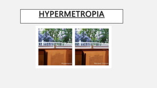
hypermetropia.pptx
- 2. OPTICS OF HYPERMETROPIA • HYPERMETROPIA also known as long-sightedness, Far-sightedness or hyperopia. • In this refractive state of eye where parallel rays of light coming from infinity are focused behind the retina with accommodation being at rest. • Is a TYPE OF AMETROPIA condition of the eye where distant objects can seen (BY USING ACCOMODATION) but near objects appear blurred. • Thus the posterior focal point (LONG FOCAL LENGTH) is behind the retina instead of on, the retina wall therefore receives a blurred image. • Minor hypermetropia (UPTO +1d ) in young patients is usually corrected by their accommodation, without any defects in vision. • But, due to this accommodative effort for distant vision, people may complain of asthenopic symptoms during prolonged reading.
- 3. • FAR …Can see up to 1 Diopter error…With accommodation (hence used the term FAR- SIGHTEDNESS) • NEAR… Not seen • WHEN ACCOMMODATION AT REST CANNOT SEE EITHER
- 4. AGE AND HYPERMETROPIA • At birth, the eyeball is relatively short, having +2 to +3 hypermetropia. • This is gradually reduced until by the age of 5-7 years, the eye is emmetropic and remains so till age of about 50years. • After this there is a tendency to develop hypermetropia again, which gradually inceases until the extreme of life by which the eye has the same =2 to +3 with which it started-SENILE HYPERMETROPIA is due to changed in the crystalline lens. • Far-sightedness is often present from birth, but children have a very flexible eye lens, which helps to compensate.
- 5. AETIOLOGICALLY, CAUSES OF HYPERMETROPIA CAN BE CLASSIFIED AS: 1.AXIAL HYPERMETROPIA: --COMMON -- WHEN THE AXIAL LENGTH OF EYEBALL IS TOO SHORT. --ABOUT 1 MM DECREASE IN AXIAL LENGTH(AP DIAMETER) A BOUT 3D OF HYPERMETROPIA --MICROPHTHALMOS AND NANOPHTHALMOS 2.CURVATURAL HYPERMETROPIA: --OCCUR WHEN CURVATURE OF LENS OR CORNEA IS FLATTER THAN NORMAL. --ABOUT 1 MM INCREASE IN RADIUS OF CURVATURE RESULTS IN 6 DIOPTERS OF HYPERMETROPIA. --CORNEA IS FLATTER IN MICROCORNEA AND CORNEA PLANA. --CONSECUTIVE HYPERMETROPIA-DUE TO SURGICALLY OVER CORRECTED MYOPIA AETIOLOGICAL CLASSIFICATION / CAUSES
- 6. 3.INDEX HYPERMETROPIA: --AGE RELATED CHANGES IN REFRACTIVE INDEX (CORTICAL SCLEROSIS, DIABETES). -- OCCASIONALLY, MILD HYPERMETROPIC SHIFT MAY BE SEEN IN ASSOCIATION WITH CORTICAL OR SUBCAPSULAR CATARACT ALSO. 4.POSITIONAL HYPERMETROPIA: --OCCUR DUE TO POSTERIOR DISLOCATION OF LENS OR I OL. --IT MAY OCCUR DUE TO TRAUMA. 7.ABSENCE OF CRYSTRALLINE LENS: CONGENITAL OR ACQUIRED APHAKIA CAUSE HIGH DEGREE HYPERMETROPIA*(+10 TO +12). IN RARE INSTANCES HYPEROPIA CAN BE DUE TO DIABETES, AND PROBLEMS WITH THE BLOOD VESSELS IN THE RETINA.
- 7. CLINICAL CLASSIFICATION Hyperopia is typically classified according to clinical appearance, its SEVERITY, OR HOW IT RELATES TO THE EYE'S ACCOMMODATIVE STATUS.There are three clinical categories of hyperopia. 1. PHYSIOLOGICAL / SIMPLE / DEVELOPMENTAL • The most common form of hypermetropia • Is caused by normal biological variations in the development of eyeball • Occurs naturally due to biological diversity 2. PATHOLOGICAL /NON-PHYSIOLOGICAL HYPEROPIA CONGENITAL- • Micropthalmos • Nanopthalmos • Microcornea • Congenital posterior dislocation of lens • Congenital aphakia
- 8. ACQUIRED- • Senile (index, Curvature), • positional, • aphakia, • CONSECUTIVE HYPERMETROPIA: occur due to surgical over correction of myopia or surgical under correction in cataract surgery. • Acquired axial (retinal detachment,CSR, orbital tumor) • Acquired curvature( inflammation,trauma) • Pseudophakic hypermetropia 3.FUNCTIONAL HYPEROPIA: • Caused by paralysis that interferes eye's ability to accommodate as seen in internal ophthalmoplegia • CN III palsy etc.
- 9. CLINICAL GRADING / CLASSIFICATION ACCORDING TO SEVERITY AMERICAN OPTOMETRIC ASSOCIATION (AOA) has defined three grades categories according to severity: • LOW: Refractive error less than or equal to +2.00 diopters (D). • MODERATE: Refractive error between +2.00 D up to +5.00 D. • HIGH: Refractive error greater than +5.00 D.
- 10. COMPONENTS OF HYPERMETROPIA Accommodation has significant role in hyperopia. Considering accommodative status, hyperopia can be classified as: • LATENT HYPEROPIA: It is the amount of hyperopia normally corrected by inherent tone of ciliary muscle (approximately +1 diopter). • MANIFEST HYPEROPIA: It is the amount of hyperopia not corrected by ciliary tone. Manifest hyperopia is further classified into two, facultative and absolute. • Facultative hyperopia: It is the part of hyperopia corrected by patient's accommodation. • Absolute hyperopia: It is the residual part of hyperopia which causes blurring of vision for distance. • TOTAL HYPERMETROPIA: It is the total amount of hyperopia which is obtained after complete relaxation of accommodation using cycloplegics like atropine. • So, TOTAL HYPEROPIA= LATENT HYPEROPIA + MANIFEST HYPEROPIA (FACULTATIVE + ABSOLUTE)
- 11. CLINICAL FEATURES-SYMPTOMS • In young patients, mild hypermetropia may not produce any symptoms. • The signs and symptoms of far-sightedness include blurry vision, frontal or fronto temporal headaches, eye strain, tiredness of eyes etc. • The common symptom is eye strain. Difficulty seeing with both eyes (binocular vision) may occur, as well as difficulty with depth perception. • The asthenopic symptoms and near blur are usually seen after close work, especially in the evening or night.
- 12. CLINICAL FEATURES- SIGNS 1. Size of eyeball may appear small as a whole especially in high hypermetropia 2. Cornea may be slightly smaller than the normal 3. Anterior chamber is comparitvely shallow 4. Retinoscopy and Auto refractrometry reveals hypermetropic refractive errors 5. A- Scan Ultrasonography(biometry) may reveal a short antero posterior length of the eyeball in Axial hypermetropia
- 13. 6. Fundus Examination reveals a small optic disc which may look more vascular with ill- defined margins and even may look more vascular with ill-defined margins and even may stimulate PAPILLITIS (though there is no swelling of the disc and so it is called PSEUDOPAPILLITIS). The retina as whole may shine due to greater brilliance of light reflections( SHOT SILK APPEARANCE-because retina is nearer to examiner).
- 14. DIAGNOSIS • A diagnosis of far-sightedness is made by utilizing either a RETINOSCOPE or an AUTOMATED REFRACTOR-objective refraction; or trial lenses in a trial frame or a PHOROPTER to obtain a subjective examination. • Ancillary tests for abnormal structures and physiology can be made via a SLIT LAMP TEST, which examines the CORNEA, CONJUNCTIVA, ANTERIOR CHAMBER, AND IRIS. • In severe cases of hyperopia from birth, the brain has difficulty in merging the images that each individual eye sees. This is because the images the brain receives from each eye are always blurred. A child with severe hyperopia can never see objects in detail. If the brain never learns to see objects in detail, then there is a high chance of one eye becoming dominant. The result is that the brain will block the impulses of the non- dominant eye. In contrast, the child with myopia can see objects close to the eye in detail and does learn at an early age to see objects in detail.
- 15. COMPLICATIONS • Recurrent Styes, Blephritis and chalazions. • Esodeviation a/k/a accommodative convergent squint. • Amblyopia -Anisotropic Amblyopia-due to unilateral refractive errors -Strabismic Amblyopia-due to squint -Ametropic Amblyopia-due to bilateral high hypermetropia • At young age, severe far-sightedness can cause the child to have double vision as a result of "over-focusing". • Primary angle closure glaucoma-hypermetropic patients with short axial length are at higher risk of dedeveloping so, routine gonioscopy and glaucoma evaluation is recommended for all hypermetropic adults.
- 16. TREATMENT • CORRECTIVE LENSES Eyeglasses or contact lenses. Eyeglasses used to correct far- sightedness have convex lenses (Converging lense) • How to identify a convex lens? 1. Thick at centre 2. Magnifies the object 3. Movement of image opposite direction to Movement of lense
- 17. PRINCIPLES OF HYPERMETROPIA CORRECTION 1. GENERAL RULES • --IMPORTANCE OF COMPLETE CYCLOPLEGIC EXAMINATION • --DO YOU PRESCRIBED ERROR < 1D only if patient is symptomatic • --SPERICAL CORRECTION should be given comfortably acceptable • --ASTIGMATISM should be fully corrected 2. FOR CHILDREN • -- <4yrs OF AGE- usually accept full cyclopegic measurements,school age reduce 1/3 • -- >4 yes of age (school Going) undercorrect and prescribe then gradually increase at interval of 6 months till accepts manifest hypermetropia. • -- IN EXOTROPHIA should be undercorrected by 1-2 Diopter • -- IN ACCOMMODATIVE CONVERGENT SQUINT– Full correction– muscle relaxed– eyes straight • --IN AMBLYOPIA—Full correction • --FOLLOW UP EVERY 6 MONTHS
- 18. AMERICAN ASSOCIATION OF OPHTHALMOLOGY -- IN CHILDREN ISOMETROPIA 0 – 1 years 1- 2 years 2 -3 years HYPERMETROPIA > Or = + 6.00 > Or = +5.00 > Or = +4.50 HYPERMETROPIA + ESOTROPIA > Or = +2.00 > Or = +2.00 > Or = +1.50 ANISOMETROPIA 0 – 1 years 1- 2 years 2 -3 years HYPERMETROPIA > Or = +2.50 > Or = +2.00 > Or = +1.50
- 19. • SURGERY There are also surgical treatments for far-sightedness as explained LASER PROCEDURES IOL IMPLANTATION NON LASER PROCEDURES PHOTOREFRACTIVE KERATECTOMY APHAKIA CORRECTION CONDUCTIVE KERATOPLASTY(CK) LASER ASSISTED IN SITU KERATOMILEUSIS (LASIK) REFRACTIVE LENS EXCHANGE(RLE) AUTOMATED LAMELLAR KERATOPLASTY(AKL) Epi-LASIK PHAKIC IOL KERATOPHAKIA LASER THERMAL KERATOPLASTY (LTK) EPIKERATOPHAKIA
- 20. LASER PROCEDURES 1.PHOTOREFRACTIVE KERATECTOMY (PRK): This is a refractive technique that is done by removal of a minimal amount of the corneal surface. Hyperopic PRK has many complications like regression effect, astigmatism due to epithelial healing, and corneal haze.Post operative epithelial healing time is also more for PRK. .
- 21. • 2.LASER ASSISTED IN SITU KERATOMILEUSIS (LASIK): Laser eye surgery to reshape the cornea, so that glasses or contactlenses are no longer needed. Excimer laser LASIK can correct hypermetropia up to +6 diopters. LASIK is contraindicated in patients with lupus and rheumatoid arthritis
- 22. 3.LASER EPITHELIAL KERATOMILEUSIS (LASEK): Resembles PRK, but uses alcohol to loosen the corneal surface. 4.EPI-LASIK : is also used to correct hyperopia. In this procedure, use of epikeratome eliminates the use of alcohol.
- 23. • 5.LASER THERMAL KERATOPLASTY (LTK): is a laser based non-destructive refractive procedure used to correct hyperopia and presbyopia. It uses Thallium-Holmium- Chromium (THC): YAG laser.
- 24. IOL IMPLANTATION 1.APHAKIA CORRECTION: High degree hypermetropia due to absence of lens (aphakia) is best corrected using intraocular lens implantation. 2.REFRACTIVE LENS EXCHANGE (RLE): A variation of cataract surgery where the natural crystalline lens is replaced with an artificial intraocular lens; the difference is the existence of abnormal ocular anatomy which causes a high refractive error. 3.PHAKIC INTRAOCULAR LENS: Phakic IOL are lenses that implanted inside eye without removing the the normal crystalline lens. Phakic IOLs can be used to correct hypermetropia up to +20 diopters.
- 25. NON LASER PROCEDURES 1.CONDUCTIVE KERATOPLASTY (CK): is a non laser refractive procedure used to correct presbyopia and low hypermetropia (+0.75D to +3.25D) with or without astigmatism (up to 0.75D). It uses radiofrequency energy to heat and shrink corneal collagen tissue. CK is contraindicated in pregnant/breastfeeding women, central corneal dystrophies and scarring, history of herpetic keratitis, type 1 diabetes etc. 2.AUTOMATED LAMELLAR KERATOPLASTY (ALK): Hyperopic automated lamellar keratoplasty (H-ALK) and Homoplastic ALK are ALK procedures that corrects low to moderate hyperopia. Poor predictability and the risk of complications limits usefulness of these procedures. 3.KERATOPHAKIA AND EPI-KERATOPHAKIA: are another two non laser surgical procedures used to correct hypermetropia. Keratophakia is a surgical technique developed by Barraquer for treating high hypermetropia and aphakia. Poor predictability and induced irregular astigmatism are complications of these procedures.