

Empfohlen
Weitere ähnliche Inhalte
Was ist angesagt?
Was ist angesagt? (20)
Andere mochten auch
Andere mochten auch (20)
Ähnlich wie Hypertrophic cardiomyopathy
Ähnlich wie Hypertrophic cardiomyopathy (20)
Kürzlich hochgeladen
Kürzlich hochgeladen (20)
Hypertrophic cardiomyopathy
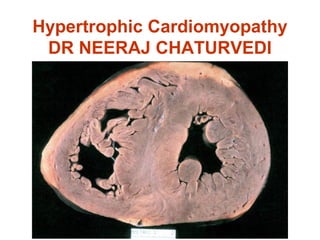
- 1. Hypertrophic Cardiomyopathy DR NEERAJ CHATURVEDI
- 2. Hypertrophic Cardiomyopathy • First described by the French and Germans around 1900 • uncommon with occurrence of 0.02 to 0.2% • a hypertrophied and non-dilated left ventricle in the absence of another disease • small LV cavity, asymmetrical septal hypertrophy (ASH), systolic anterior motion of the mitral valve leaflet (SAM)
- 3. Definition: WHO: left and/or right ventricular hypertrophy, usually asymmetric and involves the interventricular septum.
- 4. Familial HCM • First reported by Seidman et al in 1989 • occurs as autosomal dominant in 50% • 11 different genes with over 400 mutations – chromosome 14 (myosin) – chromosome 1 (troponin T) – chromosome 15 (tropomyosin) – chromosome 11 (myosin binding protein-c)
- 5. Genetic abnormality: • Autosomal dominant. • Mutations in genes for cardiac sarcomeric proteins • Recently,mutations in nonsarcomeric protein mutations also discovered (LAMP-2,PRKAG2) • ß-myosin heavy chain gene on chromosome 14.
- 7. Pathophysiology • Systole – dynamic outflow tract gradient • Diastole – impaired diastolic filling, ↑ filling pressure • Myocardial ischemia ↑ muscle mass, filling pressure, O2 demand ↓ vasodilator reserve, capillary density – abnormal intramural coronary arteries – systolic compression of arteries
- 8. • Left ventricular outflow tract gradient • ↑ with decreased preload, decreased afterload, or increased contractility. • Venturi effect: anterior mitral valve leaflets & chordae sucked into outflow tract → ↑ obstruction, eccentric jet of MR in mid-late systole.
- 12. Natural history of HCM • Mortality: 1 %/year(Higher2% in children) • Clinical course variable • Mostly asymptomatic • SCD • Progressive symptoms of heart failure • End-stage phase • Arrythmias(-AF most common) and complications(stroke,heart failure)
- 16. Risk Factors for SCD • Aborted sudden cardiac death • Sustained VT • Family history of SCD • Recurrent syncope in the young • Nonsustained VT (Holter Monitoring) • Extreme LVH(>30 mm) • Hypotensive response to exercise • End-stage phase • LV apical aneurysm
- 18. Variants of HCM: Most common location: subaortic , septal, and ant. wall. • Asymmetric hypertrophy (septum and ant. wall): 70 %. • Basal septal hypertrophy: 15- 20 %. • Concentric LVH: 8-10 %. • Apical or lateral wall: < 2 % (25 % in Japan/Asia): characteristic giant T-wave inversion laterally & spade- like left ventricular cavity: more benign.
- 26. Histology: • Myocardial fiber disarray, endocardial plaques. • Abnormal relaxation and diversely oriented myocardial fibers. • Intimal hyperplasia of intramural coronary arteries, endothelial dysfunction, myocardial perfusion defects.
- 29. EKG:
- 32. Doppler Echocardiocraphy: • Typical appearance: late-peaking signal “dagger-shaped” • Obstructive or non-obstructive • Distinguish MR and intra-cavitary obstruction
- 35. Cardiac cath: • Not necessary-replaced by echocardiography • Indicated -when planning therapy(e.g.,in severe mitral regurgitation) - excluding coronary atherosclerosis in older patients with chest pain.
- 38. Brockenbrough response • ↑ LV systolic pressure • ↓ Ao systolic pressure • ↑ gradient between LV & Ao Post PVC
- 40. HCM vs Aortic Stenosis HCM Fixed Obstruction carotid pulse spike and dome parvus murmur radiate to carotids ↑ valsalva, standing ↓ squatting, handgrip ↓ passive leg elevation systolic thrill 4th left ics 2nd right ics systolic click absent present
- 41. Other Causes of Hypertrophy • Clinical mimics – glycogen storage, infants of diabetic mothers, amyloid • Genetic – Noonan’s, Friedreich’s ataxia, Familial restrictive cardiomyopathy with disarray • Exaggerated physiologic response – Afro-Caribbean hypertension, old age hypertrophy, athlete’s heart
- 42. HCM vs Athlete’s Heart HCM Athlete + Unusual pattern of LVH + - LV cavity <45 mm + - LV cavity>55 mm _ + LA enlargement + - Bizarre ECG paterns + - Abnormal LV filling + - Female gender + - ↓thickness with deconditioning - + Family history of HCM + - Circulation 1995; 91:1596
- 44. Management • All first degree relatives: screening… echocardiography/genetic counseling • Avoid competitive athletics • Prophylactic antibiotics before medical & dental procedures • Holter x 48 hours
- 45. • β- Blockers: Propranolol 200-400 mg/d (large doses)/ Selective β- B lose selectivity at high doses: Slow HR → longer diastolic filling time → ↓ myocardial O2 consumption → ↓ myocardial ischemia & LVOT obstruction improvement in angina,exercise tolerance and syncope in 80%--sustained symptomatic improvement in 40%
- 46. Calcium channel blockers • ↓inotropy, ↓ chronotropy • improvement of diastolic relaxation. • improve exercise tolerance by 20%-30%. • Death from pulmonary edema reported in presence of severe outflow tract gradients and high diastolic filling pressures.
- 47. • Disopyramide: class I antiarrhythmic Probably acts by – increasing SVR – Negative inotropic effect – Improve balance between O2 demand and supply. • Disadvantages – Vagolytic effect – Proarrhythmic potential – Facilitate AV conduction (Dangerous in AF) – Lack of long-term sustained benefits
- 48. Strategies for Drug Therapy BJ Maron (NEJM) • Drug selection is not standardized • Based on experience and preference of individual physician • Most prefer β-blocker over verapamil as initial drug. If the patient has angina predominantly verapamil if DOE . • Some prefer verapamil - without LVOTO β-blocker/dispyramide - with LVOTO • No evidence to suggest – Verapamil + β-blocker - better than alone – Disopyramide + β-blocker - better than alone • In β-blocker non-responders verapamil should
- 49. Non-responders to Medical therapy ??? 1- Surgery (Myotomy/Myectomy) +/- MVR 2- ICD 3- DDD pacemaker 4- NSRT (alcohol septal ablation)
- 50. 1- Surgery: Septal myotomy/myectomy: • Patients < 40 years: mortality < 1 % • Patients > 65 years: mortality 10-15 % • Survival better than medically treated patients • Should be considered in: resting gradient > 50 mmHg, or refractory to medical Rx. • Young patients, particularly those with severe disease • Additional structural abnormalities affecting the mitral valve or coronary arteries. • Complications: heart block,vsd,aortic incompetence
- 51. COMPLICATIONS • Perioperative Mortality (1-5%) • Older age + NYHA class IV high risk • VSD - 3% • CHB and PPI (5%) • CVA 1-2% • LBBB-40% • MVR if done its associated complications Contd…
- 54. 2- ICD: • Previous sudden death • High risk of sudden death
- 55. 3- DDD pacemaker Substantial ↓ gradient(~ 50 %)
- 56. Effect of DDD pacemaker in HCM
- 58. Potential Mechanisms of benefit of Pacing in HCM: • RV apical pacing & maintenance of AV synchrony → abnormal pattern of septal contraction → ↓ early systolic bulging of hypertrophic subaortic septum in LVOT & ↓ Venturi forces that produce SAM. • ↑ LVOT width during systole • ↓ systolic hypercontractility: ↑ end-systolic volume → ↓ intraventricular pressure gradients & myocardial work
- 60. 4- Alcohol septal ablation (NSRT) • Controlled myocardial infarction of the basal ventricular septum to ↓ gradient. • First septal artery occluded with a balloon catheter and ETOH injected distally
- 61. Non-surgical Myocardial Reduction TASH, PTSMA, NSMR Idea borrowed from surgical myectomy (1980s) Temporary balloon occlusion of 1st large septal branch decreased LVOTO 1st TASH reported in 1995 by V. Sigwart • Modification technique – From 3-5µl 96% Alcohol --> 2ml/branch – Echocardiographic identification of target – Regularly placing a TPI to avoid intraprocedure card arrest – Use of forceful injection into target vessel 1-2cm2 radiographic contrast – Adequate balloon sizing (short length) should be used and kept inflated 5 min or longer after left alcohol.
- 62. NSRT (Non Surgical Septal Reduction Therapy) The most appropriate candidates for NSRT should meet all of the following criteria : - HCM with severe symptoms of heart failure (NYHA class III to IV) despite adequate tolerated drug therapy - An LVOT gradient 50 mmHg at rest or after exercise or >30 mmHg at rest or 60 mmHg under stress - Basal septal thickness 18 mm - NYHA class II heart failure with a resting LVOTgradient >50 mmHg or >30 mmHg at rest and 100 mmHg with stress . - Elderly or comorbidities that may increase the risk of surgical correction.
- 63. SEPTAL REDUCTION (SURGICAL / NON SURGICAL) • Qin et al (JACC 2001) • CHB 21% in PTSMA VI 7.7% myemectomy Conclusion • PTSMA / myemectomy decrease LVOTO and improve symptoms • CHB more common with PTSMA • PTSMA is less invasive shorter hospital stay • PTSMS should be the initial approach particularly in elderly and high risk • Failed PTSMA should be tried with myemectomy (1005 require PPI)
- 64. Recommendations for Athletic Activity • Avoid most competitive sports (whether or not symptoms and/or outflow gradient are present)
- 65. Recommendations for Athletic Activity • Low-risk older patients (>30 yrs) may participate in athletic activity if all of the following are absent – ventricular tachycardia on Holter monitoring – family history of sudden death due to HCM – history of syncope or episode of impaired consciousness – severe hemdynamic abnormalities, gradient ≥50 mmHg – exercise induced hypotension – moderate or sever mitral regurgitation – enlarged left atrium (≥50 mm) – paroxysmal atrial fibrillation – abnormal myocardial perfusion
- 67. TREATMENT OF ASYMPTOMATIC HCM • 75% Asymptomatic with varrying hypertrophy of LV • SCD is not correlated with the symptoms • No evidence that β - blocker / verapamil protects from SCD in HCM • No data to suggest β - blocker / verapamil prophylactically delay the progression and improve prognosis • Prospective studies are difficult as the study population is small and symptoms / SCD are infrequent • Prophylactic drug therapy to prevent progression is not JUSTIFIED • ? Asymptomatic young indicidual with marked hypertrophy and LVOTO can be tried with β -blocker / verapamil.
- 68. GENE THERAPY • HCM is the subject of intense molecular genetic research over past decade • Genetic markers for prognosis has been identified • Experimental animal models of HCM studied • Little progress in gene therapy for HCM which appears attractive
- 69. S • 25% of HCM require treatment • 15-20% are well controlled with drugs • No data conclusively shows advantage of one drug over other β- blocker/verapamil • β-blocker / verapamil should be selected depending upon the patients symptoms. • Use of Disopyramide is limited mainly because of its side effects and lack of sustained benefit. • 5% of HCM are drug refractory and require non-pharmacological approach • Surgical septal reduction / pacemaker / PTSMA all look attractive • With contrast ECHO PTSMA may be the choice. • Mid cavity obstruction pacemaker / PTSMA can be tried. • ICD has definite role in secondary prevention and should be tried in very high risk subset for primary prevention.
- 81. A 48-year-old female presents with increasing dyspnea on effort. She has been placed on large doses of beta-blockers, as her local physician has made a diagnosis of hypertrophic cardiomyopathy. Because of continued disabling symptoms and the impression of a new murmur of mitral regurgitation, she is referred to you. On your examination, the BP is 130/80 in both arms, jugular venous pressure is normal, and there is a bisferient arterial pulse. A systolic murmur is along the lower left sternal border, but extends to the apex where the murmur does seem of longer duration. The murmur increases with a Valsalva maneuver. After a premature ventricular contraction, the outflow tract gradient increases to 120 mm Hg. The chest X-ray is normal and the ECG shows severe LBBB. Echo studies reveal severe mitral regurgitation with an LV outflow gradient of 50 mm Hg. Septal thickness is 35 mm, with free wall LV thickness of 20. Systolic anterior motion of the mitral valve is noted. There is no mitral calcification or intrinsic mitral valve disease on echo. • Which one of the following statements is true regarding treatment options? A. Percutaneous septal ablation is the preferred treatment. B. Mitral valve replacement is required. C. Continue medical therapy, as the resting outflow gradient is not severe enough to consider surgery. D. Surgical myectomy alone may provide relief of symptoms, outflow obstruction, and mitral regurgitation. E. Dual-chamber pacing is the preferred therapy.
- 82. • The correct answer is D. • In this patient with classic findings of hypertrophic cardiomyopathy and disabling symptoms, the preferred therapy is surgical myectomy. Surgical myectomy performed by surgeons experienced in this operation can be done with low operative mortality and excellent relief of symptoms. In many of the patients who have mitral regurgitation as a consequence of the disturbed hemodynamics, the mitral regurgitation is also relieved by successful surgical myectomy. The resting outflow gradient is moderate and may vary at different times, but the post-extrasystolic potentiation observation confirms severe outflow tract obstruction. While there is increasing application of percutaneous septal ablation, several features in this patient should raise caution regarding this choice. The presence of LBBB makes complete heart block more likely, with septal ablation and the need for permanent pacing. Furthermore, her youth is an important consideration, as the long-term potential of a large septal scar as a substrate for lethal arrhythmias is uncertain after percutaneous septal ablation. In contrast, long-term results from a large surgical experience demonstrate not only good relief of symptoms, but some comparisons, which suggest an enhanced survival. Dual-chamber pacing has been demonstrated to be of benefit in some patients, but controlled studies have failed to document its consistent effectiveness.
- 83. A 29-year-old man, who was the defending city tennis champion, had won the title match, walked off the court, and, as he was being congratulated, collapsed. He was found to be pulseless and not breathing by an observer, who started CPR. A defibrillator arrived a few minutes later. After three shocks, he was defibrillated and resumed a slow sinus rhythm. He was taken to a local hospital and was alive on arrival, but VF occurred shortly thereafter. Further efforts at defibrillation and resuscitation ultimately failed. An uncle and an older brother had died suddenly in their early 30s. The family did not know the cause, but were told it was probably a heart attack in each instance. His wife gave permission for autopsy. • Which of the following would be most likely to be found at autopsy? A. Calcified and stenotic aortic valve. B. Gross disorganization of the cardiac muscle bundles. C. Multivessel CAD. D. Myxomatous degeneration of the mitral valve leaflets. E. Left main coronary artery arising from the right aortic sinus.
- 84. • The correct answer is B. • Calcification of the aortic cusps is the most common cause of aortic stenosis in adults, and is the result of years of mechanical stress on the valve. It is most often found in persons older that 65 years of age. Aortic stenosis accounts for an estimated 5% of the cases of sudden cardiac death (SCD). These are usually symptomatic. It does not contribute significantly to the causes of SCD in the young adult, however. In athletes 35 years of age or younger, hypertrophic cardiomyopathy is the most frequent finding at autopsy (36%). Patients are often asymptomatic, while the presence of symptoms and a family history of sudden death are markers. The characteristic microscopic pattern is disarray of myocytes in a whorled pattern and disorganization of the larger muscle bundles. Gross septal hypertrophy occurs in varying degrees, sometimes with impingement on the anterior mitral leaflet, resulting in obstruction of the LV outflow tract. Congenital coronary artery anomalies account for 19% of SCD in those 35 years and younger. Alternatively, atherosclerotic CAD is found in a reported 80% of those older than 35 years. Most CAD in the over-35-year-old victim of SCD is multivessel. In athletes of that age group, it is the cause in a reported 80%, while in those 35 years and under, only 10%. Myxomatous degeneration of the mitral valve leaflets and mitral valve prolapse raise controversy regarding the risk of sudden death. The prevalence of mitral prolapse is so great across the population that it stands to reason it will often be noted at examination. The overall incidence in the population as a whole is estimated as up to 5%. Another factor clouding the picture is the question of primary conduction system disease, a diagnosis often difficult at autopsy. Congenital coronary artery anomalies are among the leading causes of SCD in the athlete 35 years old or less, but hypertrophic cardiomyopathy is much more frequent (36% vs. 19%).
- 85. • A 78-year-old man, generally healthy and still working, comes for a consultation because he is “just not feeling well.” He is experiencing episodes of “weakness” associated with dyspnea on an irregular basis and in an unpredictable fashion. They may last from a few seconds to several minutes. Detailed history, after a myriad of questions, allows you to conclude that he probably does feel dizzy, but has never had anything suggestive of syncope, that the longer episodes may be associated with diaphoresis, and for the last 2-3 years, he has had rather typical angina on very severe effort. He hasn’t had more than 8-10 episodes of angina and the angina is distinctly different from the “spells” for which he is now consulting you. He has long been overweight, currently weighs 235 pounds, has been treated for hypertension for many years with “good control,” and has troublesome arthritis in his right shoulder—a complaint quite independent and different from the “spells” and the angina described. On physical examination, he has a BP of 130/70, and a heart rate of 62. You feel the pulse for one minute and detect 2-3 irregularities, which you cannot characterize further. On CV examination, he has no jugular venous hypertension. The point of maximal impulse is not palpable. He has a diminished second sound in the aortic area, together with a grade 3/6 systolic ejection murmur, which is heard into the neck. The murmur is faintly heard at the apex. He had no diastolic murmur and his lungs were clear, good peripheral pulses, and no bruits.
- 86. • His ECG shows LBBB with first-degree heart block. An echocardiogram is obtained, which demonstrates some LV hypertrophy with disproportionate septum versus posterior wall hypertrophy (19 mm vs. 11 mm). His EF is more than 65%. There is no systolic anterior motion of the mitral valve and the aortic valve is calcified with restricted movement and a Doppler velocity of 3 m/sec. Ambulatory ECG monitoring (Holter) was undertaken and the illustrative strips shown are representative of multiple episodes of both ectopic and conduction abnormality activity. There were no diary entries indicating symptoms during the 24-hour monitoring period.
- 87. • What would be your next step in managing this elderly patient? A. Place a DDDR pacemaker without further evaluation. B. Proceed to cardiac catheterization to evaluate aortic valve function and coronary artery anatomy. C. Perform an echocardiographic dobutamine study. D. Perform an EP study. E. Follow closely and ask the patient to keep a diary of his symptoms.
- 88. • The correct answer is A. • Many elderly patients have several—or more—important or potentially important CV conditions present, which might explain their symptoms. In this patient, mild aortic stenosis, history of hypertension with striking asymmetrical septal hypertrophy, and diastolic dysfunction and angina pectoris are present, in addition to conduction system disease including Mobitz type II AV block. A variety of ectopic beats with brief runs of VT were recorded without symptoms. The data available do not allow correlation of the symptoms he described with the arrhythmias recorded, but Mobitz type II is an indication for pacemaker placement and such should be placed to learn if his symptoms will be ameliorated. His LV hypertrophy with diastolic dysfunction makes him prone to dyspnea with very modest arrhythmias when he experiences a lack of the “booster pump action” of properly timed atrial systole. Although it can be assumed that he has some coronary disease, his LV hypertrophy could make him particularly prone to angina, which is very infrequent and does not limit him. In reference to coronary arteriography, the study would serve little purpose unless the information is needed to plan management. In this particular patient—and in virtually all people this old—symptoms should be the principal indication for arteriographic study as a prelude to revascularization. Similarly, his aortic valve disease is modest and although little is known about the progression of aortic valve disease in this age group, it can be reasonably assumed that in the absence of heavy calcification (not present in this case), progression will be slow over a period of many—certainly more than five years. An EP study will not resolve the dilemma in this patient. If placement of the pacemaker does not relieve his symptoms, then additional evaluation (almost certainly to include a full cardiac catheterization) should be undertaken.
- 89. • An 80-year-old African-American woman is admitted to the hospital under your care, having been referred for refractory heart failure. She has been hospitalized six times in the last 18 months, having responded fairly effectively to intensive heart failure treatment on each occasion. She gives a history of remote hypertension, but has required no treatment for the last three years. She denies symptoms suggestive of angina pectoris or MI. Until about five years ago, she considers herself to have been very healthy and remained active in family and church activities. Prior to her first episode of heart failure, she weighed about 175 pounds; her current weight is 142 pounds. On physical examination, she is a pleasant woman with a BP of 105/80, and regular heart rate of 110. She is suggestively pale, minimally diaphoretic, and has jugular venous hypertension to 8 or 9 cm with Kussmaul’s sign present. On cardiac examination, she has an equivocally palpable apical impulse, no RV impulse is palpable, and the heart sounds are distant with a probable third heart sound present, but no murmur. There is dullness at both lung bases to percussion and diminished breath sounds with probable underlying rales. The liver is palpable two finger breadths below the costal margin, but is not pulsatile. She has 1-2+ soft pitting edema. An echocardiogram and ECG were obtained during the admission process. The ECG is illustrated. The Echocardiogram showed striking thickening of the LV, marked dilatation of the atriae and classic "diastolic dysfunction" on MV Doppler signal.
- 91. After obtaining routine screening and laboratory studies, what will be your approach to management? A. Perform right and left heart catheterization with coronary arteriography. B. Perform endomyocardial biopsy. C. Anticipate a stable course for a few months by increasing her anti-CHF therapy. D. Perform cardiac MRI. E. Refer to hospice.
- 92. • The correct answer is E. • The course of this patient’s CHF—basically unremitting and poorly responsive to therapy since six hospitalizations have been required in less than two years— together with the striking changes on echocardiography correlated with ECG findings (low voltage and conduction detects) make the diagnosis of “senile” cardiac amyloidosis virtually certain. Although choices listed in A, B, C, and D may be performed, they are unnecessary and unimportant in planning this patient’s management. Distressingly, it can be confidently predicted that she will not stabilize and will not respond to therapy since her myocardium has been replaced by amyloid. The striking disparity between the voltage on the ECG and the thickness of the myocardium indicate an infiltrative process. In another setting, chronic constrictive pericarditis could be considered clinically, but not in this instance. Recent information adds considerable strength to the hypothesis that senile amyloidosis (at least in cases of this nature) may be related to genetic abnormalities since the amyloid deposits in senile amyloidosis have features similar to those in the familial/hereditary form. Amyloid deposits, particularly in the atrium, are present increasingly frequently after the age of 60 and are often an incidental postmortem finding. There is the probability that so-called “senile” amyloidosis has multiple etiologies and combination of genetic predisposition coupled with age-related changes. This is an active and important area of research.
- 93. A 19-year-old woman is referred after her 16-year-old brother died suddenly while playing basketball. Autopsy showed symmetric LV hypertrophy (LVH). She has been aware of the murmur since childhood, but denies any cardiac symptoms, and has always kept up with her peers. There is no other heart disease or sudden deaths in the family. Examination is remarkable for a sustained apical impulse and a mid-systolic murmur at the apex that increases with Valsalva. Her ECG shows borderline LVH. • Of the following, which is the best predictor of sudden death risk in this patient? A. Sudden death in her brother. B. Myofibrillar disarray on biopsy. C. Septal thickness of 1.8 cm. D. Paroxysmal AF on Holter monitoring. E. Resting BP 120/75, and BP at six-minute exercise 95/70.
- 94. • The correct answer is E. • In an asymptomatic patient with hypertrophic cardiomyopathy, many potential predictors of sudden death have been described, but the most widely recognized are marked LVH (>3 cm), failure to raise BP on exercise, resuscitation from sudden death, multiple sudden deaths in the kindred, and (perhaps) nonsustained VT. Biopsy is likely to show no pathology or myofibrillar disarray, but is not helpful in estimating prognosis. The answer here is therefore E.
- 95. A 21-year-old female college student with no prior history of cardiac disease or symptoms presents to the emergency room (in sinus rhythm) with the following history. While running across campus late for class, she suddenly collapsed. Fortuitously, the closest bystander was a physician (from the university hospital) who initiated CPR immediately and summoned assistance. Trained paramedics arrived within five minutes and restored sinus rhythm after three defibrillation shocks. Workup over the next three days showed normal cerebral and neurologic function. Echocardiographic study established (for the first time) the diagnosis of hypertrophic cardiomyopathy with a small left ventricular outflow gradient of 20 mm Hg due to systolic anterior motion of the mitral valve. Ventricular septal hypertrophy was marked (maximal thickness = 32 mm). • What is the most appropriate next step in management? A. Ambulatory (Holter) ECG. B. EP study with aggressive programmed stimulation protocol to induce ventricular arrhythmias. C. ICD implant for secondary prevention. D. Refer for myotomy-myectomy operation.
- 96. • The correct answer is C. • VT/VF may be recurrent in one-third of those HCM patients successfully resuscitated from cardiac arrest. ICD implant is the standard approach for secondary prevention of sudden death.
- 98. A 22-year-old man is referred for evaluation of a heart murmur. He is asymptomatic and offers no pertinent family history. Exam: Normal habitus. Heart rate 76 bpm, BP 116/76 mm Hg, respiratory rate 10/min. Jugular venous pressure 6 cm with normal wave forms. Both carotids upstroke normal. Lungs clear. LV impulse sustained. S1 normal, S2 normal split. No ejection sound. Grade 3/6 mid-peaking systolic murmur and 2nd RICS. Grade 1/6 diastolic decrescendo murmur at left sternal border. Abdomen soft, normal situs. Extremities normal. ECG: Normal sinus rhythm, increased LV voltage. • What is the most likely diagnosis? A. Bicuspid aortic valve. B. Supravalvular aortic stenosis. C. Discrete subvalvular stenosis. D. Hypertrophic obstructive cardiomyopathy.
- 99. • The correct answer is C. • The protocol describes a patient with left ventricular outflow tract obstruction and aortic regurgitation. An ejection sound should be audible in a young patient with bicuspid aortic valve disease. In patients with supravalvular aortic stenosis, the right carotid upstroke is usually more prominent than the left and there is a similar differential between right arm and left arm blood pressures. The murmur of hypertrophic obstructive cardiomyopathy is usually loudest between the lower left sternal border and the apex. Dynamic left ventricular outflow tract obstruction of this type is not accompanied by aortic regurgitation. The lack of an ejection sound as well as the presence of the diastolic murmur help to localize the obstruction to the subvalvular region.
- 100. • A 42-year-old man was admitted to the hospital after being rescued by a bystander who applied an AED device after seeing him collapse at a basketball game immediately after a winning goal was scored at the buzzer. The AED delivered a shock and the patient promptly gained consciousness. The patient had a family history of sudden death of an uncle and an older brother, and reported occasional syncope in the past. The physical examination was unremarkable. An echocardiogram and a cardiac catheterization were normal. The patient’s 12-lead ECG is shown in the figure. The patient refused cardiac EP study and an ICD.
- 101. • What therapy do you recommend? Select the best option from the five listed below. A. Beta-blockade. B. Quinidine. C. Left cervicothoracic sympathectomy. D. Avoidance of emotional excitement. E. Mexiletine.
- 102. • The correct answer is B. • This patient meets diagnostic criteria for Brugada syndrome. The diagnostic findings include symptoms of arrhythmia, family history of sudden death, absence of structural heart disease, and coved ST-segment elevation in two right precordial leads. Though long QT syndrome must be considered in a young patient with sudden death and a positive family history, even in the absence of QT prolongation on 12-lead ECG, this patient shows typical Brugada abnormalities of the ST segment in leads V1 and V2. In general, antiarrhythmic drug therapy is not effective in the Brugada syndrome. Recently, however, investigators have demonstrated that quinidine prevents both inducible and spontaneous VT and VF in these patients. If long-term efficacy of quinidine can be corroborated in larger numbers of patients, it may become an alternative to cardioverter-defibrillator implantation. Quinidine is a sodium channel blocker, and these drugs usually exacerbate the ECG characteristics and the sudden death risk in Brugada syndrome. However, quinidine also blocks the transient outward potassium current, and experimental data (and case reports) suggest this should be antiarrhythmic in Brugada syndrome.
- 103. A 68-year-old woman presents with a six-month history of gradually increasing shortness of breath on exertion, mild orthopnea, and some peripheral edema. There is no history of dizziness or syncope. She does not drink or smoke, and recent total cholesterol was 220. She is on no medications. Physical exam reveals a healthy, overweight woman who is comfortable at rest. Pulse is 82 and regular, BP is 180/90. There are a few crackles at the lung bases. The jugular venous pulse is 10 cm with positive hepatojugular reflux. Carotid pulses are full with a brisk upstroke. The point of maximal impulse is thrusting and well localized in the mid-clavicular line. There is an S4, normal S1 and S2 (which is single), and a grade 1/6 systolic ejection murmur along the left sternal border. The hepatic margin is down 2 cm and there is a 1-2+ peripheral edema. The ECG shows LV hypertrophy with repolarization abnormality. • Which of the following statements is false? A. Resting cardiac output is normal. B. A Valsalva maneuver will increase the murmur. C. On the echo-Doppler tracing of LV diastolic filling, the A wave is greater than the E wave. D. LVEF is normal.
- 104. • The correct answer is B. • This patient presents as a typical individual who has the signs and symptoms of heart failure based on diastolic dysfunction. It has been estimated that approximately one- third of individuals who develop new onset heart failure will do so on the basis of diastolic dysfunction. The usual cause is LV hypertrophy. Thus, diastolic dysfunction will be very common in the elderly who develop systolic hypertension and LV hypertrophy. They do not necessarily have a murmur, but a grade 1/6 systolic ejection murmur along the left sternal border is characteristic of elderly patients. If this patient had hypertrophic cardiomyopathy, a Valsalva maneuver might increase the murmur. However, since a Valsalva tends to decrease cardiac output, that would tend to decrease the murmur in this individual. On the echo-Doppler tracing of LV diastolic filling across the mitral valve, normally the E wave is greater than the A wave. An early change with diastolic dysfunction of the kind seen in this patient is a reduced E wave and a more prominent atrial, or A wave. This reduced E wave is in part due to delayed relaxation so that LV diastolic pressures tend to remain up throughout the early part of diastole. This reduces the filling velocity of the blood coming in from the LA. Patients with diastolic dysfunction frequently have absolutely normal or even hypernormal EFs. Although it is difficult to know how to treat such patients, in individuals who have fluid overload, a diuretic will, in fact, get rid of the excess fluid and will tend to reduce the symptoms of dyspnea.
- 105. A 23-year-old Hispanic male college basketball player collapses during a game. He is found to be in cardiac arrest and the EMT personnel cannot resuscitate him. Before being allowed to play basketball, his screening physical examination showed pectus excavatum and a grade III/VI systolic ejection murmur at the third intercostal space left sternal border. A Doppler echocardiogram showed a velocity drop across the aortic valve of 2 m/sec, LV posterior wall thickness of 1.0 cm, and septal thickness of 1.2 cm. ECG: PR interval 0.11 sec. QRS 0.10 sec. Inverted T wave in V1. • Which one of the following statistically is the most likely problem leading to his death? A. Obstructive coronary atherosclerosis. B. Hypertrophic cardiomyopathy. C. Left coronary artery arises from the anterior sinus of Valsalva. D. Congenital aortic stenosis. E. Accelerated AV conduction with rapid AF.
- 106. • The correct answer is C. • Hypertrophic cardiomyopathy is the most common etiology causing sudden death in young competitive athletes; however, the echo did not support this diagnosis. The second commonest etiology in this circumstance is anomalous origin of the left coronary artery from the anterior sinus of Valsalva. Significant aortic stenosis is not present with only a 16 mm Hg gradient across the aortic valve. Finally, the short PR interval with normal QRS is not the basis for answer E. Besides, the sudden death with Wolff- Parkinson-White is extremely rare in the cited situation. CT image shows the anomalous left coronary artery arising from the anterior sinus of Valsalva and passing posteriorly between the aortic and pulmonary arteries.
- 107. You are asked to consult on a patient hospitalized on the Psychiatry Service. She has been admitted because of severe anxiety and headaches. Neurologic evaluation is negative, with no evidence of an organic basis for her headaches. She is on an ergot alkaloid for headache. She does have a history of shortness of breath and fatigue, most of which is attributed to her emotional status. On examination, her BP is normal, and there is an increase in pulse pressure following a premature ventricular contraction and a grade III systolic murmur with a faint early diastolic sound. Echocardiographic studies document the presence of moderately severe mitral regurgitation (MR) with some thickening of the valve. • What is the pathology that is most likely to have caused the MR? A. Carcinoid heart disease. B. Ergot-induced mitral incompetence. C. Mitral stenosis. D. Hypertrophic obstructive cardiomyopathy with MR. E. Tachycardia-induced cardiomyopathy.
- 108. • The correct answer is B. • It is wise to ask all patients taking medication for headache with findings of valvular disease about the use of drugs that may contain ergot alkaloids. These agents may induce scarring of the cardiac valves.
- 109. A 42-year-old man is referred for evaluation of a systolic murmur. Your exam shows normal carotid pulses, a prominent apical impulse, an early systolic sound, and a grade III/VI mid-systolic murmur at the base. Respiration did not change the character of these auscultatory findings. After an extrasystole, the systolic murmur increased in intensity. Handgrip did not alter the systolic murmur. Valsalva decreased the intensity of the murmur, and it returned to baseline intensity after seven heart beats. • Which one of the following diagnoses is most likely? A. Congenital pulmonic stenosis. B. Innocent murmur. C. Mitral valve prolapse. D. Hypertrophic obstructive cardiomyopathy. E. Bicuspid aortic valve.
- 110. • The correct answer is E. • A systolic murmur that increases in intensity in the beat following an extrasystole is usually due to turbulent flow out of the ventricles. Mitral regurgitation is less likely because this murmur does not change following an extrasystole. The murmur of hypertrophic obstructive cardiomyopathy usually decreases with handgrip exercise. An innocent flow murmur is less likely because of the presence of an early systolic sound and grade III intensity. With pulmonic stenosis, there are characteristic changes in the intensity of the murmur and the ejection sound during respiration. The ejection sound establishes the diagnosis of an abnormal aortic valve, a bicuspid valve being the most common abnormality.
- 111. A 75-year-old Asian man is brought to the emergency department after awakening with acute shortness of breath at 3 a.m. He states that he was well until six weeks ago when he had the onset of 5-10 minutes of chest pressure when walking one flight of stairs, which went away when he stopped. He was told that he had a “faint” heart murmur five years ago and has been told on many occasions that he had normal or even “low” blood pressure. Physical exam: BP is 90/50 mm Hg. Pulse is 110/minute. Respiratory rate is 20/minute. Neck veins are 5 cm. Lungs: bilateral rales 1/2 way up posteriorly. Cardiac: Point of maximal impulse in the fifth intercostal space in the MCL and sustained. S2 is barely audible. There is a grade II/VI systolic ejection murmur at the base and an S3 gallop. On the beat after a premature ventricular contraction, the murmur with the post-extra systolic beat increases in intensity. Lab: Chest X-ray: normal heart size, LA enlargement in the left lateral view. ECG: LV hypertrophy with ST-T wave changes. • Which one of the following is the most likely diagnosis? A. Hypertrophic cardiomyopathy. B. Acute non-STEMI. C. Congestive dilated cardiomyopathy. D. Aortic stenosis. E. Acute myocarditis.
- 112. • The correct answer is D. • Unexplained LV hypertrophy (by ECG and physical examination) in a patient with acute pulmonary edema and a known heart murmur should be considered to have aortic stenosis until it is ruled out. The other diagnoses cause pulmonary edema, but the story is most likely aortic stenosis. Congestive cardiomyopathy is possible, but the LV is not dilated, just hypertrophied. This patient could have had obstructive cardiomyopathy, but that was not given as a possibility. The explanation for the increase in the intensity of the murmur in the beat after an extra systole is that with the pause after premature ventricular contraction, there is unusual time for diastolic LV filling, and with the Frank-Starling mechanism, the next ventricular contraction ejects a larger stroke volume, all of which is ejected stenotic aortic valve.