Anatomy of Eyelids & Its Clinical Correlations
•Download as PPTX, PDF•
54 likes•16,710 views

EYELIDS: Layers,Nerve Supply,Vascular Supply & Clinical Correlations

Recommended
More Related Content
What's hot
What's hot (20)
Viewers also liked
Viewers also liked (19)
Similar to Anatomy of Eyelids & Its Clinical Correlations
Similar to Anatomy of Eyelids & Its Clinical Correlations (20)
More from Sarmila Acharya
Recently uploaded
Recently uploaded (20)
Anatomy of Eyelids & Its Clinical Correlations
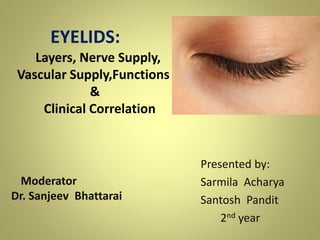
- 1. Moderator Dr. Sanjeev Bhattarai Presented by: Sarmila Acharya Santosh Pandit 2nd year EYELIDS: Layers, Nerve Supply, Vascular Supply,Functions & Clinical Correlation
- 2. Presentation Layout • Introduction • Embryology • Anatomy Layers of Eyelids Glands of Eyelids • Functions • Nerve Supply • Vascular Supply • Drainage System • Clinical Correlations
- 3. Introduction • An eyelid is a mobile, flexible, multilamellar structure that covers globe anteriorly. • Assist in distribution of tears over the anterior surface of the eyeball. • Provide protection from excessive light, desiccation and air borne foreign matter.
- 4. Embryology • Derived from surface ectoderm • Starts as a proliferation in the region future outer canthus at 4-5 weeks gestation • Mesodermal mesencyhme infiltration during 2nd month • Fusion at 10 weeks gestation • Orbicularis condensation at 12 weeks gestation • Eyelid adhesions break down during 5-6th month
- 6. The Anatomy Of The Eyelids: • Extention: – eyebrow superiorly – cheek inferiorly • Eyelid folds/sulcus: Upper eyelid: orbital part , tarsal part Formed by the insertion of the aponeurotic fibers of LPS into the skin Lower eyelid - less obvious Formed by few connections between the skin and the orbicularis oculi muscle. (with age, Nasojugal sulcus & Malar sulcus are formed)
- 7. • Canthi – Eyelids meet at medial and lateral canthi • Medial canthus: Rounded, -separated from globe by Lacus lacrimalis (tear lake) • Lateral canthus: - 5-7mm from lateral orbital margin, -60 deg with eyes wide open, -30-40 deg with eyes open in a normal way -About 2 mm above the medial canthus Epicanthus –dermal fold across the medial canthus
- 8. • Eyelid Margins - 2mm wide, round anterior border & sharp posterior border
- 9. contd… • Eyelid margin is divided into 2 portion by lacrimal papilla. 1) Lacrimal portion: -medial portion of the eyelid margin -extending from the punctum medially to medial canthal angle. -Round & devoid of lashes and glands 2) Ciliary portion: -From punctum to lateral canthal angle -Contains lashes at anterior border & sharp posterior border
- 10. contd… Gray line -Correspond histologicaly to the muscle of Riolan - relatively avascular area - Gray - Eyelashes emerge anteriorly - Posteriorly opening of meibomian gland (just anterior to mucocutaneous junction)
- 11. cont… • Interpalpebral Fissure: - the exposed zone between the upper and lower eyelid - 8 to 11 mm vertically - 27 to 30 mm horizontally - Width of the palpebral fissure is determined by the level of tonic activity in the levator palpebrae superioris and the sympathetically innervated Muller’s Muscle • Position: In primary position – Upper eyelid margin is at 1.5-2mm below the superior corneal limbus – Lower eyelid margin is at inferior corneal limbus
- 12. • Eyelashes – Arranged in 2 to 3 rows. – Upper lid: 100-150 in number and directed upward, forward and backward – Lower lid : 50-75 in number and directed forward, downwards and backward – Taper throughout the length to end in a fine sharp point – Life span : 3 to 4 months (cilia have no erector muscle)
- 13. Layers Of The Eyelids: (From front to back) • Skin • Subcutaneous Areolar Tissue • Layers of Striated Muscles • Submuscular Areolar Tissue • Fibrous Layer • Non Striated Muscles Fibres • Conjunctiva
- 14. 1. Skin: • Palpebral skin is thinnest in body (<1 mm) • Elastic & folds easily contributing to speed of mobility of upper eyelid • Nasal skin: – Smoother and more oily – Few rudimentary hairs – Many unicellular sebaceous glands (hence Xanthelasma develops on the nasal side) • Lateral skin: – Numerous sweat gland
- 15. Contd… • Epidermis – Consists of 6-7 layers of stratified squamous epithelium – Unicellular sebaceous glands & sweat glands • Dermis – Dense connective tissue – Rich network of elastic fibers, blood vessels, lymphatics & nerves
- 16. 2.Subcutaneous Areolar Tissue: • Loose connective tissue arrangement • No fat • Applied anatomy: – Fluid from oedema or haemorrhage rapidly engorges into the loose subcutaneous eyelid tissue & produce swelling of eyelids
- 17. 3. Layers Of Striated Muscles: • Consists of –Orbicularis oculi (forms thin oval sheet over eyelid) –Upper eyelid also contains LPS
- 18. The Orbicularis Oculi Muscle: • Complex striated muscle sheet • Divided anatomically into; -Orbital -Palpebral Pretarsal Preseptal
- 19. a) Orbital Orbicularis: • Origin : - Most peripheral fibers which arise from anterior part of the medial palpebral ligament & adjacent bones viz. o Upper orbital margin medial to supraorbital notch o Maxillary process of frontal bone o Frontal process of maxilla o Lower orbital margin medial to the infraorbital foramen
- 20. Orbital Orbicularis Cont: • Muscles fibres sweeps superiorly & inferiorly, covering the orbital margins in the form of ellipse & meet at the lateral palpebral raphe Superior fibres called Musculus Superciliaris get inserted into the skin of eyebrows Inferior fibres called Musculs Malaris attached to skin of cheeks • Functions: Help in forceful closure of eyelids & pull eyebrow downwards
- 21. b) Palpebral Orbicularis: • Overlies the mobile eyelid from the orbital rims to the eyelid margins • subdivided into: - preseptal -pretarsal • Function: In gentle closing of eyelid (during blinking , sleep and soft voluntary closure)
- 22. Preseptal fibres: – Arise from • Lacrimal fascia • Posterior lacrimal crest • Anterior part of medial palpebral ligament Fibers pass superiorly & inferiorly in front of the orbital septum and unite at the lateral palpebral raphe
- 23. Pretarsal fibers: – Arise from • Deep head (lacrimal fascia & posterior lacrimal crest) • And superficial head (medial palpebral ligament) Fibers pass laterally above & below, overlying the upper & lower tarsus respectively and join at lateral canthal tendon which is inserted over lateral orbital tubercule of whitnall.
- 24. Horner’s Muscle: • Prominent bundle of fibers, formed by fusion of the deep heads of the pretarsal orbicularis • Runs just behind the posterior limb of the canthal tendon • insertion – posterior lacrimal crest • Functions : - helps to maintain the posterior position of the canthal angle -tightens the eyelids against the globe during eyelid closure -aids in the lacrimal pump mechanism
- 25. • Muscle of Riolan – Small bundle of striated muscle fibers at the eyelid margin – Extension of pretarsal portion of orbicularis oculi fibers • Function: – Keep the lids in close apposition to the globe
- 26. The eyelid retractors • Upper lid 1) Levator palpabrae superioris 2) Muller’s muscle • Lower lid 1) Capsulopalpebral fascia
- 27. Levator palpebrae superioris • Major eyelid retractor • Origin: - At the apex of orbit from the under Surface of lesser wing of the sphenoid above annulus of zinn • Course and attachment: -Passes forward below the roof of the orbit, above the superior rectus -At septum orbitale,it fans out into white tendon called aponeurosis of LPS and forms medial and lateral horns
- 28. • Lateral horn: Divides the lacrimal gland into orbital & palpebral parts and inserts into superior edge of lacrimal canthal tendon • Medial horn: Passes over reflected tendon of superior oblique and fuses with medial canthal tendon Together, the two horns serve to distribute the forces of the levator muscle along the aponeurosis and the tarsal plate
- 29. Muller’s Muscle • Sympathetic accessory retractor of upper eyelid • Modulates the position of the upper and lower eyelid when the eye is open • Origin –From undersurface of the LPS • Insertion – orbital margin of the tarsal plate • Applied Anatomy: - Horner’s Syndrome (triad of ptosis, miosis & anhidrosis)
- 30. Capsulopalpebral Fascia • Fibrous sheet in the lower eyelid, that arises from the Lockwood’s ligament • Fuses with fibers of the orbital septum, forms a common fascial sheet & inserts onto the lower border of the tarsal plate
- 31. • Fine fibrous slips pass forward from this fascial sheet to the inferior conjunctival fornix , so forming the lower eyelid crease • Applied Anatomy: – Spastic Entropian (Due to disinsertion of the lower eyelid retractors from the tarsus)
- 32. 4) Submuscular Areolar Tissue • Present between orbicularis muscle & fibrous layer • Superiorly communicates with the subaponeurotic stratum of the scalp • This plane can be entered by incision at the gray line • The nerves & vessels of the eyelids also lie in this layer, and so to anaesthetise the lid, injection is made in this plane
- 33. 5) Fibrous Layer • Framework of lid • Consists of: -Central thick part Tarsal Plate -Peripheral thin part of the Septum Orbitale
- 34. Tarsal Plates • Dense fibrous tissue • Form skeleton of eyelids (gives shape and firmness) • Lateral end of tarsi attached to Whitnall’s tubercle by lateral palbebral ligament • Meibomian glands are embedded in tarsal plates
- 35. Cont.. • Medial end of tarsi attached to anterior lacrimal crest and frontal process of maxilla by medial palpebral ligament • Orbital septum & Muller’s muscle are attached at superior border of upper tarsus • Orbital septum, capsulopalpebral fascia & inferior palpebral muscle are attached to inferior border of lower tarsus
- 36. Septum Orbitale • Thin, floating membrane of connective tissue • Takes part in all movements of eyelids • Thick & strong on lateral side than medial side and upper eyelid than lower eyelid • Applied Anatomy: -Barrier to extravasation of blood / spread of infection -With age, orbitale septum weakens orbital fat herniates Dermatochalasis
- 37. The Orbital Fat: • Lies in between Orbital Septum & Levator aponeurosis(Upper Eyelid) / Capsulopalpebral Fascia (Lower Eyelid) • In the upper eyelid - medial & central fat pockets • In the lower eyelid - medial, central & lateral fat pockets • Applied Anatomy - surgically important landmark
- 38. 6) Non Striated MusclesFibres • Consists of smooth muscles fibres of Muller’s muscles which lie just deep to septum orbitale in upper & lower lid • Origin: -From the inferior terminal striated fibres of LPS in Upper Eye Lid & expansion of inferior rectus in the Lower Eye Lid -Runs vertically & gets inserted in the orbital margin of the tarsal plate • Supplied by sympathetic nerves
- 39. 7. Conjunctiva • Transparent vascularised membrane covered by a non keratinized epithelium that lines the posterior surface of the eyelids (palpebral conjuntiva) and the anterior surface of the globe (bulbar conjunctiva) • Firmly adherent to the tarsus • Small accessory lacrimal glands (Gland Of Krause & Wolfring) are located within the submucous connective tissue
- 40. Plica Semilunaris: • Pinkish crescentric fold of conjunctiva present in medial canthus • Highly vascularized & rich in goblet cells • Resemles the nictitating membrane (3rd eyelid) as in lower vertebrates
- 41. Caruncle: • Pinkish mass situated in inner canthus , just medial to Plica Semilunaris • Covered by non-keratinized stratified squamous epithelium • Actually a part of margin of lower lid which gets cut off due to development of inferior canaliculi • Contains sebaceous glands & sweat glands
- 42. Glands Of Eyelids: • Tarsal / Meibomian Glands • Gland of Zeis • Gland of Moll
- 43. Tarsal / Meibomian Glands • Modified sebaceous gland • Present on the posterior part of stroma of tarsal plate • 30 -40 no. in upper eyelid & 20-30 no. in lower eyelid • Oily secretion
- 44. Cont… • Functions: – Forms hydrophobic barrier at the margin of the eyelid,preventing spillage of tears at the lid margin – Forms oily layer of tear film over cornea &bulbar conjunctiva Retards evaporation of tears.
- 45. Gland of Zeis • Modified sebaceous glands • Attached to eyelash follicles (usually two glands with each cilium) • Sebum secretion • Functions: Prevents eyelashes from being dry & brittle
- 46. Gland of Moll • Modified sweat gland • Lies between cilia • Numerous in lower lid than upper lid
- 47. Functions Of Eyelids: I. Act to protect the anterior surface of the globe from local injury. II. Aid in regulation of light reaching the eye. III. Tear film maintenance by distributing the protective optically important tear film over the cornea during blinking. IV. Tear flow by their pumping action on the conjunctival sac and lacrimal sac.
- 48. Nerve Supply To the Eyelids: Motor Nerve Supply: motor nerves to the orbicularis oculi muscle - facial nerve (temporal & zygomatic branches) motor nerve to the levator palpebrae superioris - superior division of oculomotor nerve motor nerve to the Müller muscle - sympathetic nervous system
- 50. –Sensory Nerve Supply: ophthalmic & maxillary divisions of the trigeminal nerve • upper eyelid - supraorbital, supratrochlear & lacrimal nerves (ophthalmic division) • lateral portion of upper eyelid & zygomaticotemporal branch of the maxillary nerve • extreme medial portion of both upper & lower eyelid - infratrochlear nerve
- 51. –Sensory Nerve Supply: • lower eyelid - infraorbital nerve (maxillary division) • lateral portion of lower eyelid - zygomaticofacial branch of the maxillary nerve
- 53. Vascular supply of Eyelids Upper eyelid • Marginal Arcade – 2-3 mm from the eyelid margin; either between the tarsal plate & the orbicularis or within the tarsus Peripheral Arcade - along the upper border of tarsal between the levator aponeurosis & Müller muscle supplied by superior medial palpebral vessel • (the terminal ophthalmic artery and superior lateral palprebal Vessel from lacrimal artery)
- 54. • Lower eyelid: • By medial and lateral palpebral vessel
- 55. Venous Drainage System: • not well defined • can be divided into two portions: a superficial, or pretarsal system & a deep, or posttarsal system • mainly into several large vessels of the facial system
- 57. Lymphatic Drainage Two systems- superficial and deep system. Superficial system- drains skin and orbicularis oculi. Deep system- drain tarsi and conjunctiva. Upper lid, lateral 1/3 of lower lid and lateral canthus-> preauricular Lymph Node and deep parotid nodes-> deep cervical Lymph Node. Medial part of Upper lid, medial 2/3 of Lower Lid and medial canthus-> submandibular Lymph Node-> internal jugular vein.
- 59. Clinical corelations of eyelids Hordeolum Externum (Stye) Localized suppurative inflammation of gland of zeis at lid margin at ciliary follicle.
- 60. Hordeolum Internum • Hordeolum Internum is a suppurative inflammation of meibomian gland associated with the blockage of the duct.
- 61. Chalazion • Chalazion is chronic inflammatory granuloma of meibomian gland
- 62. Blepharitis • Blepharitis is chronic inflammation of lid margin occurring as true inflammation.
- 63. Meibomitis (posteriorBlepharitis) inflammation and obstruction of meibomian glands. Characterized by diffuse thickening of posterior border of lid margin which becomes rounded.
- 64. • Entropion Inward rolling and rotation of lid margin toward globe.
- 65. • Ectropion out rolling or outward turning of lid margin.
- 66. Lagophthalmos • Incomplete closure of the palpabral aperture when attempt is made to close the eyes.
- 67. Ptosis Drooping of upper lid usually due to paralysis or defective development of the levator palpebrae superioris (LPS)
- 68. Blepharospasm • It is the involuntary, sustained and forceful closure of the eyelids.
- 69. Symblepharon • It is a partial or complete adhesion of the palpebral conjunctiva of the eyelid to the bulbar conjunctiva of the eyeball.
- 70. Ankyloblepharon • It is an adhesion of the ciliary edges of the eyelid to each other.
- 71. Clinical correlations of eyelashes • Triachiasis Inward misdirection of cilia. • Distichiasis Extra row of cilia occupies the position of Meibomian glands which opens into their follicles.
- 72. • Madarosis Partial or complete loss of eyelashes. • Poliosis Whitening of eyelashes.
- 73. Reference • Anatomy and Physiology of eye = A.K Khurana • Comprehensive Opthalmology = A.K Khurana • Section 7 - Orbit_ Eyelids_ and Lacrimal System ( American Academy of Ophthalmology ) • Previous presentation • Internet