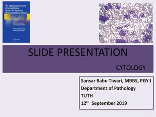
2. One Giemsa and another Pap-stained Smear of Pancreatic cytology of 28
years female.
Smears are cellular in a predominantly clean background (with few
bloody areas) and has loosely cohesive fragments of tissue with
scattered cells. Foamy histiocytes and multi-nucleated giant cells are not
seen.
The cells are monotonous with mild pleomorphism and cytoplasm is
mostly absent, scanty and granular if intact. Some of them forming
rosettes. No vacuoles are seen in the cytoplasm.
Nuclei are mostly bare; and eccentrically placed when cytoplasm is intact.
They are round to oval with regular outline. No indentations and grooves
are seen in smear.
SLIDE PRESENTATION
2
3. The chromatin is visibly coarse, stippled, evenly distributed (salt
and pepper) pattern.
Predominantly these cells have inconspicuous nucleoli whereas few
of them have 1-2 nucleoli .
Necrosis and mitosis is not seen in smear.
SLIDE PRESENTATION
3
Points in favor of NET Points not in favor of SPT
1. Cellular smear 1. Branching vessels is not seen
2. Loose cohesive fragments 2. Hyaline globules not seen
3. Eccentric nucleus 3. Nuclear indentation and grooving
not seen
4. Stippled salt and pepper chromatin 4. Foamy histiocytes not seen
5. Round to oval nuclei with regular outline 5. Multinucleated giant cells are not
observed
4. According to “The Papanicolau Reporting System”:
Satisfactory for evaluation
Neoplastic: Others
Pancreatic Neuroendocrine Neoplasm, well-
differentiated type [PanNET]
Note: Solid pseudo-papillary tumor (SPT) in a young
female of this age group cannot be ruled out. Clinical
co-relation and immuno-histochemistry is suggested.
Other possibilities:
1. Acinar cell carcinoma
2. Pancreatoblastoma
SLIDE PRESENTATION
4
14. DEVELOPMENT OF PANCREAS
14
Foregut endoderm.
Ventral pancreatic bud: Uncinate process, and part of head of pancreas
(others: Dorsal PB)
Histogenesis:
Parenchyma (basic cellular tissue): endoderm from buds, forms network of
tubules.
Acini forms at the end of these tubules
Islets develop from group of cells that separate from tubules and lies between the acini
(Neurogenin-3 is required for differentiation)
Connective tissue sheath and interlobular septa from surrounding splanchnic
mesenchyme.
15. FNA and ID brushing OF PANCREAS
15
Indications:
1. Document malignancy in malignant appearing mass
in imaging.
2. Inoperable tumors Initiation of therapy
3. Operable tumors Planning of surgery
Contra-indications:
1. Uncorrectable bleeding diasthesis.
2. GI obstruction in patients undergoing EUS-FNA.
16. FNA and ID brushing OF PANCREAS
16
Complications (approx 2%):
1. Vasovagal reaction
2. Abdominal discomfort
3. Infectious complications – bacteremia and sepsis
4. Acute pancreatitis
5. Bile peritonitis
6. Hemorrhage
7. Bowel perforation (EUS-FNA)
8. Tumor seeding along needle tract
17. The Papanicolaou Society of Cytopathology System for
Reporting Pancreatobiliary Cytology, 2015
17
I. Non-diagnostic
II. Negative (for malignancy)
I. Benign Pancreatic Tissue
II. Acute Pancreatitis
III. Chronic Pancreatitis
IV. Autoimmune Pancreatitis
V. Pseudocyst
VI. Lymphoepithelial cyst
VII. Splenule/accesory spleen
18. The Papanicolaou Society of Cytopathology System
for Reporting Pancreatobiliary Cytology
18
III. Atypical
IV. Neoplastic
I. Benign
I. Serous Cystadenoma
II. Neuroendocrine microadenoma
III. Lymphangioma
II. Other [Pre-invasive or pre-malignant]
I. Well-differentiated neuroendocrine tumor
II. Intraductal papillary mucinous neoplasm
III. Mucinous cystic neoplasm
IV. Solid-pseudopapillary neoplasm
19. The Papanicolaou Society of Cytopathology System
for Reporting Pancreatobiliary Cytology
19
V. Suspicious (for malignancy)
VI. Positive or malignant
I. Ductal adenocarcinoma of the pancreas and its variant
II. Cholangiocarcinoma
III. Acinar cell carcinoma
IV. Poorly differentiated (small and large cell)
neuroendocrine carcinoma
V. Pancreatoblastoma
VI. Lymphoma
VII. Metastatic malignancy
20. On-site Cyto-pathologists
20
Most authors agree that the presence of a cytologist
during the procedure is beneficial.
1. Higher diagnostic yield
2. Reduced number of required passes
3. Need for immunocytochemistry and flow-cytometry
Benefits to patient (RISK and COST)
23. LOW POWER
23
Cellularity (Solid > Cystic). No established
criteria of adequacy.
Smear Pattern = Background; number, size and
architecture of tissue fragments.
BG can be mucinous (Duodenum and gastric
walls, MCN, IPMN, Invasive adenocarcinoma),
clean (normal pancreas, well-differentiated
adenocarcinoma, mets), bloody (PanNET, serous
cyst adenoma, met RCC), inflammatory or dirty
and necrotic (pancreatitis, pseudocyst, abscess,
adenocarcinoma)
24. LOW POWER
24
Type of cellular elements:
Epithelial Glandular/ ductal vs Non-
glandular/ non-ductal (Acinar, neuroendocrine
or squamous cells)
Ductal, acinar and neuroendocrine cells are
normal components of pancreas.
25. INTERMEDIATE POWER
25
Assessment of architectural details can be
CONFIRMED in tissue fragments.
Benign ductal cell: “Marching band” honey-combing
Neoplastic: “Drunken-honeycomb”
Acinar cells lack honeycombing, more delicate cytoplasm
with occasional vacuolization and pyramidal, triangular or
polygonal shapes, abundant granular cytoplasm with
numerous IC zymogen granules. Eccentric round nuclei and
granular chromatin pattern and prominent nucleoli.
Islet cells: rare, discohesive tissue fragments, wispy ill-
defined amphophilic cytoplasm and oval nuclei with a
stippled chromatin pattern.
Stromal cells: Fibrous stromal fragments, fibrovascular
cores and spindle cells from stromal or mesenchymal
processes
30. HIGH POWER
30
Individual cellular details and mitoses
Nuclear size, N:C ratio, nuclear membrane
contour, distribution of chromatin, presence of
nucleoli, and mitoses.
31. 8 PATTERNS OF PANCREATIC FNAB
31
1. Inflammatory cells predominating with or without epithelial
tissue fragments.
2. Mucinous background.
3. Dirty or necrotic background predominating.
4. Predominantly cohesive epithelial or ductal-type tissue
fragments.
5. Loosely cohesive tissue fragments with predominantly
dispersed single cells.
6. Epithelial proliferations with fibro-vascular stroma or cores
within epithelial-tissue fragments.
7. Squamous cell predominating.
8. Stromal fragments with or without epithelial tissue
fragments.
55. PANCREATIC NEUROENDOCRINE NEOPLASM
(PanNEN)
55
Prevalence: 2-5% among pancreatic tumors
Incidence: <1 case per 100,000 person-years
M=F; 30-60 years
KRAS mutation like in PDA
Associated with MEN I, VHL, NF1, Tuberous Sclerosis
[PanNET only]
Risk factors:
• Family history of cancer
• Smoking
• Alcohol consumption
• Obesity and diabetes
56. PANCREATIC NEUROENDOCRINE NEOPLASM
(PanNEN)
56
PanNEN well differentiated [PanNET]
PanNEN poorly differentiated [PanNEC]
• Functioning (Insulinoma, Glucagonoma, Gastrinoma,
VIPoma, serotonin, ACTH, GHRH, PTHrP, CCK)
• Nonfunctioning (PP, somatostatin and chromogranin)
• Microadenomas (<5cm): usually non functioning
• Previously F>NF, Now NF (60%) >F
57. PANCREATIC NEUROENDOCRINE TUMOR(PanNET)
57
Minimal to moderate atypia lacking necrosis and
expressing Synaptophysin and Chromogranin.
Low, intermediate or high-grade
Mitoses/2 mm3 Ki-67 Proliferative
Index
G1 <2 <3%
G2 2-20 3-20 %
G3 >20 >20%
59. PANCREATIC NEUROENDOCRINE CANCER (PanNEC)
59
Poorly-differentiated high-grade NEN, composed of
highly atypical small cells or intermediate to large cells
expressing the neuroendocrine markers.
Mitoses >20/mm3 and Ki-67 >20%
Not associated with genes involved in PanNET, whereas
one case was found to have BRCA1 mutation.
TP53 mutation and inactivation of RB1/p16 pathway is
common.
60. PANCREATIC NEUROENDOCRINE CANCER (PanNEC)
60
Usually a/w other non-neuroendocrine types (PDA and
Acinar cell carcinoma) .
When each component accounts for >30%, Mixed
Neuroendocrine-non-neuroendocrine carcinoma
(MiNEC) is applicable
Subtypes:
1. Large cell NEC
2. Small cell NEC
62. SOLID PSEUDOPAPILLARY TUMOR
62
• Low-grade malignant pancreatic tumor
• Preference to tail of pancreas
• Predominantly in adolescence girls and young
women ( mean age: 28 years, 7-79 years)
• 30% of all pancreatic neoplasm in patients <40
years.
• Rare association with FAP (B-catenin pathway)
71. PANCREATOBLASTOMA
71
Malignant epithelial neoplasm of the pancreas showing
predominantly acinar differentiation with squamoid nests.
25% of pancreatic neoplasm in 1st decade.
Approx 40 cases have been reported in patients between 18-78
years.
Association with 11p LOH, Beckwith-weidemann syndrome and FAP
Essential diagnostic criteria: Multiple lines of differentiation
including acinar, endocrine and sometimes ductal differentiation;
squamoid nests