4. • Inadequate oxygen delivery to meet
metabolic demands
• Results in global tissue hypo-perfusion and
metabolic acidosis
• Shock can occur with a normal blood
pressure and hypotension can occur
without shock
5. • Shock characterized as:
I. Pallor
II. Sweating
III. Cold and calm skin
IV. Peripheral cyanosis
V. Tachycardia with thready pulse
VI. Persistent hypoperfusion
6.
7.
8.
9. Stages of shock
Three major stages:
1. Nonprogressive stage: (sometimes called the
compensated stage): normal circulatory
compensatory mechanisms eventually cause full
recovery without help from outside therapy
• “Reversible stage during which compensatory
mechanisms are effective and homeostasis is
maintained”
• Clinical presentation begins to reflect the body’s
response to the imbalance of oxygen supply and
demand
10. • At this stage, the body is able to compensate
for the changes in tissue perfusion. If the
underlying cause is corrected, the patient will
recover with little to no residual effects.
• If the body is unable to compensate the body
will enter the progressive stage of shock
Compensated shock
11. 2. Progressive stage/Uncompensated: without
therapy the shock becomes steadily worse
until death.
• This stage of shock begins when the body’s
compensatory mechanisms fail.
• Aggressive interventions are need to prevent
the development of multiple organ dysfunction
syndrome (MODS)
• Continued decreased cellular perfusion and
resulting alerted capillary permeability are the
distinguishing features of this stage
12. • Irreversible/refractory stage: shock progressed to
such an extent that all forms of known therapy are
inadequate to save the person's life, even though,
for the moment, the person is still alive.
• Final stage of shock
• Decreased perfusion from peripheral
vasoconstriction and decreased cardiac output
exacerbate anaerobic metabolism
• Lactic acid accumulates and contributes to an
increased capillary permeability and dilation of the
capillaries
• Increased capillary permeability allows for fluid and
plasma to leave the vascular space and move to the
interstitial space
13. • Blood pools in the capillary beds secondary to
constricted veins and dilated arteries
• Loss of intravascular volume leads to
worsening of hypotension and tachycardia
resulting in a decrease in coronary blood flow
• Decreased coronary blood flow results in
decreased cardiac output
• Cerebral blood flow cannot be maintained and
cerebral ischemia results
Irreversible Shock
14.
15.
16.
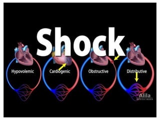
17. Types of shock
A. Aetiological Classification
I. Hypovolemic shock
II. Cardiogenic shock
III. Septic shock
IV. Anaphylactic shock
V. Neurogenic shock
25. Cardiogenic shock
• Due to failure of myocardial pump because of intrinsic
myocardial damage or extrinsic obstruction to
outflow:
Etiologies: some causes of cardiogenic shock
• AMI (acute myocardial infarction)
• Sepsis
• Myocarditis
• Myocardial contusion
• Aortic or mitral stenosis, HCM
• Acute aortic insufficiency
26. Pathophysiology of cardiogenic shock
• Often after ischemia, loss of LV (left
ventricle) function
• Lose 40% of LV = clinical shock ensues
• CO (cardiac output) reduction = lactic
acidosis, hypoxia
• Stroke volume is reduced
• Tachycardia develops as compensation
• Ischemia and infarction worsens
27. Clinical features
I. Chest pain
II. Central cyanosis
III. Increased JVP
IV. Sweating
V. Pallor
VI. Hypotension
VII.Tachycardia (Tachycardia/Bradicardia)
28. Investigation
1. ECG: Evidence of myocardial infarction and ischemia
2. Routine tests: CPK-MB, LDH, AST, Troponin-I
3. Chest-Xray
Treatment
a. Establish ABC
b. High flow oxygen
c. I.V line open
d. ECG Monitoring
e. Monitor Vitals, urine-output
f. Transfer pt. to CCU
g. Inotropic agents: Dopamine, dobutamine
29. • Sepsis: systemic inflammatory response to a
documented or suspected infection
• Septic Shock: presence of sepsis with
hypotension despite fluid resuscitation along
with the presence of tissue perfusion
abnormalities.
• Caused by systemic microbial infection mainly
by GM-ve bacteria (endotoxic shock),also can
occur with GM+ve and fungal infection
•
Septic Shock
32. Clinical features
I. History of infection/ surgery, with medical
conditions like DM, HIV infection
II. Fever, chills rigor
III. Hypotension
IV. Oedema
V. Skin hot and flushed
VI. Bounding pulse
VII.Oliguria, anuria
VIII.Dyspnoea
38. Anaphylactic Shock
• Anaphylaxis – a severe systemic
hypersensitivity reaction characterized by
multisystem involvement
• IgE mediated
• Anaphylactoid reaction – clinically
indistinguishable from anaphylaxis, do not
require a sensitizing exposure
• Not IgE mediated
39. Anaphylactic shock
• It is initiated by generalized IgE mediated hypersensitivity
reaction leading to systemic vasodilatation and increased
vascular permeability
Allergic substance enters the organism
Systemic release of mediators (histamines,
bradykinin)
vasodilation
Venous pooling of blood and hypotension,also
bronchoconstriction and skin rashes may be
manifested
p
a
t
h
o
p
h
y
s
i
o
l
o
g
y
40. Clinical features
• First- Pruritus, flushing, urticaria appear
•Next- Throat fullness, anxiety, chest tightness,
shortness of breath and lightheadedness
•Finally- Altered mental status, respiratory
distress and circulatory collapse
46. Neurogenic Shock
• Occurs after acute spinal cord injury
• Sympathetic outflow is disrupted leaving unopposed
vagal tone
• Results in hypotension and bradycardia
• Spinal shock- temporary loss of spinal reflex activity
below a total or near total spinal cord injury (not the
same as neurogenic shock, the terms are not
interchangeable)
47. • Loss of sympathetic tone results in warm and
dry skin
• Shock usually lasts from 1 to 3 weeks
• Any injury above T1 can disrupt the entire
sympathetic system
• Higher injuries = worse paralysis
Neurogenic Shock