2. OBJECTIVES
• By the end of this lecture the student should be able to
• Describe the anatomy of the esophagus: extent, length,
parts, strictures, relations, blood supply, innervation and
lymphatics.
2
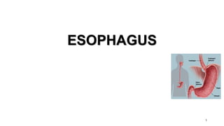
3. ESOPHAGUS
• Tubular structure about 25
cm long.
• Begins at the level of C6.
• Pierces the diaphragm atT10.
• It is divided into 3 parts:
• 1- Cervical.
• 2- Thoracic.
• 3- Abdominal.
• Proximal and distal- oncosurgery
Abdominal
thoracic
Cervical
3
4. 4
CERVICAL PART
• Posteriorly
• Post: Vertebral column.
• Laterally:
• Lat: Lobes of the
thyroid gland.
• Anteriorly:
• Ant: Trachea and the
recurrent laryngeal
nerves.
RELATIONS
5. 5
THORACIC PART
• In the thorax, it passes
downward and to the left
through superior then to
posterior mediastinum
• At the level of the sternal angle,
the aortic arch pushes the
esophagus again to the midline.
7. POSTERIOR RELATIONS – Thoracic duct
• Bodies of the thoracic vertebrae
• Thoracic duct
• Azygos vein
• Right posterior intercostal arteries
• Descending thoracic aorta (at the
lower end)
7
8. LATERAL RELATION
• On the Right side:
• Right mediastinal pleura
• Terminal part of the azygos vein.
• On the Left side:
• Left mediastinal pleura
• Left subclavian artery
• Aortic arch
• Thoracic duct
8
9. ESOPHAGUS AND LEFT ATRIUM
• close relationship
• What is the clinical
application?
• A barium swallow will
help the physician to
assess the size of the
left atrium (dilation).
10. 10
RELATIONS IN THE ABDOMEN
• In the Abdomen, the esophagus
descends for 1.3 cm and joins the
stomach.
• Anteriorly, left lobe of the liver.
• Posteriorly, left crus of the
diaphragm.
• Fibers from the right crus of the
diaphragm form a sling around the
esophagus.
• At the opening of the diaphragm, the
esophagus is accompanied by:
– The two vagi
– Branches of the left gastric vessels
– Lymphatic vessels.
11. ESOPHAGEAL
CONSTRICTIONS
• The esophagus has 3 anatomic
constrictions.
• The first is at the junction with the
pharynx(pharyngeoesophageal
junction).
• The second is at the crossing with
the aortic arch and the left main
bronchus.
• The third is at the junction with the
stomach.
• They have a considerable clinical
importance.
• Why?
12. 1. They may cause difficulties in
passing an endoscope.
2. In case of swallowing of caustic
liquids (mostly in children), this
is where the burning is the
worst and strictures develop.
3. The esophageal strictures are a
common sites of the
development of esophageal
carcinoma.
4. In this picture what is the
importance of the scale?
13. 13
ARTERIAL SUPPLY
• Upper third by the
inferior thyroid
artery.
• The middle third by
the thoracic aorta.
• The lower third by the
left gastric artery.
14. VENOUS
DRAINAGE
• The upper third drains
in into the inferior
thyroid veins.
• The middle third into
the azygos veins.
• The lower third into
the left gastric vein,
which is a tributary of
the portal vein.
• NB. Esophageal
varices.
15. LYMPH DRAINAGE
• The upper third is drained into the
deep cervical nodes.
• The middle third is drained into the
superior and inferior mediastinal
nodes.
• The lower third is drained in the celiac
lymph nodes in the abdomen.
15
16. NERVE SUPPLY
• It is supplied by sympathetic fibers from the
sympathetic trunks.
• The parasympathetic supply comes form the
vagus nerves.
• Inferior to the roots of the lungs, the vagus
nerves join the sympathetic nerves to form
the esophageal plexus.
• The left vagus lies anterior to the esophagus.
• The right vagus lies posterior to it.
16
17. 17
CARDIAC ORIFICE
• It is the site of the gastro-
esophageal sphincter.
• It is a physiological rather
than an anatomical,
sphincter.
• Consists of a circular layer
of smooth muscle (under
vagal and hormonal
control).
• Function:
• Prevents (GER)
regurgitation (reflux)
• NB. Notice the abrupt
mucosal transition from
esophagus to stomach (Z-
line)
18. Microscopic anatomy
• 2 layers of muscles- longitudinal and circular.
• Lined by non keratinizing stratified squamous epithelium.
• Squamocolumnar jn.
21. • Causes can be grouped into:
• Intrinsic – due to inflammation, fibrosis or neoplasia
• Extrinsic – due to external compression
• Disruption of peristalsis
22. Proximal and mid esophagus
• Caustic ingestion (acid or alkali)
• Malignancy
• Radiation therapy
• Infectious esophagitis - Candida, herpes simplex virus (HSV),
cytomegalovirus (CMV), human immunodeficiency virus (HIV)
• AIDS and immunosuppressed patients
• Diseases of the skin - Pemphigus vulgaris, benign mucous membrane
(cicatricial) pemphigoid, epidermolysis bullosa dystrophica
• Idiopathic eosinophilic esophagitis
• Extrinsic compression
• Squamous cell carcinoma
24. • Heartburn
• Dysphagia, odynophagia
• Food impaction
• Weight loss
• Chest pain
• Poor nutritional status
• Patients with collagen vascular diseases -- joint abnormalities,
calcinosis, telangiectasias, sclerodactyly, or rashes
• Virchow node
25. Corrosive injury
• Accidental or suicidal.
• The type of agent, its concentration and the volume ingested
determine the extent of damage.
• Pathophysiology:
• Alkalis cause liquefaction that leads to fibrous scarring.
• Acids cause coagulative necrosis with eschar formation, and this coagulum
limit penetration to deeper layers.
• Acids cause more gastric damage because of intense pylorospasm with
pooling in the antrum.
26. Treatment
• Supportive
• Feeding jejunostomy until patient starts swallowing saliva.
• Repeated endoscopy and dilation.
• Esophageal replacement for very long or multiple strictures. Why not
resection?
29. • Usually iatrogenic or due to ‘barotrauma’.
• Spontaneous perforation is a life-threatening condition.
• Iatrogenic perforation can be managed conservatively.
• Can be pathological
• Due to penetrating injury or foreign body
30. Barotrauma (spontaneous perforation,
Boerhaave syndrome)
• occurs classically when a person vomits against a closed glottis.
• The pressure in the oesophagus increases rapidly, and the
• oesophagus bursts at its weakest point in the lower third, sending
• a stream of material into the mediastinum and often the
• pleural cavity as well. The condition was first reported by
• Boerhaave, who reported the case of a grand admiral of the
• Dutch fleet who was a glutton and practised autoemesis.
• Boerhaave syndrome is the most serious type of perforation
32. Clinical presentation
• Severe pain in the chest or upper abdomen following a meal or a bout
of drinking
• SOB
• Sometimes misdiagnosed as MI, perforated peptic ulcer or
pancreatitis
• Rigidity of the upper abdomen
• Dec. breath sounds
• Dullness on percussion, subcutaneous emphysema
• In late cases sepsis is present
33. Investigations
• CXR - air in the mediastinum, pleura or peritoneum.
• Pleural effusion
• A barium swallow
• CECT
34. Iatrogenic injury
• Most common cause of esophageal perforation.
• Most common site is the cricopharyngeus.
• Factors associated with increased risk are including large anterior
cervical osteophytes, the presence of a pharyngeal pouch and
mechanical causes of obstruction
• It may follow biopsy
• Patients undergoing therapeutic endoscopy have a 10 times greater
perforation risk than those undergoing diagnostic endoscopy.
35. Treatment
• Aim is to limit mediastinal contamination and infection
• The decision between operative and non-operative management
rests on four factors:
1 the site of the perforation (cervical vs. thoracoabdominal oesophagus)
2 the event causing the perforation (spontaneous vs. instrumental)
3 underlying pathology (benign or malignant)
4 the status of the oesophagus before the perforation (fasted and empty vs.
obstructed with a stagnant residue).
36. • Indications for non-operative management include:
• pain that is readily controlled with opiates;
• absence of crepitus, diffuse mediastinal gas, hydropneumothorax or
pneumoperitoneum;
• no evidence of widespread extravasation of contrast material;
• no evidence of on-going luminal obstruction or a retained foreign body.
• patients who have remained clinically stable despite diagnostic delay.
• The principles of non-operative management are hyperalimentation,
nasogastric suction and broad-spectrum intravenous antibiotics.
37. • Surgical management is indicated when:
• Patients are unstable with sepsis or shock
• Have evidence of a heavily contaminated mediastinum, pleural space
or peritoneum.
• Surgery can be a primary repair, creation of an external fistula or
resection.
38. Key points
• Most perforations are iatrogenic.
• Surgical emphysema is pathognomonic.
• Complications are mediastinitis and sepsis.
• Treatment is both conservative or surgical but requires specialised
care.