Fetal surgery is a clinical reality
•
2 gefällt mir•1,966 views

Fetal surgery is a clinical reality

Empfohlen
Empfohlen
Weitere ähnliche Inhalte
Andere mochten auch
Andere mochten auch (20)
Ähnlich wie Fetal surgery is a clinical reality
Ähnlich wie Fetal surgery is a clinical reality (20)
Mehr von South Dakota Pain Capable Unborn Child Protection Act
Mehr von South Dakota Pain Capable Unborn Child Protection Act (20)
Kürzlich hochgeladen
Kürzlich hochgeladen (20)
Fetal surgery is a clinical reality
- 1. Seminars in Fetal & Neonatal Medicine 15 (2010) 58–67 Contents lists available at ScienceDirect Seminars in Fetal & Neonatal Medicine journal homepage: www.elsevier.com/locate/siny Fetal surgery is a clinical reality Jan A. Deprest a, *, Roland Devlieger a, Kasemsri Srisupundit a, Veronika Beck a, Inga Sandaite b, Silvia Rusconi a, Filip Claus b, Gunnar Naulaers a, Marc Van de Velde c, Paul Brady d, Koen Devriendt d, Joris Vermeesch d, Jaan Toelen e, Marianne Carlon e, Zeger Debyser e, Luc De Catte a, Liesbeth Lewi a a Division Woman and Child, University Hospital Gasthuisberg, Leuven, Belgium b Division Medical Imaging, University Hospital Gasthuisberg, Leuven, Belgium c Division Anesthesiology and Critical Care Medicine, University Hospital Gasthuisberg, Leuven, Belgium d Centre of Medical Genetics, University Hospital Gasthuisberg, Leuven, Belgium e Laboratory for Molecular Medicine, Virology and Gene Therapy, Faculty of Medicine, Leuven, Belgium s u m m a r y Keywords: An increasing number of fetal anomalies are being diagnosed prior to birth, some of them amenable to Congenital diaphragmatic hernia fetal surgical intervention. We discuss the current clinical status and recent advances in endoscopic and Fetal pain open surgical interventions. In Europe, fetoscopic interventions are widely embraced, whereas the Fetal surgery uptake of open fetal surgery is much less. The indications for each access modality are different, hence Fetoscopy they cannot substitute each other. Although the stage of technical experimentation is over, most Monochorionic twins interventions remain investigational. Today there is level I evidence that fetoscopic laser surgery for twin-to-twin transfusion syndrome is the preferred therapy, but this operation actually takes place on the placenta. In terms of surgery on the fetus, an increasingly frequent indication is severe congenital diaphragmatic hernia as well as myelomeningocele. Overall maternal safety is high, but rupture of the membranes and preterm delivery remain a problem. The increasing application of fetal surgery and its mediagenicity has triggered the interest to embark on fetal surgical therapy, although the complexity as well as the overall rare indications are a limitation to sufficient experience on an individual basis. We plead for increased exchange between high volume units and collaborative studies; there may also be a case for self-regulation. Inclusion of patients into trials whenever possible should be encouraged rather than building up casuistic experience. Ó 2009 Elsevier Ltd. All rights reserved. 1. Introduction to fetal therapy, a subsequent further referral to a highly specialized unit may be required, as many therapies cannot be offered locally As ultrasound screening programmes become more widely with a reasonably sufficient experience. implemented and qualitatively improved, an increasing number of This issue of Seminars in Fetal and Neonatal Medicine describes anomalies will be diagnosed prior to birth. When fetal malforma- the area of expertise of one of the world’s largest centers in fetal tions, genetic diseases or conditions acquired in utero are assessment and therapy. The Children’s Hospital of Philadelphia suspected, those patients should be referred to fetal care centers. (CHOP) is a ‘quaternary’ care unit, i.e. it typically accepts patients These have the necessary equipment and the required skills for from secondary but mainly tertiary referring hospitals offering more accurate prenatal evaluation, and are located in institutions review for an invasive procedure. As such, this issue has become an with multidisciplinary teams familiar with the perinatal manage- interesting collection of reviews on modern fetal therapy. Fetal ment of those conditions. They will make a prognostic evaluation therapy has recently received much attention, in part thanks to and define all potential options open to the family. When it comes groups like CHOP who have pushed at the frontiers of knowledge. Historically fetal surgery was mainly an American pediatric surgery enterprise, pioneered by Michael Harrison at the University of California at San Francisco (UCSF). Being also an important * Corresponding author. Address: Division Woman and Child, Department postnatal pediatric surgery training center, the seed of fetal surgery Women, University Hospitals Leuven, B-3000 Leuven, Belgium. Tel.: þ32 16344215; fax: þ32 16344205. spread as his trainees dispersed over North America. Thus, UCSF E-mail address: jan.deprest@uzleuven.be (J.A. Deprest). probably trained the vast majority of clinicians at some stage in 1744-165X/$ – see front matter Ó 2009 Elsevier Ltd. All rights reserved. doi:10.1016/j.siny.2009.10.002
- 2. J.A. Deprest et al. / Seminars in Fetal & Neonatal Medicine 15 (2010) 58–67 59 their fetal surgery career, with Scott Adzick as one of the earliest but there are no officially endorsed wider initiatives. For instance, and most prominent research fellows. in the EU healthcare is still a responsibility of the individual (national) European member states. Therefore any effort to regulate 2. Conditions for fetal surgery and centers offering it quality of care but also optimal access for patients will ultimately be at a national level, because the EU itself is not empowered to do so. Programmes such as those from CHOP and UCSF have inspired many others, including ourselves, to explore the avenues of fetal 3. In-utero endoscopy: differences in approach between therapy. Also, the mediagenicity of fetal surgery is currently Europe and the USA tempting an increasing number of hospitals and clinicians to open centers for comprehensive fetal care throughout the world. As fetal The growing availability of video-endoscopic surgery in the medicine specialists, we intuitively applaud this increased interest 1990s and miniaturization of cameras as well as fiberendoscopes for the fetal patient, but there are certainly downsides to this. introduced the concept of endoscopic fetal surgery. A firm boost to First, most prenatal conditions can await therapy until after fetoscopic surgery was given by the European Commission when birth, but there are rare conditions that require prenatal interven- the ‘Eurofoetus’ research and development project was granted. tion to save the life of the fetus, or prevent permanent organ It funded a consortium of selected European fetal medicine units damage. This can be achieved by correcting the malformation, by and one endoscopic instrument-maker to design new endoscopes arresting the progression of the disease, or by treating some of the and instruments.3 We, at our institution, were able to develop immediately life-threatening effects of the condition, delaying safely our novel operating skills in an ovine model by optimal more definitive repair until after birth. By definition we are dealing cross-fertilization of a pediatric surgeon, Francois Luks, and the with uncommon conditions, and very few patients may opt for fetal primary author, a gynaecologic endoscopic surgeon.4 According to therapy. This inherently means that there is a case-load limitation, the IFMSS guidelines, we then moved to clinical application, by and this is even so for any center offering advanced fetal diagnosis. doing Europe’s first successful umbilical cord ligation, unaware of Second, most therapies described herein require access to the an almost simultaneously performed successful procedure done uterus and/or fetus, hence are invasive. They carry a substantial risk by Ruben Quintero and colleagues at Wayne State University and/or may fail, so that the claimed benefits of the intervention (Detroit).5 At that time, the scene was set for minimal access to the must be weighed against the inherent risk for complications. This fetal patient. Given the general assumption that fetoscopy signifi- balance was worded more than 25 years ago by the International cantly reduces the risk for ruptured membranes and preterm Fetal Medicine and Surgery Society (IFMSS) (Box 1).1 A small group labour, a number of teams started reconsidering fetal surgery. of pioneers in the field of fetal intervention drafted guidelines for Remarkably, the revival of fetoscopy initially focused on surgery novel interventions, designed to save the life or the organ function, of the placenta and umbilical cord in complicated monochorionic but which are in the early phase without any proven benefit. At that twin pregnancies, and to a lesser extent the fetal membranes time, these interventions typically required maternal laparotomy, (Table 1).6 This indication was initially not on the shortlist of fetal and partial exteriorization of the fetus through a stapled hyster- surgery targets. The placenta not being the usual habitat of the otomy. Even as today the fetus can be minimally invasively pediatric surgeon prompted the need for involvement of fetal accessed, these consensus guidelines have remained a reference, medicine specialists. Although this may initially have caused some and in our opinion, should remain so. Where fetal intervention has friction, the full participation of obstetricians has probably lowered not been formally evaluated, we believe the fetus should not be the threshold for fetal surgical intervention. Another boost to operated except within a formal trial, in a select number of centers fetoscopy was that complications of monochorionic twinning are that fully comply with these guidelines. Who and where that would much more common than any other typical indication for fetal be is certainly a matter of debate, and it is nearly impossible to surgery. In Europe, the Eurofoetus project climaxed with the judge in an evidence-based way. In an effort to offer maximal successful execution of a randomized trial on laser coagulation for quality and also make further clinical research viable, it will have to twin-to-twin transfusion syndrome (TTTS). Whereas this operation be agreed upon by the fetal medicine community. In North America was originally pioneered in the USA by Julian DeLia, its wider this could be in organizations such as NAFTNeT,2 whereas for other clinical implementation in the USA was much slower at the areas of the world this is much less clear. Initiatives such as Euro- ‘traditional’ fetal surgery centers. By contrast, Ruben Quintero, who foetus are focused around one disease or instrument development, is an obstetrician by training, initially did most of these operations in the USA and as such advanced much the knowledge about this condition just as Kypros Ncolaides, Yves Ville and Kurt Hecher did Box 1. Criteria for fetal surgeryf in Europe. In fact, the successful Eurofoetus trial removed many questions about maternal safety as well as feasibility of fetoscopic 1. Accurate diagnosis and staging possible, with exclusion surgery, hence has been pivotal for the wide clinical acceptance of of associated anomalies. fetoscopy. Another factor that may have contributed to an initially 2. Natural history of the disease is documented, and slower uptake of fetoscopy in the USA may be the delay in regu- prognosis established. latory approval of novel surgical instrumentation. Though these 3. Currently no effective postnatal therapy. 4. In-utero surgery proven feasible in animal models, regulations are obviously there to protect the patient from both an reversing deleterious effects of the condition. overzealous industrial and potentially also medical community, it is 5. Interventions performed in specialized multidisciplinary at times difficult to understand from a European perspective why fetal treatment centers within strict protocols and subsequent subtle variations of the same instrument (an endo- approval of the local ethics committee with informed scope) have to go through such a long regulatory process. As consent of the mother or parents. a side-effect, this may even feed already present suspicions that the technique is controversial and unsafe. There are fewer regulatory issues in Europe, without – as far as we know today – compromising patient safety. Regulatory issues have to our knowledge also partly contributed to limited clinical activity in yet f Adapted from Harrison et al.1 another potential indication for fetal surgery, i.e. congenital
- 3. 60 J.A. Deprest et al. / Seminars in Fetal & Neonatal Medicine 15 (2010) 58–67 Table 1 Indications and rationale for in-utero surgery on the fetus, placenta, cord or membranesa Pathophysiology Rationale for in-utero therapy Surgery on the fetus: 1. Congenital diaphragmatic hernia Pulmonary hypoplasia and pulmonary hypertension Timely reversal of pulmonary hypoplasia and prevention of pulmonary hypertension 2. Lower urinary tract obstruction Progressive renal damage by obstruction Urinary diversion prevents obstructive uropathy and Pulmonary hypoplasia by oligohydramnios restores amniotic fluid volume 3. Sacrococcygeal teratoma High output cardiac failure by arteriovenous Cessation of steal phenomenon shunting Reversal of cardiac failure; Fetal anemia by tumor growth and/or Prevention of polyhydramnios bleeding within a tumor 4. Thoracic space-occupying lesions Pulmonary hypoplasia (space-occupying mass) Prevention of pulmonary hypoplasia and cardiac failure Hydrops by impaired venous return (mediastinal compression) 5. Neural tube defects Damage to exposed neural tube;cerebrospinal Covering exposed spinal cord, cessation of leakage fluid leak, leading to Chiari malformation and preventing hydrocephaly and reversing cerebellar herniation hydrocephalus 6. Cardiac malformations Critcal lesions causing irreversible hypoplasia or damage Prevention of hypoplasia or arrest of progression of damage Surgery on the placenta, cord or membranes: 7. Chorioangioma High output cardiac failure by arteriovenous shunting Prevention of cardiac failure and hydrops fetoplacentalis and polyhydramnios 8. Amniotic bands Progressive constrictions causing irreversible neurological Prevention of limb deformities and function loss or vascular damage 9. Abnormal monochorionic twinning: Twin-to-twin transfusion Intertwin transfusion leads to oligopolyhydramnios sequence, Bichorionization stops intertwin transfusion, reverses hemodynamic changes; obstetric complications (preterm labor cardiac failure, preventing neurological damage, and rupture of the membranes) delaying delivery (amniodrainage) Fetus acardiacus and discordant Discordant anomalies: where one fetus can be a threat to the Fetocide to improve chances of the other fetus; avoidance of anomalies other, or to avoid termination of entire pregnancy termination of entire pregnancy a Adapted from Deprest et al.6 diaphragmatic hernia. Although all the pioneering work has been function in selected fetuses.11–13 This is described in detail by done in the USA, by the teams of UCSF, Boston, CHOP, and Buffalo, Adzick.14 The National Institutes of Health (NIH) decided to sponsor this clinical indication is currently much more explored in Europe the Management Of Myelomeningocele Study (MOMS) randomized than in the USA. Conversely, Europe has failed to embrace open trial, which involves three centers (San Francisco, Nashville and fetal surgery to the same degree as in the USA. This has traditionally Philadelphia) (www.spinabifidamoms.org). The primary outcome been explained by concerns about maternal safety and side-effects, is death or the need for shunting by the age of 1 year. The set-up of as well as the risk for preterm delivery. The latter is in reality not this trial may have a number of major benefits. First, it may a valid objection, as outcomes from open fetal surgery in larger unequivocally demonstrate whether we should operate on a fetus series compare favorably to what has been published a decade for this condition (or not). Second, it is conceptually interesting as earlier and remained the general perception (Table 2).7,8 Despite the trial explores the role of fetal surgery in a condition that is not that, open fetal surgery is rarely done in Europe, with the exception of operations on placental support.9,10 This is in our opinion not Table 2 scientifically supported, therefore at the least open to criticism – as Obstetrical and short-term outcomes in the Children’s Hospital of Philadelphia we will touch on at the end of this contribution. This issue of (CHOP)7 and Vanderbilt University8 series on myelomeningocele repair Seminars covers nearly the entire range of fetal intervention. In this CHOP (n ¼ 51) Vanderbilt (n ¼ 178) article we summarize each of these, with work from CHOP as well Gestation at surgery 23þ0 (20þ0 to 25þ4) (19–30); later 26 weeks as others. (weeks) þ4 þ4 a Gestation at delivery 34 (25 to 37) 33þ5 (25–38) (weeks) 4. Myelomeningocele Chiari malformation graded as either (moderate/severe) Although myelomeningocele (MMC) may be a non-lethal Before surgery 14%/86% congenital defect, it does lead to significant lifelong morbidity and After surgery 100%/0% 7%/0% burden. This condition should now be detected as early as the first, Postnatal shunt 46% (21 weeks) 46% (12 weeks) or at the latest in the second, trimester. Also its extent or location, (postnatal age) which are the most important prognostic factors, can be easily Perinatal losses 3/51 (6%) (prematurity) 5/178 (2.8%) (not specified) Length of hospital stay 4 days 3.3 days (3–7) determined. In view of the prognosis, many pregnancies are Oligohydramnios 25% early on; terminated, and at this point there is not much hope for an 30% readmission rate improved postnatal management strategy eventually altering Delivery 30 weeks 5/47 (10.6%)b 11.8% b outcome. All hope is therefore focused on prenatal intervention to Delivery 32 weeks (40/47) 85% (not specified) Maternal complications None reported, including 9 (5.1%) mild pulmonary improve outcome. Experimental work in animal models has shown dehiscence or rupture; edema; that this is possible. Early clinical evidence from both Vanderbilt 1 amniotic fluid leak 1 bowel obstruction; University and CHOP (Table 2) underscored this hypothesis.7,8 In through hystertomy 4 (2.2%) dehiscence, their experience timely prenatal microsurgical layered repair asymptomatic in 3 reversed hindbrain herniation, decreased the need for shunting, a Includes all patients. b improved leg and bladder function, as well as later cognitive Denominator is survivors only.
- 4. J.A. Deprest et al. / Seminars in Fetal Neonatal Medicine 15 (2010) 58–67 61 lethal, hence does not even appear in the initial shortlist (Box 1 and Late cases without hydrops are delivered in optimal conditions, Table 1). The inherent side-effect is that an occasional preterm with attendants prepared for the management of respiratory delivery may lead to neonatal death in a condition that is in essence distress. In cases of hydrops their outcome is poorer, and the CHOP non-lethal, and unlikely to cause preterm birth per se.7 Third, it has group proposes resection while on placental circulation (EXIT-to- prevented an unfettered promulgation of this type of operation. CCAM resection).10 Macrocystic masses can be punctured or Fourth, it shows that centers of excellence, although they might shunted and both procedures are minimally invasive. The CHOP have competing interests, can collaborate effectively. In that experience group is probably the largest, with 23 shunted cases at respect, the assignment of patients to geographical areas was a mean of 21–22 weeks. Shunting reduced CCAM volume by an interesting. Some other additional interesting aspects of this trial average of 70% and it can also reverse hydrops. Survival rate was are that it is overseen and outcomes are evaluated by independent 74% and other smaller series confirm this.25,26 In another chapter, assessors, and that investigators are blinded to the outcomes for the Adzick discusses the use of open fetal surgery for CCAM.27 Fetal duration of the trial. Also some safety and standardization lobectomy for more solid masses has been shown to be both precautions are taken, such as defining an upper limit of maternal feasible and effective. Adzick reports on 24 cases operated between body mass index, in an effort to minimize maternal risks. Due to 21 and 31 weeks. Half of them survived and developed normally.28 a less than expected recruitment rate of the 200 patients, the trial is Overall, the ideal fetal intervention remains a matter of debate. only expected to be completed in 2010 but the results should be Interestingly, prenatal steroids were shown to resolve hydrops worth waiting for. fetalis and it would seem logical to try this treatment first.29,30 The MOMS trial has also closed the backdoor: major fetal care Along the same lines, shunts are also used for significant centers have agreed that no fetal surgery outside of the trial would thoracic effusions, which may have a wide etiologic range, in an be offered within the USA for the duration of the trial. This certainly effort to prevent pulmonary hypoplasia. Bebbington et al. reviewed is the ideal scenario for an effective trial. In other countries where the CHOP experience (n ¼ 27), with an overall survival rate of 70%. termination of pregnancy is less available (or not at all), patients It is important to realize that the need for insertion of a second may be more tempted to offers for fetal surgery. It is important that shunt because of dislodgment shunt is as high as one in three. This this form of experimental fetal surgery should also there be con- may increase the risk for preterm membrane rupture, leading ducted using the same stringent selection criteria and follow-up. potentially to pretem delivery, which is the most important nega- As an unanticipated side-effect, this study apparently tempo- tive prognostic factor.31 In reality, neither appropriate case selec- rized the clinical development of minimally invasive techniques to tion nor optimal fetal intervention are truly known. Shunting is perform this operation in the USA. However, one already needs to only one of a multitude of interventions including also single as think a step further. The potential of endoscopic coverage of the well as serial thoracentesis, combinations and pleurodesis using defect is currently explored by several teams, experimentally and a variety of products. In a recent review by Deurlo and Devlieger all also clinically.15,16 So far this is a multiple port procedure, with these were shown to be equally effective.32 potentially a higher risk for membrane rupture and early delivery, LUTO is a descriptive term for a heterogeneous group of hence neonatal death. It is also technically different from a layered conditions, of which posterior urethral valves is by far the most repair and does therefore not avoid the requirement of a shunt. common. The typical ultrasound ‘key hole sign’ can, however, also be caused by conditions such as stenosis of the urethral meatus, 5. In-utero shunting anterior valves, urethral atresia, ectopic insertion of a ureter or even (peri)vesical tumors. Female fetuses very often have more complex Shunts are devices that surgically connect two spaces, so that cloacal malformations. Any obstructive lesion will cause compen- fluid can freely drain from one to another. Their size and satory hypertrophy of the bladder wall smooth muscle, vesico- introduction method compare very much to what operators do ureteral reflux and loss of normal renal function. Resulting when performing fetoscopy. In this respect, it was surprising to see oligohydramnios will further cause pulmonary hypoplasia, how people who were widely using these devices 10 years earlier ultimately making the condition lethal. One of the potential were often criticizing the use of fetoscopy, though the use of shunts solutions, irrespective of the cause, is to create urinary diversion, was at least as debatable. Apart from that, it should be remembered which will salvage renal function when this was within reasonable that these are only devices, so that their merits are completely limits prior to the procedure. The exact way to determine renal dependent on the indication for its use. For instance, they have been function ahead of time, and predict ultimate outcome, remains to historically used for draining isolated hydrocephaly. Later on, be demonstrated.17,33,34 Landmark work comes from Johnson and a moratorium on this indication was agreed on because prenatal Biard at CHOP, who were able to determine the long-term outcome shunting did not alter outcome. Their further use in cases of urinary of in-utero-treated patients. They concluded that the actual type of tract obstruction and thoracic pathology is reviewed by Mann et al.17 urinary obstruction diagnosed postnatally was predictive of Congenital cystic adenomatoid malformation of the lung long-term renal outcome. In essence, posterior urethral valves do (CCAM) is an overgrowth of terminal bronchioles which act as much better, whereas babies with urethral atresias or the ‘prune a space-occupying lesion. If large enough, it causes pulmonary belly’ phenotype do less well.35 Second, despite favorable prenatal hypoplasia and mediastinal shift, with subsequent hydrops fetalis estimation of kidney function, up to half of survivors may end up and polyhydramnios. Growth, which is maximal around 28 weeks, with chronic renal insufficiency.36,37 This underscores the urgent can be followed by longitudinal measurement of the lesion. need for better anatomical and functional prenatal evaluation Crombleholme et al. proposed to use the proportion of the mass of methods than we currently have. Also it means that urinary the lesion divided by the head circumference (CCAM volume ratio: diversion is a salvage solution, not guaranteeing rescue of organ CVR) to plan follow-up and/or predict fetal demise.18 After function. 28 weeks the majority will decrease in size. Planned delivery and Two future strategies should change this. Kilby et al. proposed neonatal resection will be sufficient, with excellent long-term a randomized trial of the value of fetal intervention (http://www. outcomes.19–22 Only a small subset cause fetal problems, with fetal pluto.bham.ac.uk) with currently available means. Also we need hydrops being the best predictor of death. When CVR is 1.6, risk to explore the possibility of better anatomical evaluation, e.g. using for hydrops is 80% (range: 15–75%). In this group intervention in-utero endoscopy. At present the equipment available is less than seems justified.23,24 For only a minority this will be fetal surgery. ideal. Even in the hands of the pioneers it was not possible to
- 5. 62 J.A. Deprest et al. / Seminars in Fetal Neonatal Medicine 15 (2010) 58–67 distinguish between urethral valves and atresia.38 Their size is still experience at San Francisco.64 Removal of the balloon was prenatal too large and it is difficult to direct the endoscope to the bladder either by fetoscopy or ultrasound-guided puncture, intrapartum by neck, being the area of interest. However, the advantage of fetal EXIT (ex-utero intrapartum therapy) or postnatal either by cystoscopy is that it can be extended to a therapeutic procedure. tracheoscopy or percutaneous puncture. Delivery took place at Both fetoscopic antegrade catheterization, hydro- or laser ablation a median of 35.3 weeks. Again, it was before 34 weeks in 30.9% of urethral valves have been described.38–40 cases. In 204 (97.1%) cases the babies were live born and 98 (48.0%) were discharged from the hospital alive. On the basis of the 6. Congenital diaphragmatic hernia relationship between survival and O/E LHR in expectantly managed fetuses with CDH, as reported in the antenatal CDH registry, In this issue Hedrick reviews the issue of prenatal assessment we estimated that in fetuses with left CDH treated with FETO the and therapy for isolated congenital diaphragmatic hernia (CDH).41 survival rate increased from 24.1% to 49.1% and in right CDH CDH is a surgically correctable defect, but the essential problem is survival increased from 0% to 35.3% (P 0.001).65 Operation the coexisting pulmonary hypoplasia. This causes ventilatory time was dependent on surgeon experience. It also predicted insufficiency and pulmonary hypertension (PHT) in the neonatal the occurrence of PPROM. The strongest predictors of outcome period. Prenatal referral, high case load and advanced neonatal care were O/E LHR prior to the procedure, PPROM and gestational age at improve survival but the condition is still lethal in around 30% of delivery. cases (Table 3).42–49 Prenatal prediction of poor outcome creates In earlier work in smaller series, we showed that increase in a clinical need to counsel parents on prenatal options, including lung area or volume after FETO is an independent predictor of termination of pregnancy or prenatal intervention to trigger lung survival.66 Short-term morbidity in survivors is comparable to what growth. Fetal imaging techniques are used to measure lung size and is expected in fetuses with moderate hypoplasia managed vascular development or reactivity. The best validated prediction postnatally.67 Whereas a later FETO would theoretically reduce the method today is the use of the lung-to-head ratio (LHR), which risk for preterm birth, it needs to be balanced against a lesser lung involves standardized two-dimensional ultrasound measurement response. This was demonstrated experimentally and clinically, so of the contralateral lung on a cross-section through the chest.50 that it seems that later occlusion would be reserved for milder When expressed as a proportion to what is normally expected cases.68,69 These observations were the basis for the start of [observed/expected (O/E) LHR] prediction is independent of a randomized trial in Europe, currently looking at the value of FETO gestational age and technique used.51 When O/E LHR 15% the in moderate hypoplasia, but soon also for severe cases. This will be fetus has extreme pulmonary hypoplasia and virtually no chance of a multicenter trial, which has required the drafting of a consensus survival. Fetuses with O/E LHR ¼ 15–25% have severe pulmonary protocol for postnatal management by Tibboel et al., which in itself hypoplasia and their predicted survival is around 15%. Above that, is already an achievement.70 survival increases to 60% or more (Figure 1).52 MRI volumetry is very likely to become more accurate, hence better predictive.53–55 It 7. Interventions in monochorionic twins also allows quantification of the degree of liver herniation.56 Measurement of pulmonary vessels, their resistance and reactivity Monochorionic (MC) twins account for 70% of monozygous are currently evaluated as predictors of PHT.57–59 twins and 20% of all twins. Compared with dichorionic twins, they Fetal tracheal occlusion (TO) entraps lung liquid, leading to are at increased risk for fetal loss, preterm delivery and perinatal stretch-induced growth of airways and vessels. The experimental mortality and morbidity. A number of complications are amenable background on effects of TO was recently summarized elsewhere.60 to fetal intervention either on the cord or placenta. As these are all Our clinical experience with percutaneous fetoscopic endoluminal possible through minimal access, these interventions became tracheal occlusion (FETO) started in 2004, prompting abandonment quickly accepted. As a result the general knowledge about MC twins of other occlusion methods (Figure 2).61,62 We recently reported the has increased greatly. Herein we summarize briefly the current entire experience (n ¼ 210) of the FETO consortium (Leuven, state of affairs; more extensive reviews can be found elsewhere.71,72 Barcelona and London).63 FETO was offered to severe cases, and TTTS complicates 9% of MC twin pregnancies.73 It is an ultra- occassionally in a number of cases with associated anomalies. FETO sound diagnosis based on the following criteria: polyuric poly- was performed at a median gestation of 27.1 (range: 23.0–33.3) hydramnios in the sac of the recipient twin (defined as deepest weeks. The median duration of FETO was 10 (range: 3–93) min. vertical pocket of 8 cm prior to 20 weeks and 10 cm between 20 Spontaneous prelabor rupture of membranes (PROM) prior to 37 and 26 weeks; in the USA 8 cm is the single cut-off parameter) weeks (preterm PROM, or PPROM) occurred in 47.1% cases at combined with oligouric oligohydramnios in the donor sac (deep- a median of 30 days after FETO. A more informative number is that est vertical pocket 2 cm). The immediate threat to pregnancy is preterm PROM occurred within 3 weeks in 16.7% cases. Although polyhydramnios-induced PPROM and preterm delivery. In view of still clinically highly relevant, this is much less than in the earlier the poor survival rates with expectant management, there is little disagreement that therapy should be offered. For years therapy therefore consisted in repetitive amniodrainages with a total Table 3 survival rate of 61%, gestational age at delivery around 28 weeks Recent series on postnatal outcome of isolated congenital diaphragmatic hernia and morbidity of around 20%.74 As the condition is caused by Publication No. of cases TOP rate Survival rate specific patterns of anastomoses which run at the placental surface, Stege et al. (2003)42 185 NA 70% the logical therapy consists of fetoscopic identification and ablation Gallot et al. (2007)43 314 7% 63% of all visible anastomoses. This should be done as sparingly as Sartoris et al. (2006)45,a 244 NA 70% possible without being at the expense of leaving anastomoses, Hedrick et al. (2007)46 89 NA 66% which cause recurrence, intrauterine fetal death or fetal anemia.75– Datin-Dorriere et al. (2008)47 ` 99 20% 63% 78 Mettauer et al. (2009)48,a 147 – 77% Fetoscopic laser is performed percutaneously under local or loco- Grushka et al. (2009)49,a 121 – 81% regional anesthesia and requires only a short hospital stay. A TOP, termination of pregnancy; NA, not available. randomized trial demonstrated better results with laser than a Some units report survival rates after transfer of the neonate, and therefore do amnioreduction, both in terms of gestational age at delivery (33.3 not include the hidden mortality. vs 29.0 weeks), survival till 6 months (76% vs 51%; relative
- 6. J.A. Deprest et al. / Seminars in Fetal Neonatal Medicine 15 (2010) 58–67 63 100 90 extreme severe moderate mild 80 Survival rate (%) 70 60 50 40 30 20 10 0 15 15-25 26-35 36-45 ≥46 O/E LHR (%) liver in thorax (‘up’) liver in abdomen (‘down’) irrespective of liver position Figure 1. Survival rates of fetuses with isolated left-sided congenital diaphragmatic hernia, depending on measurement of the observed/expected lung:head ratio (O/E LHR) measurements and liver position as in the antenatal congenital diaphragmatic hernia registry. Reproduced with permission from Deprest et al.50 risk ¼ 1.49) and neurologic outcome.3 IUFD after laser can be pre- to the experience from leading centers in Boston (USA), London dicted preoperative by abnormal Doppler.79 Preoperative short- (UK) and Linz (Austria). The potential of these interventions has ened cervix is predictive for premature delivery.80 Postoperative revived interest such that even a new society was proposed.99 Key patients should be watched for anemia, persisting transfusion limitations at this moment are appropriate and timely selection of syndrome and cardiac problems.81,82 Brain lesions are the main patients and the lack of purpose-designed instruments. concern, but some can be detected by ultrasound in 80% of cases, Tran has described feto-maternal anesthetic techniques. Only and magnetic resonance imaging identifies another 20%.83 a limited number of procedures today require general anesthesia.100 A number of discussions are ongoing regarding this condition. This provides perfect feto-maternal anesthesia as well as uterine The first considers whether the current staging system, based on relaxation. The vast majority of fetal interventions are being done the presence of fluid discrepancy (stages I–II) and haemodynamic under local anesthesia, usually with intravenous maternal sedation. impact without (stage III) or with (stage IV) hydrops should be Loco-regional anesthesia is also used and particularly useful in the changed. Survival is indeed stage dependent.84,85 Assessment of viable period, as one can proceed immediately with urgent cardiac function is now being suggested to be incorporated into abdominal delivery in cases of fetal distress. During the previable a staging system because it would better reflect the pathophysi- period, in-utero resuscitation is the only option, and requires ology of the condition.86–90 However, it is unclear whether that will excellent teamwork between anaesthesiologists and the fetal change therapy at all. Also the place for invasive therapy for stage I surgeon. This chapter also touches on the newly developing field of disease is currently at stake, and a trial has been announced. fetal pain relief. The requirement for pain relief in the neonatal MC twins are more prone to structural anomalies, more than 80% period informs those caring for the antenatal patient, as all being discordant. Patients may prefer selective fetocide in such anatomical and functional features required for pain sensation are cases. Another anomaly is twin reversed arterial perfusion sequence present in the gestational age period during which the fetal surgeon (acardiac fetus). The normal ‘pump’ twin perfuses the anomalous works.101 As a consequence, ways to manage fetal pain and the parasitic mass, leading to congestive heart failure and hydrops in stress response during invasive fetal interventions are required.102 50%. Fetal death puts the healthy one at risk, due to feto-fetal As a side-effect it may also reduce fetal movements, which can be hemorrhage over the anastomoses. In those cases controlled feto- helpful for some procedures or diagnostic assessment.103 cide can be done by obliteration of arterial and venous flow, most often with ultrasound-guided bipolar cord coagulation with 9. Future challenges and innovations by transcontinental instruments comparable in size to those used for TTTS. Cord occlu- communication sion has a 78–84% survival rate.91,92 Pre- and perinatal losses of the healthy twin are mainly due to cord entanglement through iatro- An important chapter in this issue is that by Roybal et al. on the genic rupture of the intertwine membrane or early delivery future non-surgical approach, using stem cell and gene therapy.104 following PPROM, also responsible for 7% developmental problems Surgery is always an invasive solution, typically only partly solving in survivors. An effect of operator experience was demonstrated, the problem. Similarly, we would like to draw attention to another showing decrease in PPROM and morbidity after 40 procedures. cellular approach, which is the use of tissue engineering in the Alternative energy sources such as laser, monopolar or radio- perinatal period. The amniotic fluid is an evident source of rapidly frequency energy may be used through 14–18 G needles.93–95 They expanding fetal cells, wherein multipotent mesenchymal stem cells work effectively in TRAP where low flow conditions are present, have been demonstrated.105,106 As amniocentesis is typically part of where all energy modalities show comparable outcomes.96,97 The the initial assessment, these cells are readily available. Several experience for other indications is less so that some centers will not groups are currently exploring the potential of these stem cells as offer those when there are two pumping hearts.98 well as other tissue engineering techniques. It might be possible to engineer homologous ‘biological’ grafts while the fetal patient 8. Other indications and fetal pain relief awaits postnatal therapy during further pregnancy. One example is diaphragmatic reconstruction. Rather than using a synthetic graft, Because of space constraints, the percutaneous interventions which does not grow with the infant and is basically non-func- such as fetal valvuloplasty or cardiac septostomy will not be dis- tional, one could aim at a more functional diaphragmatic cussed. For advances in this exciting field of fetal medicine, we refer replacement.107
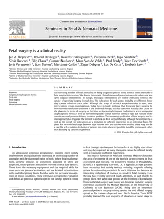
- 7. 64 J.A. Deprest et al. / Seminars in Fetal Neonatal Medicine 15 (2010) 58–67 Figure 2. Left: fetoscopic images of balloon insertion. The catheter, loaded with the balloon, is inserted; the balloon is inflated between carina and vocal cords and then detached. Middle, lower: ultrasound image of the balloon in place. Middle, upper two images: balloon is being retrieved by fetoscopic extraction, using a 1 mm forceps. Right: schematic drawing of cannula insertion towards the mouth (upper corner). Reproduced with permission from Gucciardo et al.62 One of the initial assumptions with the introduction of operative As mentioned earlier, open fetal surgery is rarely done in fetoscopy was that minimization of uterine and membrane trauma Europe. To us, the dogmatic denial of open fetal surgery reflects would obviate the problem of iatrogenic preterm prelabor rupture rather an attitude of the physician than one of the patient. In our of the membranes (iPPROM). Unfortunately this has not material- view such denial is not justified. Moreover, if the NIH-sponsored ized so far and we actually referred to it as the Achilles’ heel of ‘MOMS’ trial demonstrates decreased morbidity in fetuses with fetoscopic surgery.5 Many researchers have spent time in trying to MMC undergoing surgical repair, open fetal surgery will inevitably solve this problem. Strategies explored are: (i) prevention, by using have to be placed on the European agenda once more. Along the the most appropriate access method, the use of smaller and fewer same lines, we have to permanently question the indications or instruments, or sealing the defect on the way out of the uterus; criteria for intervention. Dogmatism indeed prevents overzealous (ii) treatment: using membrane sealing agents to arrest amnior- application of invasive therapy, but it may arrest innovation. rhexis in the absence of infection.108 As to the former, we are Flexibility and reconsideration can force breakthroughs. Debates currently investigating the use of an acellular collagen matrix to be between opponents and proponents are only apparent conflicts, inserted through the cannula. It acts as a sponge, which, after and these days they focus around indications such as CDH.41 Both absorption of body fluids, can increase in diameter so that it sides agree that the clinical problem of CDH fetuses is far from over-sizes the initial defect as well as locking the membranes solved. It is therefore important that scientists from different against the uterine wall, while being held in place by the ‘schools’ liaise, always questioning themselves and working contracting myometrium.109 It is hoped that the matrix allows cells together on this and other obstacles for fetal surgery. If this to invade and trigger or stimulate a true membrane wound-healing happens at the level of the most senior research groups, it is more process. As membranes heal poorly spontaneously, adjuncts may likely to succeed, rather than by an exclusive attitude which forces have to be used, e.g. platelets.110,111 The EU-sponsored EuroSTEC every new researcher or team to first reinvent the wheel. We project pursues additional strategies (www.euroSTEC.eu), such as therefore might have to lobby for the concept of transcontinental the use of purpose-designed membrane-derived matrices, funding for collaborative research between centers of excellence on eventually seeded with cells, which might improve in growth.112,113 both sides of the ocean – and further. For the latter, therapeutic sealing clinically is done by the injection The practice of fetal surgery today floats on a permanent of platelets, which are programmed to adhere to (epithelial) conflict between what is optimal quality and how this can be membrane defects.114 Clinical trials will have to show that these guaranteed, versus that of sufficient numbers of patients as well as strategies are effective at all. fetal care centers.115 Again it needs to balance between the drive
- 8. J.A. Deprest et al. / Seminars in Fetal Neonatal Medicine 15 (2010) 58–67 65 for innovation and alternative approaches questioning the dogmas at the Children’s Hospital of Philadelphia possible. This grant and its of the past. We need a balance of appropriate regulation and content were independent from the assignment to this paper. concern for patient protection, versus sufficient space for enthu- siastic new scientists and clinicians who may be the pioneers of References the future. It is our personal belief that such balance is best made within a few leading centers, which have the necessary means and 1. Harrison MR, Filly RA, Golbus MS, et al. Fetal treatment. N Engl J Med case load, as well as sufficient senior people, who are prepared to 1982;307:1651–2. guide their young researchers along paths that seem today far 2. Johnson MP. The North American Fetal Therapy Network (NAFTNet): a new approach to collaborative research in fetal diagnosis and therapy. Semin Fetal from the beaten track. This will not only save on resources but Neonatal Med 2010;15:52–7. advance the field much faster, ultimately serving the fetal patient 3. Klaritsch P, Albert K, Van Mieghem T, et al. Instrumental requirements for earlier in time. Other more mainstream fetal procedures, where minimal invasive fetal surgery. Br J Obstet Gynaecol 2009;116:188–97. 4. Deprest J, Luks F, Peers K, et al. Intra-uterine endoscopic creation of urinary sufficient turnover can be expected such that quality and training tract obstruction in the fetal lamb: a model for fetal surgery. Am J Obstet can be guaranteed, should be taught at those institutions, and Gynecol 1995;172:1422–6. brought closer to the patients in areas where these services are 5. Deprest J, Evrard V, Van Schoubroeck D, Vandenberghe K. Fetoscopic cord currently unavailable. ligation. Lancet 1996;348:890–1. ´ 6. Deprest JA, Done E, Van Mieghem T, Gucciardo L. Fetal surgery for anesthe- siologists. Curr Opin Anaesthesiol 2008;21:298–307. 7. Johnson MP, Adzick NS, Rintoul N, et al. Fetal myelomeningocele repair: short- term clinical outcomes. Am J Obstet Gynecol 2003;189:482–7. 8. Bruner J, Tulipan N. Intra-uterine repair of spina bifida. Clin Obstet Gynecol Practice points 2005;48:942–55. 9. Bouchard S, Johnson P, Flake A, et al. The EXIT procedure: experience and Fetal surgery is only required in a very small minority of outcome in 31 cases. J Pediatr Surg 2002;37:418–26. patients. 10. Liechty K. Ex utero intrapartum therapy. Semin Fetal Neonatal Med Open fetal surgery as well as operative fetoscopy has 2010;15:34–9. 11. Sutton LN, Adzick NS, Bilaniuk LT, Johnson MP, Crombleholme TM, Flake AF. limited maternal risks. Improvement in hindbrain herniation by serial fetal MRI following fetal Fetal surgery requires highly specialized skills, based on surgery for myelomeningocele. J Am Med Assoc 1999;282:1826–31. extensive experimental work and clinical experience, 12. Bruner JP, Tulipan N, Paschall RL, et al. Intrauterine repair of myelomeningo- and is only offered within strict protocols and by multi- cele, ‘hindbrain restoration’ and the incidence of shunt-dependent hydro- disciplinary teams. cephalus. J Am Med Assoc 1999;282:1819–25. 13. Danzer E, Gerdes M, Zarnow DM, Bebbington M, Adzick NS, Johnson MP. Preschool neurodevelopmental outcome of children following fetal myelo- meningocele closure. Am J Obstet Gynecol 2008;199:S15. 14. Adzick NS. Fetal myelomeningocele: natural history, pathophysiology and in-utero intervention. Semin Fetal Neonatal Med 2010;15:9–14. 15. Bruner JP, Tulipan NB, Richards WO, Walsh WF, Boehm FH, Vrabcak EK. In utero repair of myelomeningocele: a comparison of endoscopy and hyster- Research directions otomy. Fetal Diagn Ther 2000;15:83–8. 16. Kohl T, Herin R, Heep A, et al. Percutaneous fetoscopic patch coverage of spina Fetoscopy may have reduced preterm labor as bifida aperta in the human – early clinical experience and potential. Fetal compared to open fetal surgery, but the problem of Diagn Ther 2006;21:185–93. 17. Mann S, Johnson P, Wilson RD. Fetal thoracic and bladder shunts. Semin Fetal iatrogenic PPROM remains. There is a clinical need for Neonatal Med 2010;15:28–33. methods to address this problem. 18. Crombleholme TM, Coleman B, Hedrick H, et al. Cystic adenomatoid malfor- There is a need for formal trials to determine the place of mation volume ratio predicts outcome in prenatally diagnosed cystic adeno- fetal surgery for most indications. matoid malformation of the lung. J Pediatr Surg 2002;37:331–8. Given the rarity of diseases, collaborative studies are 19. Calvert JK, Boyd PA, Chamberlain PC, et al. Outcome of antenatally suspected congenital cystic adenomatoid malformation of the lung: 10 years’ experience required. 1991–2001. Arch Dis Child Fetal Neonatal Ed 2006;91:F26–8. Long-term outcomes for most conditions remain 20. Kamata S, Usui N, Kamiyama M, et al. Long-term outcome in patients with unknown. prenatally diagnosed cystic lung disease: special reference to ventilation and The future lies in non-surgical treatment of diseases, perfusion scan in the affected lung. J Pediatr Surg 2006;41:2023–7. and will be based on the use of cells or engineered 21. Kunisaki SM, Barnewolt CE, Estroff JA, et al. Large fetal congenital cystic adenomatoid malformations: growth trends and patient survival. J Pediatr tissues and gene therapy. Surg 2007;42:404–10. 22. Ierullo AM, Ganapathy R, Crowley S, et al. Neonatal outcome of antenatally diagnosed congenital cystic adenomatoid malformations. Ultrasound Obstet Gynecol 2005;26:150–3. 23. Davenport M, Warne SA, Cacciaguerra S, et al. Current outcome of antenally Acknowledgements diagnosed cystic lung disease. J Pediatr Surg 2004;39:549–56. 24. Wilson RD, Hedrick HL, Liechty KW, et al. Cystic adenomatoid malformation of The clinical fellows (Leonardo Gucciardo, Tim Van Mieghem, the lung: review of genetics, prenatal diagnosis, and in utero treatment. Am J Med Genet A 2006;140:151–5. Philip De Koninck) are thanked for their contribution to our fetal 25. Wilson RD, Baxter JK, Johnson MP, et al. Thoracoamniotic shunts: fetal surgery programme. treatment of pleural effusions and congenital cystic adenomatoid malforma- tions. Fetal Diagn Ther 2004;19:413–20. Conflict of interest statement 26. Knox EM, Kilby MD, Martin WL, Khan KS. In utero pulmonary drainage in the management of primary hydrothorax and congenital cysti lung lesion: None declared. a systematic review. Ultrasound Obstet Gynecol 2006;28:726–34. 27. Adzick NS. Open fetal surgery for life-threatening fetal anomalies. Semin Fetal Funding sources Neonatal Med 2010;15:1–8. Our research has been funded by the European Commission 28. Adzick NS. Management of fetal lung lesions. Clin Perinatol 2003;30:481–92. (EuroSTEC, 6th Framework LSHC-CT-2006-037409, Marie Curie 29. Tsao K, Hawgood S, Vu L, et al. Resolution of hydrops fetalis in congenital cystic adenomatoid malformation after prenatal steroid therapy. J Pediatr Surg Programme MEST CT2005 019707), the Fonds voor Weten- 2003;38:508–10. schappelijk Onderzoek Vlaanderen (FWO; 1.8.012.07.N.02) and the 30. Peranteau WH, Wilson RD, Liechty KW, et al. Effect of maternal betametha- Instituut voor Wetenschap en Technologie (IWT/070715). The sone administration on prenatal congenital cystic adenomatoid malformation growth and fetal survival. Fetal Diagn Ther 2007;22:365–71. University of Pennsylvania, the FWO and the University Hospitals 31. Bebbington M, Rosner M, Wilson RD, Mann S, Johnson M. Perinatal outcomes Leuven are thanked for supporting a grant making a research period with fetal chest shunts. Am J Obstet Gynecol 2008;192:S138.
- 9. 66 J.A. Deprest et al. / Seminars in Fetal Neonatal Medicine 15 (2010) 58–67 32. Deurloo K, Devlieger R, Lopriore E, Klumper F, Oepkes D. Isolated fetal 60. Khan PA, Cloutier M, Piedboeuf B. Tracheal occlusion: a review of obstructing hydrothorax with hydrops: a systematic review of prenatal treatment options. fetal lungs to make them grow and mature. Am J Med Genet C Semin Med Genet Prenat Diagn 2007;27:893–9. 2007;145:125–38. 33. Nicolini U, Spelzini F. Invasive assessment of fetal renal abnormalities: 61. Deprest J, Gratacos E, Nicolaides KH, et al. Fetoscopic tracheal occlusion (FETO) urinalysis, fetal blood sampling and biopsy. Prenat Diagn 2001;21:964–9. for severe congenital diaphragmatic hernia: evolution of a technique and 34. Morris RK, Quinlan-Jones E, Kilby M, et al. Systematic review of accuracy of preliminary results. Ultrasound Obstet Gynecol 2004;24:121–6. fetal urine analysis to predict poor postnatal renal function in cases of ´ 62. Gucciardo L, Deprest J, Done E, et al. Prediction of outcome in isolated congenital urinary tract obstruction. Prenat Diagn 2007;27(10):900–11. congenital diaphragmatic hernia and its consequences for fetal therapy. Best 35. Biard JM, Johnson MP, Carr MC, et al. Long-term outcomes in children treated Pract Res Clin Obstet Gynaecol 2008;22:123–38. by prenatal vesicoamniotic shunting for lower urinary tract obstruction. ´ 63. Jani JC, Nicolaides KH, Gratacos E, et al. Severe diaphragmatic hernia treated by Obstet Gynecol 2005;106:503–8. fetal endoscopic tracheal occlusion. Ultrasound Obstet Gynecol 2009;34:304–10. 36. Clark TJ, Martin WL, Divakaran TG, et al. Prenatal bladder drainage in the 64. Harrison MR, Keller RL, Hawgood SB, et al. A randomized trial of fetal endo- management of fetal lower urinary tract obstruction: a systematic review and scopic tracheal occlusion for severe fetal congential diaphragmatic hernia. meta-analysis. Obstet Gynecol 2003;102:367–82. N Engl J Med 2003;349:1916–24. 37. Holmes N, Harrison MR, Baskin LS. Fetal surgery for posterior urethral valves: 65. Jani J, Nicolaides KH, Gratacos E, et al. Fetal lung-to-head ratio in the long-term postnatal outcomes. Pediatrics 2001;108:E7. prediction of survival in severe left-sided diaphragmatic hernia treated by 38. Quintero RA, Johnson MP, Romero R, et al. In-utero percutaneous cystoscopy in fetal endoscopic tracheal occlusion (FETO). Am J Obstet Gynecol 2006;195: the management of fetal lower obstructive uropathy. Lancet 1995;346(8974): 1646–50. 537–40. 66. Peralta CFA, Jani J, Van Schoubroeck D, Nicolaides KH, Deprest J. Fetal lung 39. Welsh A, Agarwal S, Kumar S, et al. Fetal cystoscopy in the management of volume after endoscopic tracheal occlusion in the prediction of postnatal fetal obstructive uropathy: experience in a single European center. Prenat outcome. Am J Obstet Gynecol 2008;198:60.e1–5. Diagn 2003;23:1033–41. 67. Jani J, Nicolaides K, Gratacos E, et al. Short term neonatal morbidity in severe 40. Quintero RA, Hume R, Smith C, et al. Percutaneous fetal cystoscopy and endo- left sided diaphragmatic hernia treated by tracheal occlusion before 30 weeks. scopic fulguration of posterior urethral valves. Am J Obstet Gynecol 1995;172: Am J Obstet Gynecol 2007;197:554. S161. 206–9. 68. Deprest J, Jani J, Gratacos E, et al. Deliberately delayed and shortened feto- 41. Deprest JA, Hyett JA, Flake AW, Nicolaides K, Gratacos E. Current controversies scopic tracheal occlusion – a different strategy after prenatal diagnosis of life- in prenatal diagnosis. 4: Should fetal surgery be done in all cases of severe threatening congenital diaphragmatic hernias. J Pediatr Surg 2006;41:1345–6. diaphragmatic hernia? Prenat Diagn 2009;29:15–9. 69. Cannie MM, Jani JC, De Keyzer F, Allegaert K, Dymarkowski S, Deprest J. 42. Stege G, Fenton A, Jaffray B. Nihilism in the 1990s. The true mortality of CDH. Evidence and patterns in lung response after fetal tracheal occlusion: clinical Pediatrics 2003;112:532–5. controlled study. Radiology; 2009 Jun 9 [Epub]. 43. Gallot D, Boda C, Ughetto S, et al. Prenatal detection and outcome of 70. Deprest JA, Gratacos E, Nicolaides K, et al. Changing perspectives on the congenital diaphragmatic hernia: a French registry-based study. Ultrasound perinatal management of isolated congenital diaphragmatic hernia in Europe. Obstet Gynecol 2007;29:276–83. Clin Perinatol 2009;36:329–47. ix. 44. Javid P, Jaksic T, Skarsgard E, Lee S, Canadian Neonatal Network. Survival rate 71. Lewi L, Jani J, Deprest J. Invasive interventions in complicated multiple preg- in congenital diaphragmatic hernia: the experience of the Canadian Neonatal nancies. Clin Obstet Gynecol North Am 2005;32:105–26. Network. J Pediatr Surg 2004;39:657–60. 72. El Kateb A, Ville Y. Update on twin-to-twin transfusion syndrome. Best Pract 45. Sartoris J, Varnholt V, Dahlheim D, Schaible T. CDH in Mannheim – algorithm Res Clin Obstet Gynaecol 2008;22:63–75. and results. Monatsschr Kinderheilkd 2006;153:717. 73. Lewi L, Jani J, Blickstein I, Huber A, et al. The outcome of monochorionic 46. Hedrick HL, Danzer E, Merchant A, et al. Liver position and lung-to-head ratio diamniotic twin gestations in the era of invasive fetal therapy: a prospective for prediction of extracorporeal membrane oxygenation and survival in isolated cohort study. Am J Obstet Gynecol 2008;199:514.e1–8. left congenital diaphragmatic hernia. Am J Obstet Gynecol 2007;197:422.e1–4. 74. Skupski DW, Gurushanthaiah K, Chasen S. The effect of treatment of twin– 47. Datin-Dorriere V, Rouzies S, Taupin P, et al. Prenatal prognosis in isolated twin transfusion syndrome on the diagnosis to delivery interval. Twin Res congenital diaphragmatic hernia. Am J Obstet Gynecol 2008;198:80.e1–5. 2002;5:1–4. 48. Mettauer NL, Pierce CM, Cassidy JV, Kiely EM, Petros AJ. One year survival in 75. Quintero RA, Ishii K, Chmait RH, et al. Sequential selective laser photocoag- congenital diaphragmatic hernia, 1995–2006. Arch Dis Child 2009;94:407. ulation of communicating vessels in twin-twin transfusion syndrome. 49. Grushka JR, Laberge JM, Puligandla P, Skarsgard E, the Canadian Pediatric J Matern Fetal Neonatal Med 2007;20:763–8. Surgery Network. Effect of hospital case volume on outcome in congenital 76. Stirnemann JJ, Nasr B, Quarello E, et al. A definition of selectivity in laser diaphragmatic hernia: the experience of the Canadian Pediatric Surgery coagulation of chorionic plate anastomoses in twin-to-twin transfusion Network. J Pediatr Surg 2009;44:873–6. syndrome and its relationship to perinatal outcome. Am J Obstet Gynecol 50. Jani J, Keller RL, Benachi A, et al. Prenatal prediction of survival in isolated 2008;198:62.e1–6. left-sided diaphragmatic hernia. Ultrasound Obstet Gynecol 2006;27:18–22. 77. Robyr R, Lewi L, Salomon LJ, et al. Prevalence and management of late fetal 51. Jani J, Nicolaides KH, Keller RL, et al, on behalf of the antenatal-CDH-Registry complications following successful selective laser coagulation of chorionic group. Observed to expected lung area to head circumference ratio in the plate anastomoses in twin-to-twin transfusion syndrome. Am J Obstet Gynecol prediction of survival in fetuses with isolated diaphragmatic hernia. Ultra- 2006;194:796–803. sound Obstet Gynecol 2007;30:67–71. 78. Lewi L, Jani J, Cannie M, et al. Intertwin anastomoses in monochorionic 52. Deprest JA, Flemmer AW, Gratacos E, Nicolaides K. Antenatal prediction of placentas after fetoscopic laser coagulation for severe twin-to-twin trans- lung volume and in-utero treatment by fetal endoscopic tracheal occlusion in fusion syndrome: is there more than meets the eye? Am J Obstet Gynecol severe isolated congenital diaphragmatic hernia. Semin Fetal Neonatal Med 2006;194:790–5. 2009;14:8–13. 79. Cavicchioni O, Yamamoto M, Robyr R, et al. Intrauterine fetal demise following 53. Cannie M, Jani J, Meersschaert J, et al. Prenatal prediction of survival in laser treatment in twin-to-twin transfusion syndrome. Br J Obstet Gynaecol isolated diaphragmatic hernia using observed to expected total fetal lung 2006;113:590–4. volume determined by magnetic resonance imaging based on either 80. Robyr R, Boulvain M, Lewi L, et al. Cervical length as a prognostic factor for gestational age or fetal body volume. Ultrasound Obstet Gynecol 2008;32: preterm delivery in twin-to-twin transfusion syndrome treated by fetoscopic 633–9. laser coagulation of chorionic plate anastomoses. Ultrasound Obstet Gynecol 54. Jani J, Cannie M, Done E, et al. Relationship between lung area at ultrasound 2005;25:37–41. examination and lung volume assessment with magnetic resonance imaging 81. Muratore CS, Carr SR, Lewi L, et al. Survival after laser surgery for twin-to-twin in isolated congenital diaphragmatic hernia. Ultrasound Obstet Gynecol transfusion syndrome: when are they out of the woods? J Pediatr Surg 2007;30:855–60. 2009;44:66–9. 55. Cannie M, Jani J, De Keyzer F, et al. The use of fetal body volume at magnetic 82. Lopriore E, Deprest J, Slaghekke F, et al. Placental characteristics in mono- resonance imaging to accurately quantify fetal relative lung volume in fetuses chorionic twins with and without twin anemia–polycythemia sequence. with suspected pulmonary hypoplasia. Radiology 2006;241:847–53. Obstet Gynecol 2008;112:753–8. 56. Cannie M, Jani J, Chaffiotte C, Vaast P, et al. Quantification of intrathoracic liver 83. Quarello E, Molho M, Ville Y. Incidence, mechanisms, and patterns of fetal herniation by magnetic resonance imaging and prediction of postnatal cerebral lesions in twin-to-twin transfusion syndrome. J Matern Fetal Neonatal survival in fetuses with congenital diaphragmatic hernia. Ultrasound Obstet Med 2007;20:589–97. Gynecol 2008;32:627–32. 84. Quintero RA, Morales WJ, Allen MH, et al. Staging of twin–twin transfusion 57. Sokol J, Shimizu, Bohn D, et al. Fetal pulmonary artery diameter syndrome. J Perinatol 1999;19:550–5. measurements as a predictor of morbidity in antenatally diagnosed 85. Huber A, Diehl W, Bregenzer T, et al. Stage-related outcome in twin-twin congenital diaphragmatic hernia: a prospective study. Am J Obstet Gynecol transfusion syndrome treated by fetoscopic laser coagulation. Obstet Gynecol 2006;195:470–7. 2006;108:333–7. 58. Ruano R, Aubry MC, Barthe B, et al. Quantitative analysis of pulmonary 86. Anderson BL, Sherman FS, Mancini F, Simhan HN. Fetal echocardiographic vasculature by 3D-power Doppler ultrasonography in isolated congenital findings are not predictive of death in twin–twin transfusion syndrome. diaphragmatic hernia. Am J Obstet Gynecol 2006;195:1720–8. J Ultrasound Med 2006;25:455–9. 59. Done E, Gucciardo L, Van Mieghem T, et al. In utero oxygen reactivity in 87. Rychik J, Tian Z, Bebbington M, et al. The twin–twin transfusion syndrome: fetuses with congenital diaphragmatic hernia and correlations with postnatal spectrum of cardiovascular abnormality and development of a cardiovascular respiratory function. Am J Obstet Gynecol 2007;197(65):S30. score to assess severity of disease. Am J Obstet Gynecol 2007;197:392.e1–8.
- 10. J.A. Deprest et al. / Seminars in Fetal Neonatal Medicine 15 (2010) 58–67 67 88. Michelfelder E, Gottliebson W, Border W, et al. Early manifestations and 102. Fisk NM, Gitau R, Teixeira JM, Giannakopoulos X, Cameron AD, Glover VA. spectrum of recipient twin cardiomyopathy in twin–twin transfusion Effect of direct fetal opioid analgesia on fetal hormonal and hemodynamic syndrome: relation to Quintero stage. Ultrasound Obstet Gynecol 2007;30: stress response to intrauterine needling. Anesthesiology 2001;95:828–35. 965–71. 103. Van de Velde M, Van Schoubroeck D, Lewi LE, et al. Remifentanil for fetal 89. Ville Y. Twin-to-twin transfusion syndrome: time to forget the Quintero immobilization and maternal sedation during fetoscopic surgery: a random- staging system? Ultrasound Obstet Gynecol 2007;30:924–7. ized, double-blind comparison with diazepam. Anesth Analg 2005;101:251–8. ´ 90. Van Mieghem T, Klaritsch P, Done E, et al. Assessment of fetal cardiac function 104. Roybal JL, Santore MT, Flake AW. Stem cell and genetic therapies for the fetus. before and after therapy for twin-to-twin transfusion syndrome. Am J Obstet Semin Fetal Neonatal Med 2010;15:46–51. Gynecol 2009;200:400.e1–7. 105. Kaviani A, Guleserian K, Perry TE, Jennings RW, Ziegler MM, Fauza DO. Fetal 91. Robyr M, Yamamoto M, Ville Y. Selective feticide in complicated mono- tissue engineering from amniotic fluid. J Am Coll Surg 2003;196:592–7. chorionic twin pregnancies using ultrasound-guided bipolar cord coagulation. ¨ ´ 106. Gucciardo L, Lories R, Ochsenbein-Koble N, Done E, Zwijsen A, Deprest J. Fetal Br J Obstet Gynaecol 2005;112:1344–8. mesenchymal stem cells: isolation, properties and potential use in peri- 92. Lewi L, Gratacos E, Ortibus E, et al. Pregnancy and infant outcome of 80 natology and regenerative medicine. Br J Obstet Gynaecol 2009;116:166–72. consecutive cord coagulations in complicated monochorionic multiple 107. Kunisaki SM, Fuchs JR, Kaviani A, et al. Diaphragmatic repair through fetal pregnancies. Am J Obstet Gynecol 2006;194:782–9. tissue engineering: a comparison between mesenchymal amniocyte- and 93. Sepulveda W, Sebire NJ. Acardiac twin: too many invasive treatment options myoblast-based constructs. J Pediatr Surg 2006;41:34–9 [discussion 34–9]. – the problem and not the solution. Ultrasound Obstet Gynecol 2004;24: 108. Devlieger R, Millar LK, Bryant-Greenwood G, Lewi L, Deprest JA. Fetal 387–9. membrane healing after spontaneous and iatrogenic membrane rupture: 94. Lee H, Wagner AJ, Sy E, et al. Efficacy of radiofrequency ablation for twin- a review of current evidence. Am J Obstet Gynecol 2006;195:1512–20. reversed arterial perfusion sequence. Am J Obstet Gynecol 2007;196:459.e1–4. 109. Luks FI, Deprest JA, Peers KH, Steegers EA, Van Der Wildt B. Gelatin sponge 95. Moise Jr KJ, Johnson A, Moise KY, Nickeleit V. Radiofrequency ablation for plug to seal fetoscopic port sites: technique in ovine and primate models. Am J selective reduction in the complicated monochorionic gestation. Am J Obstet Obstet Gynecol 1999;181:995–6. Gynecol 2008;198:198.e1–5. 110. Gratacos E, Sanin-Blair J, Lewi L, et al. An histological study of fetoscopic 96. Quintero RA, Chmait RH, Murakoshi T, et al. Surgical management of twin membrane defects to document membrane healing. Placenta 2006;27:452–6. reversed arterial perfusion sequence. Am J Obstet Gynecol 2006;194:982–91. 111. Liekens D, Lewi L, Jani J, et al. Enrichment of collagen plugs with platelets and 97. Hecher K, Lewi L, Gratacos E, Huber A, Ville Y, Deprest J. Twin reversed arterial amniotic fluid cells increases cell proliferation in sealed iatrogenic membrane perfusion: fetoscopic laser coagulation of placental anastomoses or the defects in the foetal rabbit model. Prenat Diagn 2008;20:503–7. umbilical cord. Ultrasound Obstet Gynecol 2006;28:688–91. ¨ 112. Ochsenbein-Kolble N, Jani J, Verbist G, et al. Closure of fetal membrane defects 98. Rossi AC, D’Addario V. Umbilical cord occlusion for selective feticide in by amniotic cell in-growth in native amniotic and synthetic scaffolds in the complicated monochorionic twins: a systematic review of literature. Am J rabbit model. Am J Obstet Gynecol 2007;196:263–7. Obstet Gynecol 2009;200:123–9. 113. Mallik AS, Fichter MA, Rieder S, et al. Fetoscopic closure of punctured fetal 99. Jacobs JP, Rychik J, Tulzer G, et al. A vision for an International Society for Fetal membranes with acellular human amnion plugs in a rabbit model. Obstet and Perinatal Cardiovascular Disease. Curr Opin Pediatr 2008;20:532–7. Gynecol 2007;110:1121–9. 100. Tran KM. Anesthesia for fetal surgery. Semin Fetal Neonatal Med 2010;15:40–5. 114. Lewi L, Van Schoubroeck D, Van Ranst M, et al. Successful patching of 101. Van de Velde M, Jani J, De Buck F, Deprest J. Fetal pain perception and pain iatrogenic rupture of the fetal membranes. Placenta 2004;25:352–6. management. Semin Fetal Neonatal Med 2006;11:232–6. 115. Chescheir NC. Maternal–fetal surgery. Obstet Gynecol 2009;113:717–31.