Practical Histopathology and cytopathology
Histopathological examination is used to provide diagnostic information that is important for timely diagnosis of disease to determine treatment plan. Fresh tissue is extremely fragile & subject to autolysis.
Andheri East ^ (Genuine) Escort Service Mumbai ₹7.5k Pick Up & Drop With Cash...
Practical histopathology and cytopathology dr.ameer
1. Practical Histopathology Assistant Lecturer Dr. Ameer S. Alfatlawi
ameers226@uowasit.edu.iqWasit UniversityCollege of MedicineMSc. Ameer S. Alfatlawi
Practical Histopathology and cytopathology
Introduction:
Histopathological examination is used to provide diagnostic information that is
important for timely diagnosis of disease to determine treatment plan. Fresh tissue is
extremely fragile & subject to autolysis.
Terminology:
• Biopsy: examination of tissue taken from living body (gross & microscopic
examination).
• Autopsy: examination of dead body
• Disease: any abnormality in the structure or function of an organ or tissue.
Types of biopsy:
1. Incisional biopsy: a portion of tissue from a large lesion is taken-only diagnostic.
2. Excisional biopsy: the entire lesion is removed with a margin of adjacent normal
tissue-diagnostic & therapeutic.
3. Punch biopsy: by biopsy forceps in the uterus ,cervix, oral cavity, esophagus.
4. Core needle biopsy: by wide bore needle used percutaneously for sampling of
internal organs.
5. Curettage biopsy: for diagnosis of uterine diseases.
Handling of Specimen
Specimen should be transport in glass , plastic or material container or in the
plastic bag in 10% formalin. If formalin is not available at hand ,place the specimen in
refrigerator at 4C slow down autolysis. the container should have an opening larger
enough so that the tissue can be removed easily after it has hardened by fixation.
2. Practical Histopathology Assistant Lecturer Dr. Ameer S. Alfatlawi
ameers226@uowasit.edu.iqWasit UniversityCollege of MedicineMSc. Ameer S. Alfatlawi
Fresh material is needed for the following purpose:
1. Frozen section
2. Immunocytochemistry
3. Cytological examination
4. Microbiological
5. Chromosome analysis
6. Research purpose
7. Museum display
General principles for gross examination:
• Proper identification and orientation of the specimen.
• Place the specimen on a cutting board & record all the following data:
1. Type of specimen
2. Dimension (in centimeters)
3. Weight (in grams)
4. Shape
5. Consistency
6. Surgical margins whether included or not involved by the tumor.
Histopathological techniques
• Deals with tissue deals with preparation of tissue for histopathological examination.
• The aim of these technique is to preserve microscopic anatomy of tissues and to cut tissue in very thin
sections (4-5 microns) this is achieved by passing tissue in a series of process.
Tissue processing can be done manually or mechanically & includes the following processes:
1.Fixation
3. Practical Histopathology Assistant Lecturer Dr. Ameer S. Alfatlawi
ameers226@uowasit.edu.iqWasit UniversityCollege of MedicineMSc. Ameer S. Alfatlawi
2.Dehydration
3.Cleaning
4.Embedding
5.Cutting
6.Staining
Fixation of tissue:
Any tissue removed from the body starts decomposing immediately because of loss of
blood supply and oxygen, accumulation of products of metabolism, action of autolytic
enzymes and putrefaction by bacteria. This process of decomposition is prevented by
fixation. Fixation is the method of preserving cells and tissues in life-like conditions as
far as possible. During fixation, tissues are fixed in complete physical and partly
chemical state. Most fixatives act by denaturation or precipitation of cell proteins or by
making soluble components of cell insoluble. Fixative produces the following effects:
i. Prevents putrefaction and autolysis.
ii. Hardens the tissue which helps in section cutting.
iii. Makes cell insensitive to hypertonic or hypotonic solutions.
iv. Acts as a mordant.
v. Induces optical contrast for good morphologic examination.
An ideal fixative has the following properties:
i. It should be cheap and easily available.
ii. It should be stable and safe to handle.
iii. It should cause fixation quickly.
iv. It should cause minimal loss of tissue.
4. Practical Histopathology Assistant Lecturer Dr. Ameer S. Alfatlawi
ameers226@uowasit.edu.iqWasit UniversityCollege of MedicineMSc. Ameer S. Alfatlawi
v. It should not bind to the reactive groups in tissue.
vi. It should give even penetration.
vii. It should retain the normal colour of the tissue.
Fixation is usually the first stage in a multistep process to prepare a sample of
biological material for microscopy or other analysis.
fixative acts minimizing the loss of cellular components, including proteins,
peptides, mRNA, DNA, and lipids, prevents the destruction of macromolecular
structures such as cytoplasmic membranes, smooth endoplasmic reticulum, rough
endoplasmic reticulum, nuclear membranes, lysosomes, and mitochondria and
extracellular molecules, maintaining macromolecular structures and protecting tissues
from destruction by microorganisms.
Good fixative is most important in the production of satisfactory results in
histopathology
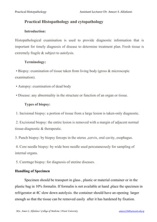
Small intestine well preserved Autolyzed Small intestine
5. Practical Histopathology Assistant Lecturer Dr. Ameer S. Alfatlawi
ameers226@uowasit.edu.iqWasit UniversityCollege of MedicineMSc. Ameer S. Alfatlawi
10% neutral buffered form(NBF) is most commonly used fixative.
It is prepared by mixing 40% formaldehyde gas in 100 w/v of distilled
water.
• The resultant mixture is 100% formalin. Routinely, 10% formalin is used which is
prepared by mixing 10ml of 100% formalin in 90ml of distilled water.
Mechanism of action:
• It forms cross links between amino acids of proteins thereby making them insoluble.
• It fixes 4 mm thick tissue in 8 hours .
• The fixative should be 10 times more in volume then the specimen.
Advantages:
• Rapid penetration
• Easy availability & cheap
• Does not overharden the tissue
• Fixes lipids for frozen sections
Disadvantages:
• Irritant to the nose, eyes and mucous membranes
• Formation of precipitate of paraformaldehyde (which can be
prevented by adding 11- 16% methanol)
• Formation of black formalin pigment (acid formaldehyde hematin).
Types of fixatives
• Physical methods:
– Heating, Microwaving, Freeze drying
Chemical methods
• Coagulatant fixatives :(Precipitate and coagulate protein)
– Alcohols and acetone
– Picric acid and trichloro acetic acid
• Cross linking fixatives:
– Formaldehyde and glutaraldehyde
– Other aldehydes (chloral hydrate, glyoxal)
6. Practical Histopathology Assistant Lecturer Dr. Ameer S. Alfatlawi
ameers226@uowasit.edu.iqWasit UniversityCollege of MedicineMSc. Ameer S. Alfatlawi
– Metal salts (Mercuric and zinc chloride, osmium tetroxide)
• Compount fixatives.
Other Fixatives:
• Glutaraldehyde and Osmium Tetraoxide
• B5 fixative:
– stock solution: Mercuric chloride, sodium acetate & distilled water
– Add 2ml of formaldehyde to 20ml of stock solution
• Zenker's fluid
• Bouin’s Fluid
Decalcification:
• It is the process of removal of the calcium salts from the specimen.
• The various agents used for decalcifying are:
• Nitric acid
• Hydrochloric acid
• Formic acid
• Picric acid
• Acetic acid
• Citric
Factors affecting the quality of fixation:
1. Size and thickness of piece of tissue.
2. Tissue covered by large amount of mucous and blood fix slowly.
3. Fatty and lipomatous tissue fix slowly.
4. pH : Should be kept in the physiological range, between pH 4-9. The pH for
the ultrastructure preservation should be at pH 7.2–7.4 (i.e. neutral buffered formalin).
5. Osmolarity :Hypertonic solutions give rise to cell shrinkage, Hypotonic
solutions result in cell swelling and poor fixation.
7. Practical Histopathology Assistant Lecturer Dr. Ameer S. Alfatlawi
ameers226@uowasit.edu.iqWasit UniversityCollege of MedicineMSc. Ameer S. Alfatlawi
6. Temperature : Hot formalin will fix tissues faster , care is required to avoid
cooking the specimen.
7. Concentration of fixative.
8. Volume of the Fixative: At least 15-20 times greater than tissue volume.
9. Time interval from of removal of tissues to fixation: The faster you can get
the tissue and fix it, the better.
10- Duration of fixation : Most fixatives, such as NBF, will penetrate tissue to
the depth of approximately 1 mm in one hour.
Dehydration:
This is a process in which water from cells and tissues is removed so that this space is
subsequently taken up by wax. Dehydration is carried out by passing the tissues
through a series of ascending grades of alcohol: 70%, 80%, 95% and absolute alcohol.
If ethyl alcohol is not available then methyl alcohol, isopropyl alcohol or acetone.
Clearing
This is the process in which alcohol from tissues and cells is removed and is
replaced by a fluid in which wax is soluble and it also makes the tissue
transparent. Xylene is the most commonly used clearing agent. Toluene,
benzene (it is carcinogenic), chloroform (it is poisonous) and cedar wood
oil (it is expensive and very viscous) can also be used as clearing agent.
Impregnation
This is the process in which empty spaces in the tissue and cells after
removal of water are taken up by paraffin wax. This hardens the tissue
8. Practical Histopathology Assistant Lecturer Dr. Ameer S. Alfatlawi
ameers226@uowasit.edu.iqWasit UniversityCollege of MedicineMSc. Ameer S. Alfatlawi
which helps in section cutting. Impregnation is done in molten paraffin wax
which has the melting point of 56oC (54-62oC).
https://www.youtube.com/watch?v=7-LIbAWPc-g
https://www.youtube.com/watch?v=fIs6PlCDeRE
Embedding and blocking
Embedding of tissue is done in molten wax. Wax blocks are conventionally
prepared using metallic L (Leuckhart’s mould); nowadays plastic moulds of
different colours for blocking are also available (. The moulds are placed
over a smooth surfaced glass tile. Molten wax is poured in the cavity in the
moulds. The processed tissue pieces are put into wax with number tag and
examining surface facing downward. Wax is allowed to solidify. After
solidification, if L-moulds are used they are removed while plastic mould
remains with the wax block. In either case, each block contains a tissue
piece carrying a identification label. Embedding and blocking can also be
performed in a special instrument called embedding centre. It has a wax
reservoir, heated area for steel moulds, wax dispenser, and separate hot and
cold plates for embedding and blocking.
Paraffin wax is used for embedding of tissue which form tissue blocks after
cooling the it can be trimmed into thin sections (4-5 microns) using microt-
ome, the sections are placed on glass slides and become ready for staining.
9. Practical Histopathology Assistant Lecturer Dr. Ameer S. Alfatlawi
ameers226@uowasit.edu.iqWasit UniversityCollege of MedicineMSc. Ameer S. Alfatlawi
10. Practical Histopathology Assistant Lecturer Dr. Ameer S. Alfatlawi
ameers226@uowasit.edu.iqWasit UniversityCollege of MedicineMSc. Ameer S. Alfatlawi
Routine staining (H & E): is done with haematoxylin and eosin (H&E): is the
most widely used stain in histopathology.
Nuclei appear dark blue
Collage and cytoplasm appear pink
Keratin appears pink to red
Procedure for Staining:
Sections are first deparaffinised (removal of wax) by placing the slide in a
jar of xylene for 10-15 minutes. As haematoxylin is a water-based dye, the
sections before staining are rehydrated which is done by passing the
sections in a series of descending grades of alcohol and finally bringing the
section to water.
Place the slide in haematoxylin stain for 8-10 minutes.
Rinse in water.
Differentiation (i.e. selective removal of excess dye from the
section) is done by putting the slide in a solution of 1% acid
alcohol for 10 seconds.
11. Practical Histopathology Assistant Lecturer Dr. Ameer S. Alfatlawi
ameers226@uowasit.edu.iqWasit UniversityCollege of MedicineMSc. Ameer S. Alfatlawi
Rinse in water.
Blueing (i.e. bringing of required blue colour to the section) is
done by putting the section in Scott’s tap water (containing
sodium bicarbonate and magnesium sulfate) or saturated
solution of lithium carbonate for 2-10 minutes.
Counterstain with 1% aqueous solution of eosin for 1- 3
minutes.
Rinse in tap water.
Before mounting, the sections have to be dehydrated which is
done by passing the sections in a series of ascending grades of
alcohol and finally cleared in xylene, 2-3 dips in each solution.
Mount in DPX (dextrene polystyrene xylene) or
Canada balsam.
Hematoxylin and Eosin (H&E) staining
•Place slides containing paraffin sections in a slide holder (glass
or metal)
•Deparaffinize and rehydrate sections:
Xylene I 5 minutes (blot excess xylene before going into
ethanol)
Xylene II 5 minutes
100% ethanol 2 minutes
100% ethanol 2 minutes
90% ethanol 2 minutes
70% ethanol 2 minutes
12. Practical Histopathology Assistant Lecturer Dr. Ameer S. Alfatlawi
ameers226@uowasit.edu.iqWasit UniversityCollege of MedicineMSc. Ameer S. Alfatlawi
50% ethanol 2 minutes
deionized H2O 5 minutes
•While sections are in water, skim surface of hematoxylin with a
Kim wipe to remove oxidized particles. Blot excess water from
slide holder before going into hematoxylin.
Hematoxylin staining:
Hematoxylin 3 minutes
Tap water 5 minutes (to allow stain to develop)
Acid ethanol 8-12x (fast Dip) (to de-stain)
Tap water Rinse 2 x 1´
Note :Blot excess water from slide holder before going into
eosin.
Eosin staining and dehydration:
Eosin 1 minutes
50% ethanol 2 minutes
70% ethanol 2 minutes
90% ethanol 2 minutes
100% ethanol 2 minutes (blot excess ethanol before going into
xylene)
100% ethanol 5 minutes
Xylene 2 minutes
Xylene 2 minutes
Dry
13. Practical Histopathology Assistant Lecturer Dr. Ameer S. Alfatlawi
ameers226@uowasit.edu.iqWasit UniversityCollege of MedicineMSc. Ameer S. Alfatlawi
Handling procedures for surgical pathology tissue specimens:
SPECIMEN REQUEST PROCEDURE TEST
Tissue Routine Microscopy Fix in 10% Formalin H&E Stained Slide Review
Tissue Frozen Section for
Diagnosis and/or
Margins
Notify pathology lab in
advance, send fresh
immediately
Frozen section
consultation
Tissue Immunohistochemistry for
tumor/tissue typing
Fix in 10% Formalin Immunohistochemical
stained slide review
Tissue Electron microscopy
for tumor/tissue typing
Notify pathology lab in
advance, send fresh
immediately
Electron microscopic
analysis Reference
Laboratory
Tissue Chromosomal Analysis Notify pathology lab in
advance, send fresh in a
sterile container
immediately
Chromosome analysis
Reference Laboratory
Tissue Special stain for
Organism or
Cellular Product
Fix in 10% Formalin Fungus-PAS/Silver
Methenamine Stain
AFB /Leprosy-AFB FITE
stain
Breast
biopsy
Hormone Receptor
Analysis
Prognostic/Predictiv
e Marker Analysis
Fix in 10% Formalin
immediately, minimum 6
hours and no longer than 72
hours
Immunohistochemical
analysis quantification
Bone
Marrow
Biopsy
Routine Microscopy Fix in 10% Formalin H&E Stained slide review
Kidney
Biopsy
Electron Microscopy
Immunofluorescenc
e
Light Microscopy
Notify pathology lab in
advance, send fresh
immediately
EM/Immunofluorescence
Light Microscopy
Special Stains
Lymph Node R/O Lymphoma Send fresh immediately Special fixation
Touch Preparations
Flow Cytometry
Lymph Node Culture for Infectious
Disease/Microbiolog
y
Notify pathology lab in
advance, send fresh in
sterile container
immediately
Culture
Skin
Biopsy
(mucosa,
lung)
Immunofluorescence
for immune mediated
disease
Notify pathology lab in
advance, send fresh
immediately
Special holding medium
immunofluorescence
Reference Laboratory
Skeletal
Muscle
Biopsy
Enzyme
Histochemistry for
disease typing
Notify pathology lab in
advance, send fresh
immediately
Enzyme Histochemistry
14. Practical Histopathology Assistant Lecturer Dr. Ameer S. Alfatlawi
ameers226@uowasit.edu.iqWasit UniversityCollege of MedicineMSc. Ameer S. Alfatlawi
Cytology: Is the study of cell (normal or diseased altered cell) obtained from various
sites of the body. It allows rapid diagnosis often within minutes. Cells examined by
this process are collected by one of the following methods: 1. Exfoliated cell 2. Cells
removed by brushing or scraping 3. Removal of cells from deep tissue (by aspiration)
Indications of cytopathology
1. Diagnosis of malignancy. 2. Diagnosis of precancerous changes e.g. cervical atypia.
3. Detection of inflammation and pathogenic agents e.g. fungal or parasitic infection of
vagina 4.Study of hormonal patterns and gonadal function e.g. examination of
squamous cells in vaginal smear which are under influence of ovarian hormones to
assess ovarian function in infertility. 5. Identification of sex chromosome in newborn
with ambiguous genitalia, buccal smear is used as a source of cells.
Limitations of cytology
1. The nature of lesion is not so obvious as in a histological section. 2. Difficulty in
identification of the exact site of lesion e.g. malignant squamous cells shaded in
sputum may arise from buccal mucosa, pharynx, larynx and bronchi. 3. The size
of lesion cannot be approximated by cytology.
Technique of cytology:
is sprayed on a slide directly.
into the mass (while the mass is fixed in between fingers and thumb) and is
moved in different directions while applying negative pressure onto the syringe
with continuous suction of material throughout the process, then the needle
should be withdrawn gently from the mass.
2. Smearing collected material on a glass slide .
3. Immediate immersion of the slide in a fixative (95% ethanol) to avoid dryness
of material. 4. Applying a stain. 5. Examine under the microscope.
15. Practical Histopathology Assistant Lecturer Dr. Ameer S. Alfatlawi
ameers226@uowasit.edu.iqWasit UniversityCollege of MedicineMSc. Ameer S. Alfatlawi