SlideShare a Scribd company logo
1 of 167
Download to read offline
Preoperative planning in
Veterinary Orthopaedics
CAPT. DR. RAVI RAIDURG
M.V.Sc (Surgery), PhD
Associate Professor & Head,
Department of Surgery & Radiology, Veterinary College,
Vinoba Nagar, Shivamogga 577 204, Karnataka, India
Phone: +919449827183, E mail: raviraidurg@gmail.com 1
raviraidurg@gmail.com
2
PROGENY PPAK WEBINAR SERIES
S.No Webinar Topics
1 Preoperative planning in Veterinary Orthopaedics
2 Evolution of Internal Fixation in Veterinary Orthopaedics
3 Methods of fracture repair in Veterinary Orthopaedics
4 Plate Osteosynthesis – Terminologies and Instrumentation
5
Open Reduction and Internal Fixation (ORIF) for long bone
fracture repair in dogs
6
Management of long bone diaphyseal fractures with minimal
invasive plate osteosynthesis (MIPO)in dogs
raviraidurg@gmail.com
3
PROGENY PPAK WEBINAR SERIES
For every fracture,
there are more than one method of
fixation that will lead to acceptable
results
raviraidurg@gmail.com
4
For every fracture,
there are more than one method of
fixation that will lead to acceptable
results
and
many more that will lead to
unacceptable results
raviraidurg@gmail.com
5
Preoperative planning
in Veterinary Orthopaedics
Aim : Introduce the concept of “ Preoperative planning in
Veterinary Orthopaedics”
Learning Objectives
At the end of this session you should be able to:
• Describe Fracture healing (Primary vs Secondary bone healing)
• Describe Fracture Classification
• Describe AO Fracture Classification of Long bones (eg AO 32 A3
???)
• Derive a Fracture Patient Assessment Score (FPAS) and describe
factors (mechanical, biological and clinical) which support the score.
• List Preoperative planning in MIO (Minimally Invasive
Osteosynthesis)
6
raviraidurg@gmail.com
S.
No
Topic
Time
(min)
Slides
1 Introduction 10 01-12
2 Case presentations 15 13 - 39
3 Fracture healing
20
40 – 47
4 Fracture classification 48 – 59
5 AO Fracture classification of long bones 60 – 68
6 FPAS (Fracture Patient Assessment Score)
30
70 – 79
7 Treatment guidelines based on FPAS 80 – 140
8
Preoperative planning in MIO
(Minimally Invasive Osteosynthesis)
10 141 – 158
9 Summary 05 159 – 160
raviraidurg@gmail.com
7
Preoperative planning
in Veterinary Orthopaedics
SESSION OUTLINE (90 Min)
Cases presented to Veterinary
College Shivamogga (2015 – 2018)
Humerus
raviraidurg@gmail.com
8
Case example
14 Year old, 5 kg dog (SPITZ) ie
GERIATRIC DOG
Diaphyseal long oblique /
Spiral fracture
raviraidurg@gmail.com
9
How could I fix this
1. POP cast (Conservative
method)
2. Wiring
3. External fixator (ESF)
4. Internal fixator
IMP
IMP + wiring
IMP + Thomas splint
ILN (Interlocking Nails)
TENS (Titanium elastic nail system)
PO (Plate osteosynthesis ie Bone plating)
Results of the Poll
raviraidurg@gmail.com
10
Case example
14 Year old, 5 kg dog (SPITZ) ie
GERIATRIC DOG
Diaphyseal long oblique /
Spiral fracture
raviraidurg@gmail.com
11
How could I fix this
Internal fixator
1. IMP
2. IMP + wiring
3. ILN (Interlocking Nails)
4. Lag Screw with Plate osteosynthesis (Bone plating)
Results of the Poll
raviraidurg@gmail.com
12
Case No 1
13
raviraidurg@gmail.com
Humerus
Case No 4946_25 Nov 2015
raviraidurg@gmail.com
14
A 14 Year old, 5 kg dog ie GERIATRIC DOG
Diaphyseal long oblique / Spiral fracture
Humerus Case No 4946_25 Nov 2015
A 14 Year old, 5 kg dog ie GERIATRIC DOG
Diaphyseal long oblique / Spiral fracture
raviraidurg@gmail.com
15
Humerus Case No 4946_25 Nov 2015
A 14 Year old, 5 kg dog ie GERIATRIC DOG
Diaphyseal long oblique / Spiral fracture
raviraidurg@gmail.com
16
Humerus Case No 4946_25 Nov 2015
A 14 Year old, 5 kg dog ie GERIATRIC DOG
Diaphyseal long oblique / Spiral fracture
raviraidurg@gmail.com
17
Humerus Case No 4946_25 Nov 2015
A 14 Year old, 5 kg dog ie GERIATRIC DOG
Diaphyseal long oblique / Spiral fracture
raviraidurg@gmail.com
18
Humerus Case No 4946_25 Nov 2015
A 14 Year old, 5 kg dog ie GERIATRIC DOG
Diaphyseal long oblique / Spiral fracture
raviraidurg@gmail.com
19
6 th Week Post Operative
Humerus Case No 4946_25 Nov 2015
A 14 Year old, 5 kg dog ie GERIATRIC DOG
Diaphyseal long oblique / Spiral fracture
raviraidurg@gmail.com
20
6 th Week Post Operative
Case No 2
21
raviraidurg@gmail.com
Humerus
Case No 8698 dated 07 Dec 2015
raviraidurg@gmail.com
22
A 05 mth, 45 kg dog ie JUVENILE DOG
Diaphyseal Transverse fracture with longitudinal slit in distal fragment
Humerus_ Case No 8698 dated 07 Dec 2015
A 05 mth, 45 kg Great Dane Pup ie JUVENILE DOG
Diaphyseal Transverse fracture with longitudinal slit in
distal fragment
raviraidurg@gmail.com
23
raviraidurg@gmail.com
24
Humerus_ Case No 8698 dated 07 Dec 2015
A 05 mth, 45 kg Great Dane Pup ie JUVENILE DOG
Diaphyseal Transverse fracture with longitudinal slit in
distal fragment
Humerus_ Case No 8698_GD_07 Dec 2015
A 05 mth, 45 kg dog ie JUVENILE DOG
Diaphyseal Transverse fracture with longitudinal slit
in distal fragment
raviraidurg@gmail.com
25
raviraidurg@gmail.com
26
Humerus_ Case No 8698 dated 07 Dec 2015
A 05 mth, 45 kg Great Dane Pup ie JUVENILE DOG
Diaphyseal Transverse fracture with longitudinal slit in
distal fragment
raviraidurg@gmail.com
27
Humerus_ Case No 8698 dated 07 Dec 2015
A 05 mth, 45 kg Great Dane Pup ie JUVENILE DOG
Diaphyseal Transverse fracture with longitudinal slit in
distal fragment
raviraidurg@gmail.com
28
Humerus_ Case No 8698 dated 07 Dec 2015
A 05 mth, 45 kg Great Dane Pup ie JUVENILE DOG
Diaphyseal Transverse fracture with longitudinal slit in
distal fragment
raviraidurg@gmail.com
29
10 th Post Operative day
Humerus_ Case No 8698 dated 07 Dec 2015
A 05 mth, 45 kg Great Dane Pup ie JUVENILE DOG
Diaphyseal Transverse fracture with longitudinal slit in
distal fragment
raviraidurg@gmail.com
303 rd Week Post Operative day
Humerus_ Case No 8698 dated 07 Dec 2015
A 05 mth, 45 kg Great Dane Pup ie JUVENILE DOG
Diaphyseal Transverse fracture with longitudinal slit in
distal fragment
raviraidurg@gmail.com
31
3 rd Week Post Operative day
Humerus_ Case No 8698 dated 07 Dec 2015
A 05 mth, 45 kg Great Dane Pup ie JUVENILE DOG
Diaphyseal Transverse fracture with longitudinal slit in
distal fragment
raviraidurg@gmail.com
32
17 th Week Post Operative day
Humerus_ Case No 8698 dated 07 Dec 2015
A 05 mth, 45 kg Great Dane Pup ie JUVENILE DOG
Diaphyseal Transverse fracture with longitudinal slit in
distal fragment
Humerus_ Case No 8698_GD_07 Dec 2015
A 05 mth, 45 kg dog ie JUVENILE DOG
Diaphyseal Transverse fracture with longitudinal slit
in distal fragment
raviraidurg@gmail.com
33
30th Month Post Operative day
Case No 3
34
raviraidurg@gmail.com
Humerus
Case No 10148 dated 27 June 2016
raviraidurg@gmail.com
35
A 2 ½ Year old, 22 kg Mudhol Hound dog
Transverse fracture in distal fragment
raviraidurg@gmail.com
36
Humerus
Case No 10148 dated 27 June 2016
raviraidurg@gmail.com
37
Humerus
Case No 10148 dated 27 June 2016
raviraidurg@gmail.com
38
Humerus
Case No 10148 dated 27 June 2016
raviraidurg@gmail.com
39
Humerus
Case No 10148 dated 27 June 2016
Preoperative planning in Veterinary
Orthopaedics
40
raviraidurg@gmail.com
Preoperative planning
in Veterinary Orthopaedics
Learning Objectives
At the end of this session you should be able to:
• Describe Fracture healing
(Primary vs Secondary bone healing)
• Describe Fracture Classification
• Describe AO Fracture Classification of Long bones (eg AO 32 A3 ???)
• Derive a Fracture Patient Assessment Score (FPAS) and describe factors
(mechanical, biological and clinical) which support the score.
• List Preoperative planning in MIO (Minimally Invasive Osteosynthesis)
41
raviraidurg@gmail.com
FRACTURE HEALING
PRIMARY BONE HEALING
SECONDARY BONE HEALING
42
raviraidurg@gmail.com
FRACTURE HEALING
PRIMARY BONE HEALING
• Rigidly stabilised fracture fragments with direct
contact between them heal by migration of ‘cutting
cones’ (comprising osteoclasts and osteoblasts)
from one fragment to the other, resulting in
reformation of lamellar bone (Haversian
remodelling) without any requirement for precursor
tissue or callus.
• The lack of a callus phase can be very important in
some cases, particularly when joints are affected.
43
raviraidurg@gmail.com
FRACTURE HEALING
PRIMARY BONE HEALING
• ‘Gap healing’, in which rigidly stabilised fragments
are separated by a gap of up to 1 mm, is a slight
variation of this mechanism.
• The space is initially filled by lamellar bone
orientated at 90° to the long axis of the bone,
which enables the cutting cones to migrate across
the gap and reorientate the lamellar bone.
44
raviraidurg@gmail.com
FRACTURE HEALING
PRIMARY BONE HEALING
• Clinical union takes significantly longer to achieve with primary bone healing than
secondary bone healing, and therefore relies on implants for longer.
• Primary bone healing can only be obtained with the use of internal fixation due to the
requirement for absolute rigidity with less than 2 per cent strain at the fracture site.
45
raviraidurg@gmail.com
FRACTURE HEALING
SECONDARY BONE HEALING
• Fragments separated by a gap of more than 1 mm or with an interfragmentary strain
greater than 2 per cent will undergo secondary bone healing.
• This involves the formation of tissues (ie, callus) that can cope with the strain occurring
at the fracture site. As increased stability is conferred by the callus, the next tissue in the
sequence forms, which increases stability still further and, in turn, allows the next tissue
to form, until bone tissue can once again fill the fracture gap.
46
raviraidurg@gmail.com
SECONDARY BONE HEALING
raviraidurg@gmail.com
47
Preoperative planning
in Veterinary Orthopaedics
Learning Objectives
At the end of this section you should be able to:
• Describe Fracture healing (Primary vs Secondary bone healing)
• Describe Fracture Classification
• Describe AO Fracture Classification of Long bones (eg AO 32 A3 ???)
• Derive a Fracture Patient Assessment Score (FPAS) and describe factors
(mechanical, biological and clinical) which support the score.
• List Preoperative planning in MIO (Minimally Invasive Osteosynthesis)
48
raviraidurg@gmail.com
Fracture classification
■ Cause
■ Open or closed
■ Extent of bone damage
■ Number and position of fracture lines
■ Direction
■ Location
■ Forces acting on the fracture
■ Stability
■ Degree of soft tissue damage
raviraidurg@gmail.com
49
Fracture classification
CAUSE OF THE FRACTURE
Intrinsic
■ MUSCULAR (eg, tibial tuberosity avulsion secondary to quadriceps muscle
contraction).
■ PATHOLOGICAL (eg, secondary to a primary bone neoplasm).
■ STRESS (a multiple low-grade trauma can eventually result in a fracture).
Extrinsic
• Extrinsic fractures are caused by external trauma (eg, a
• road traffic accident).
raviraidurg@gmail.com
50
Fracture classification
OPEN/CLOSED FRACTURE
• Radiographs should be examined for signs of gas shadows within the soft tissue
planes that may suggest communication with the external environment.
• Open fractures should be graded
raviraidurg@gmail.com
51
Fracture classification
■ OPEN VERSUS CLOSED
• Open fractures are more challenging to treat due to the risks of infection and the
potential for damage to the soft tissues surrounding the bone. The importance of
soft tissue damage is reflected in the grading system used to describe these
fractures:
GRADE 1. The skin has been punctured by a sharp bone fragment that has usually
retracted back beneath the surface. There is typically only mild soft tissue damage
associated with the relatively small wound;
GRADE 2. The skin lesion is due to extrinsic trauma leaving a variable-sized soft
tissue deficit and more severe damage to the surrounding tissues. Foreign material may
be present within the wound;
GRADE 3. There is extensive loss of multiple tissue types due to extrinsic trauma. The
fracture is usually fragmented due to the high-energy nature of the injury.
Neurovascular injury is likely and could cause complications.
raviraidurg@gmail.com
52
Fracture classification
EXTENT OF BONE DAMAGE
Incomplete
■ GREENSTICK. Young animals can fracture one cortex, with the opposite cortex
undergoing plastic deformation rather than fracture (like snapping a green stick).
■ FISSURE. Radiographs should be examined carefully for fissures leading away from
a fracture line, as they can greatly increase the complexity of the repair or progress to a
full fracture if not addressed. A fissure is often associated with comminuted fractures.
■ DEPRESSED. Depressed fractures usually occur in the skull.
Complete
• Complete fractures involve both cortices of the bone.
raviraidurg@gmail.com
53
Fracture classification
NUMBER AND POSITION OF THE FRACTURE LINES
■ SIMPLE. One fracture line without small fragments of bone.
■ SEGMENTAL. Two or more fracture lines that do not communicate with each other.
■ COMMINUTED. Two or more fracture lines that do communicate with each other.
This type of fracture is usually associated with high-energy trauma and therefore
extensive soft tissue damage.
raviraidurg@gmail.com
54
Fracture classification
raviraidurg@gmail.com
55
DIRECTION OF FRACTURE LINES
■ TRANSVERSE. The fracture line is perpendicular to the long axis of the bone.
■ OBLIQUE. Generally, a fracture that is 1·5 to two times longer than the diameter of the
bone (or longer) is considered oblique. The point when a short oblique fracture is
classified as a transverse fracture is a unclear.
■ SPIRAL. The fracture line curves around the bone (usually due to a twisting force)
creating a complex three-dimensional fracture.
Fracture classification
raviraidurg@gmail.com
56
FRACTURE LOCATION
■ DIAPHYSEAL. Cortical bone is affected
and the
fracture can be described as proximal, mid or
distal.
■ METAPHYSEAL. Cancellous bone
situated towards the ends of the bones
(metaphyses) has differing characteristics of
implant holding and will often heal more
quickly once stabilised than diaphyseal bone.
■ ARTICULAR. Once a joint suffers a
fracture, a degree of secondary osteoarthrosis
is inevitable, but the extent of the
degeneration and effect on joint mobility can
be minimised by appropriate and prompt
fracture management.
■ CONDYLAR.
Fracture classification
raviraidurg@gmail.com
57
FRACTURE LOCATION
■ PHYSEAL. Fractures involving the physis
of skeletally immature bones are classified
according to the Salter-Harris system.
Smooth implants that cross the physis
perpendicular to the direction of growth and
occupy as little of the physeal area as
possible are ideal for repair in order to
preserve as much growth potential as
possible. Fracture location and configuration
usually dictate implant orientation away from
this ideal, with the result that implants are
removed in very immature animals to allow
continued growth. Unfortunately, despite
gentle handling, growth disturbances are
common following physeal injury and
owners should be made aware of this.
Forces acting on Bone
Bending Torsional Axial compression
Axial tension Shearing
Preoperative planning
in Veterinary Orthopaedics
Learning Objectives
At the end of this section you should be able to:
• Describe Fracture healing (Primary vs Secondary bone healing)
• Describe Fracture Classification
• Describe AO Fracture Classification of Long
bones (eg AO 32 A3 ???)
• Derive a Fracture Patient Assessment Score (FPAS) and describe factors
(mechanical, biological and clinical) which support the score.
• List Preoperative planning in MIO (Minimally Invasive Osteosynthesis)
59
raviraidurg@gmail.com
AO
FRACTURE CLASSIFICATION OF
LONG BONES
60
raviraidurg@gmail.com
1958 AO/ASIF
(Arbeitsgemeinschaft fur osteosynthesefragen)
Maurice Müller, Martin Allgöwer, Hans Willenegger,
Robert Schneider and Robert Mathys (Switzerland)
• Plate and screw osteosynthesis.
• A group of Swiss orthopaedic surgeons formed the
Arbeitsgemeinschaft fur osteosynthesefragen (AO), also
known as the Association for the Study of Internal Fixation
(ASIF).
• The principles for fracture management developed by the AO
group defined the standard of care for fracture
61
62
1958
AO founded in Biel (Bienne)
November 6, 1958, Hotel Elite
Biel (Bienne), Switzerland
63
1958 AO
Founders
Arbeitsgemeinschaft für Osteosynthesefragen
Maurice Müller
Zürich
1918−2009
Martin Allgöwer
Chur
1917−2007
Walter Bandi
Interlaken
1912−1997
Robert Schneider
Grosshöchstetten
1912−1990
Hans Willenegger
Liestal
1910−1998
Fracture classification
raviraidurg@gmail.com
64
• The first symbol of the alpha-numeric code represents
the fractured bone, humerus is coded as 1, Radius –
Ulna as 2, femur as 3 and tibia which was coded as 4.
• The second symbol represents “location of the fracture”
ie Proximal as 1, middle diaphyseal as 2 and Distal as 3
• Simple (A), wedge (B) and complex fractures (C).
• A1 incomplete fractures.
• A2 indicates diaphyseal simple oblique fractures,
• A3 indicates diaphyseal simple transverse fractures,
• B1 indicates diaphyseal one reducible wedge,
• B2 indicates diaphyseal several reducible wedge
• B3 indicated diaphyseal non-reducible wedge.
• C1 indicates Complex reducible wedge
• C 2 indicates Complex segmental wedge,
• C3 indicates Complex non reducible wedge,
AO 32 A3
raviraidurg@gmail.com
65
AO
FRACTURE CLASSIFICATION OF LONG BONES
raviraidurg@gmail.com
66
AO
FRACTURE CLASSIFICATION OF LONG BONES
raviraidurg@gmail.com
67
AO
FRACTURE CLASSIFICATION OF LONG BONES
raviraidurg@gmail.com
68
AO
FRACTURE CLASSIFICATION OF LONG BONES
Preoperative planning
in Veterinary Orthopaedics
Learning Objectives
At the end of this section you should be able to:
• Describe Fracture healing (Primary vs Secondary bone healing)
• Describe Fracture Classification
• Describe AO Fracture Classification of Long bones (eg AO 32 A3 ???)
• Derive a Fracture Patient Assessment Score
(FPAS) and describe factors (mechanical, biological and clinical) which
support the score.
• List Preoperative planning in MIO (Minimally Invasive Osteosynthesis)
69
raviraidurg@gmail.com
FPAS
(Fracture Patient Assessment Score)
70
raviraidurg@gmail.com
FPAS
• The FPAS is a simple 1 -10 scoring system that is determined
by evaluating pertinent mechanical, biological, and clinical
factors to determine the relative needs of the fracture case.
• The “10” end of the scale indicates ideal conditions (i.e. good
news) and the “1” end represents the most challenging
conditions (i.e. bad news) .
• Acknowledging that each and every orthopedic patient is
different, it is advisable to derive a FPAS for each patient prior
to treatment.
raviraidurg@gmail.com
71
FPAS
raviraidurg@gmail.com
72
1 ☺10
Marked instability Moderate instability Mild instability
MECHANICAL
Non-Load-Sharing Partial Load-Sharing Ideal Load-Sharing
Large Patient Size Medium Patient Small Patient Size
FACTORS
Multi-limb dysfunction Moderate multi-limb Single limb dysfunction
CLINICAL
FACTORS
Unwilling to follow-up Questionable followup Willing/able to follow-up
Owner
Unable to restrict activity Willing to try Able to restrict activity
Intolerant of activity restrict Somewhat tolerant Tolerant of activ. restr..
Patient
Rambunctious Active, but responsive Calm
Intolerant of treatments Difficult to treat Easy to treat
BIOLOGICAL
FACTORS
Severe tissue injury Moderate tissue injury Minimal tissue injuryLocal Factors
Comminuted Transverse/Oblique Spiral Greenstick
Open Fx – Type III Type II Type I Closed Fracture
Marked instability Moderate instability Mild instability
Previous radiation Tx
Systemic Geriatric Mature Adult Immature Patient
Factors Debilitated/ill Compensated illness Healthy
Second-hand smoke?
FPAS
(Fracture Patient Assessment Score)
MECHANICAL FACTORS
73
raviraidurg@gmail.com
FPAS MECHANICAL FACTORS
raviraidurg@gmail.com
74
The amount of loading that a given
fixation device must withstand is
influenced by the following factors:
• palpable stability of the fracture zone,
•
• the number of dysfunctional limbs,
•
• the patient’s coordination and agility
•
• the patient’s weight,/size, and
•
• the degree of “load-sharing”
anticipated between the fixation
device and the bony column.
1 ☺10
Marked instability Moderate instability Mild instability
MECHANICAL
Non-Load-Sharing Partial Load-Sharing Ideal Load-Sharing
Large Patient Size Medium Patient Small Patient Size
FACTORS
Multi-limb dysfunction Moderate multi-limb Single limb dysfunction
CLINICAL
FACTORS
Unwilling to follow-up Questionable followup Willing/able to follow-up
Owner
Unable to restrict activity Willing to try Able to restrict activity
Intolerant of activity restrict Somewhat tolerant Tolerant of activ. restr..
Patient
Rambunctious Active, but responsive Calm
Intolerant of treatments Difficult to treat Easy to treat
BIOLOGICAL
FACTORS
Severe tissue injury Moderate tissue injury Minimal tissue injuryLocal Factors
Comminuted Transverse/Oblique Spiral Greenstick
Open Fx – Type III Type II Type I Closed Fracture
Marked instability Moderate instability Mild instability
Previous radiation Tx
Systemic Geriatric Mature Adult Immature Patient
Factors Debilitated/ill Compensated illness Healthy
Second-hand smoke?
raviraidurg@gmail.com
75
Fig 1: Ideal load-sharing Fig 2: Non-load-sharing Fig 3: Partial load-sharing
The mechanical FPAS shifts downward (toward 1) when
there is multi-limb dysfunction, a large, clumsy patient,
and/or the anticipation of a non-load-sharing
osteosynthesis.
FPAS
(Fracture Patient Assessment Score)
CLINICAL FACTORS
76
raviraidurg@gmail.com
FPAS CLINICAL FACTORS
raviraidurg@gmail.com
77
• Poor patient/owner compliance leads to excessive
patient activity and/or insufficient physical
therapy.
• Fracture treatment plans should be made with
possibility of poor exercise restriction kept in
mind.
• Poor anticipated compliance shifts the FPAS
downward. Since poor patient/owner compliance
invariably increases the mechanical demand upon
the fixation, the clinical FCAS is averaged with
the mechanical FPAS to derive a total mechanical
score.
• Sometimes owner compliance concerns cause
us to avoid the use of External Skeletal
Fixation.
• In attempt to shift the FPAS toward 10, I spend
extra time with pet owners explaining the
importance of exercise restriction as well as the
potential consequences of failing to do so
(especially in challenging fracture cases).
1 ☺10
Marked instability Moderate instability Mild instability
MECHANICAL
Non-Load-Sharing Partial Load-Sharing Ideal Load-Sharing
Large Patient Size Medium Patient Small Patient Size
FACTORS
Multi-limb dysfunction Moderate multi-limb Single limb dysfunction
CLINICAL
FACTORS
Unwilling to follow-up Questionable followup Willing/able to follow-up
Owner
Unable to restrict activity Willing to try Able to restrict activity
Intolerant of activity restrict Somewhat tolerant Tolerant of activ. restr..
Patient
Rambunctious Active, but responsive Calm
Intolerant of treatments Difficult to treat Easy to treat
BIOLOGICAL
FACTORS
Severe tissue injury Moderate tissue injury Minimal tissue injuryLocal Factors
Comminuted Transverse/Oblique Spiral Greenstick
Open Fx – Type III Type II Type I Closed Fracture
Marked instability Moderate instability Mild instability
Previous radiation Tx
Systemic Geriatric Mature Adult Immature Patient
Factors Debilitated/ill Compensated illness Healthy
Second-hand smoke?
FPAS
(Fracture Patient Assessment Score)
BIOLOGICAL FACTORS
78
raviraidurg@gmail.com
FPAS BIOLOGICAL FACTORS
raviraidurg@gmail.com
79
• Bone healing is influenced by
• local factors(fracture zone):
open fractures, severe soft tissue
disruption, and previous local
radiation fields.
• Systemic factors: old age,
cachexia, and systemic illness.
• Each of these local and systemic
factors will shift the biological
FPAS toward 1. Conversely, bone
healing is more rapid in closed
fractures with minimal soft tissue
injury, and in young, healthy,
well-nourished patients; each of
which shifts the FPAS toward 10.
1 ☺10
Marked instability Moderate instability Mild instability
MECHANICAL
Non-Load-Sharing Partial Load-Sharing Ideal Load-Sharing
Large Patient Size Medium Patient Small Patient Size
FACTORS
Multi-limb dysfunction Moderate multi-limb Single limb dysfunction
CLINICAL
FACTORS
Unwilling to follow-up Questionable followup Willing/able to follow-up
Owner
Unable to restrict activity Willing to try Able to restrict activity
Intolerant of activity restrict Somewhat tolerant Tolerant of activ. restr..
Patient
Rambunctious Active, but responsive Calm
Intolerant of treatments Difficult to treat Easy to treat
BIOLOGICAL
FACTORS
Severe tissue injury Moderate tissue injury Minimal tissue injuryLocal Factors
Comminuted Transverse/Oblique Spiral Greenstick
Open Fx – Type III Type II Type I Closed Fracture
Marked instability Moderate instability Mild instability
Previous radiation Tx
Systemic Geriatric Mature Adult Immature Patient
Factors Debilitated/ill Compensated illness Healthy
Second-hand smoke?
TREATMENT GUIDELINES
BASED ON PRE-OPERATIVE FPAS
80
raviraidurg@gmail.com
81
raviraidurg@gmail.com
FPAS 1 – 3
raviraidurg@gmail.com
82
FPAS
1 – 3
1 ☺10
Marked instability Moderate instability Mild instability
MECHANICAL
Non-Load-Sharing Partial Load-Sharing Ideal Load-Sharing
Large Patient Size Medium Patient Small Patient Size
FACTORS
Multi-limb dysfunction Moderate multi-limb Single limb dysfunction
CLINICAL
FACTORS
Unwilling to follow-up Questionable followup Willing/able to follow-up
Owner
Unable to restrict activity Willing to try Able to restrict activity
Intolerant of activity restrict Somewhat tolerant Tolerant of activ. restr..
Patient
Rambunctious Active, but responsive Calm
Intolerant of treatments Difficult to treat Easy to treat
BIOLOGICAL
FACTORS
Severe tissue injury Moderate tissue injury Minimal tissue injuryLocal Factors
Comminuted Transverse/Oblique Spiral Greenstick
Open Fx – Type III Type II Type I Closed Fracture
Marked instability Moderate instability Mild instability
Previous radiation Tx
Systemic Geriatric Mature Adult Immature Patient
Factors Debilitated/ill Compensated illness Healthy
Second-hand smoke?
AO 32 C1
• Cases in which the overall FCAS =1-3 require an orthopedist who
is an expert (5-10 years of frequent use) with the chosen fixation
system.
• Fixation systems considered are bone plates, interlocking nails
(IN), or plate + IM pin combinations, ESF +/- IM pin.
Percutaneous application of an internal fixator is a consideration.
• There is no room for technical or judgment error or excessive soft
tissue manipulation.
• A careful balance between mechanical stability and preservation of
soft tissue integrity is vital as dictated by the mechanical and
biological FPAS.
• There is significant risk of complications requiring surgical
revision even with logical use of the FPAS, error-free application of
the fixation, and attentive postoperative care.
raviraidurg@gmail.com
83
FPAS 1 - 3
• When mechanical FCAS = 1-3, strict attention must be paid to maximizing
fixation strength. The fixation device must be modern and its application
must use flawless technique.
• When biologic FCAS = 1 -3 or preoperative radiographs suggest that
anatomic fracture reconstruction is unlikely or extremely cumbersome
(non-reconstructable), the surgical priorities shift from anatomic
reconstruction to spatial alignment, preservation of soft tissue viability and
rigid fixation capable of withstanding the stresses of non-load-sharing
fixation for the estimated healing period.
• Closed (percutaneous) approaches are desired and can often be applied to
the radius and tibia utilizing a hanging limb position.
• Such applications to the humerus and femur are challenging; Open But Do
Not Touch (OBDNT) applications are used if needed. Cancellous bone
grafting is employed in open approaches.
• Attentive convalescent care and monitoring is critical.
raviraidurg@gmail.com
84
FPAS 1 - 3
FPAS 1 - 3
Case no 10295
Femur_26 July 2016 AO 32C1
raviraidurg@gmail.com
85
FPAS 1 - 3
Case no 10295, 26 July 2016, Femur
raviraidurg@gmail.com
86
Preoperative Planning
FPAS 1 - 3
Case no 10295, 26 July 2016, Femur
Pre Op Post Op
raviraidurg@gmail.com
87
Post Op 03 Weeks Post Op
raviraidurg@gmail.com
88
FPAS 1 - 3
Case no 10295, 26 July 2016, Femur
89
raviraidurg@gmail.com
FPAS 4 – 6FPAS 4 – 6
raviraidurg@gmail.com
90
FPAS
4– 6
1 ☺10
Marked instability Moderate instability Mild instability
MECHANICAL
Non-Load-Sharing Partial Load-Sharing Ideal Load-Sharing
Large Patient Size Medium Patient Small Patient Size
FACTORS
Multi-limb dysfunction Moderate multi-limb Single limb dysfunction
CLINICAL
FACTORS
Unwilling to follow-up Questionable followup Willing/able to follow-up
Owner
Unable to restrict activity Willing to try Able to restrict activity
Intolerant of activity restrict Somewhat tolerant Tolerant of activ. restr..
Patient
Rambunctious Active, but responsive Calm
Intolerant of treatments Difficult to treat Easy to treat
BIOLOGICAL
FACTORS
Severe tissue injury Moderate tissue injury Minimal tissue injuryLocal Factors
Comminuted Transverse/Oblique Spiral Greenstick
Open Fx – Type III Type II Type I Closed Fracture
Marked instability Moderate instability Mild instability
Previous radiation Tx
Systemic Geriatric Mature Adult Immature Patient
Factors Debilitated/ill Compensated illness Healthy
Second-hand smoke?
AO 13 A1
• Cases in which the overall FPAS = 4-6 require an orthopedist with considerable
experience (3-5 years of frequent use) with the chosen fixation system.
• Fixation systems considered are bone plates, interlocking nails (IN), plate + IM
pin combinations and ESF +/- IM pin fixation. Minor technical error or slightly
excessive soft tissue manipulation often causes failure.
• The balance between mechanical stability and preservation of soft tissue integrity is
important and is not forgiving in these patients. Attentive postoperative care is
important. There is risk of implant complications, but revision surgeries are
uncommon if logical application of the FPAS, error-free application, and
thoughtful convalescent care are employed.
raviraidurg@gmail.com
91
FPAS 4 - 6
• When mechanical FPAS = 4-6, the need for fixation strength cannot be ignored.
• When biological FPAS = 4-6, preservation of fracture zone viability remains a
priority.
• Surgical approaches may be percutaneous or OBDNT if anatomic reconstruction is
not a goal.
• Open approaches can be used if it will aid anatomic reconstruction (and thereby
load-sharing) of reconstructable fractures. Cancellous grafting is employed
whenever open approaches are used.
raviraidurg@gmail.com
92
FPAS 4 - 6
FPAS 4 - 6
Case no 15503_21 Apr 2018 AO 13 A1
raviraidurg@gmail.com
93
FPAS 4 – 6
Case no 15503_21 Apr 2018_Humerus
Case no 15503_21 Apr 2018 AO 13 A1
raviraidurg@gmail.com
94
FPAS 4 – 6
Case no 15503_21 Apr 2018_Humerus
Craniolateral approach Fractured ends
raviraidurg@gmail.com
95
FPAS 4 – 6
Case no 15503_21 Apr 2018_Humerus
IMP in situ LCP on caudal aspect
raviraidurg@gmail.com
96
FPAS 4 – 6
Case no 15503_21 Apr 2018_Humerus
raviraidurg@gmail.com
97
FPAS 4 – 6
Case no 15503_21 Apr 2018_Humerus
Post Op_18 hrs_22.04.2018 4th Month Post Op_21.08.2018
raviraidurg@gmail.com
98
FPAS 4 – 6
Case no 15503_21 Apr 2018_Humerus
AO 13 A1
raviraidurg@gmail.com
99
raviraidurg@gmail.com
100
FPAS 7 – 9
raviraidurg@gmail.com
101
FPAS
7– 9
1 ☺10
Marked instability Moderate instability Mild instability
MECHANICAL
Non-Load-Sharing Partial Load-Sharing Ideal Load-Sharing
Large Patient Size Medium Patient Small Patient Size
FACTORS
Multi-limb dysfunction Moderate multi-limb Single limb dysfunction
CLINICAL
FACTORS
Unwilling to follow-up Questionable followup Willing/able to follow-up
Owner
Unable to restrict activity Willing to try Able to restrict activity
Intolerant of activity restrict Somewhat tolerant Tolerant of activ. restr..
Patient
Rambunctious Active, but responsive Calm
Intolerant of treatments Difficult to treat Easy to treat
BIOLOGICAL
FACTORS
Severe tissue injury Moderate tissue injury Minimal tissue injuryLocal Factors
Comminuted Transverse/Oblique Spiral Greenstick
Open Fx – Type III Type II Type I Closed Fracture
Marked instability Moderate instability Mild instability
Previous radiation Tx
Systemic Geriatric Mature Adult Immature Patient
Factors Debilitated/ill Compensated illness Healthy
Second-hand smoke?
• Cases in which the overall FPAS = 7-9 require an orthopedist
with a thorough academic training and some clinical
experience (1-2 years) with the chosen fixation system.
• Fixation systems considered are bone plates, interlocking nails
(IN), intramedullary pin + full cerclage wire, plate + IM pin
combinations and ESF +/- IM pin fixation.
• Minor technical errors or slightly excessive soft tissue
manipulation are usually tolerated.
• The balance between mechanical stability and preservation of
soft tissue integrity is important, but somewhat forgiving.
raviraidurg@gmail.com
102
FPAS 7 - 9
• When mechanical FPAS = 7-9, fixation strength is often not
the great priority.
• When biological FPAS = 7-9, a full open approach is used
when it will aid anatomic reconstruction of reconstructable
fractures (to achieve load-sharing); otherwise, closed or
OBDNT approaches are used with non-reconstructable
fractures.
• Cancellous grafting is often employed with many open
surgical approaches, but may be less critical in these cases.
raviraidurg@gmail.com
103
FPAS 7 - 9
FPAS 7 - 9
Case no 17633_11.01.2019
raviraidurg@gmail.com
104
FPAS 7 - 9
Case no 17633 dt 11.01.2019 Preoperative Plan
raviraidurg@gmail.com
105
FPAS 7 – 9
Case no 17633_11.01.2019_Mandible
3.5 mm, 06 hole Recon LCP
raviraidurg@gmail.com
106
FPAS 7 – 9
Case no 17633_11.01.2019_Mandible
WIRING
raviraidurg@gmail.com
107
FPAS 7 – 9
Case no 17633_11.01.2019_Mandible
3.5 mm, 06 hole Recon LCP
raviraidurg@gmail.com
108
Ravi Raidurg., Chandrashekharappa M and Naveen M
(2020): Plate osteosynthesis for mandibular body
fracture repair in a Mudhol Hound using Locking
Reconstruction Plate. Ind J Vet Surg: 40 (2): 144.
raviraidurg@gmail.com
109
raviraidurg@gmail.com
110
FPAS 10
raviraidurg@gmail.com
111
FPAS
10
1 ☺10
Marked instability Moderate instability Mild instability
MECHANICAL
Non-Load-Sharing Partial Load-Sharing Ideal Load-Sharing
Large Patient Size Medium Patient Small Patient Size
FACTORS
Multi-limb dysfunction Moderate multi-limb Single limb dysfunction
CLINICAL
FACTORS
Unwilling to follow-up Questionable followup Willing/able to follow-up
Owner
Unable to restrict activity Willing to try Able to restrict activity
Intolerant of activity restrict Somewhat tolerant Tolerant of activ. restr..
Patient
Rambunctious Active, but responsive Calm
Intolerant of treatments Difficult to treat Easy to treat
BIOLOGICAL
FACTORS
Severe tissue injury Moderate tissue injury Minimal tissue injuryLocal Factors
Comminuted Transverse/Oblique Spiral Greenstick
Open Fx – Type III Type II Type I Closed Fracture
Marked instability Moderate instability Mild instability
Previous radiation Tx
Systemic Geriatric Mature Adult Immature Patient
Factors Debilitated/ill Compensated illness Healthy
Second-hand smoke?
AO 32 A2
• Cases in which the overall FPAS = 10 are ideal for building
experience in fracture treatment or in getting comfortable with a
new fixation system.
• Fixation systems considered are bone plates, interlocking nails (IN),
ESF or intramedullary pin + full cerclage wire combinations.
• An academic training background is presumed, but little previous
clinical experience is required because minor technical error or
excessive soft tissue manipulations are almost uniformly tolerated.
The balance between mechanical stability and preservation of soft
tissue integrity is not particularly critical and novice surgeons may
opt for relatively “mechanical” or “biological” fixations with little
fear of failure.
raviraidurg@gmail.com
112
FPAS 10
• When mechanical FPAS = 10, success can achieved even with less
than ideal fixation devices and systems.
• When biological FPAS = 10, a full open approach can safely be used
for anatomic reduction of reconstructable fractures (which is the
case for most of these patients).
• Extensive surgical time and tissue manipulations, which commonly
occur with less experienced surgeons, are tolerated for these
patients.
• Bone grafting is not necessary for these cases. Because many of
these patients are skeletally immature, care should be taken to
preserve physeal function and to avoid manipulations that may risk
soft tissue contractures (ie, quadriceps contracture). Additionally,
early restoration of limb function is vital for normal skeletal
development in dogs < 6 months of age.
raviraidurg@gmail.com
113
FPAS 10
FPAS 10
Case no 16059_04 July 2018 AO 32 A2
raviraidurg@gmail.com
114
FPAS 10
Case no 16059_04 July 2018 AO 32 A2
raviraidurg@gmail.com
115
FPAS 10
Case no 16059_04 July 2018 AO 32 A2
raviraidurg@gmail.com
116
FPAS 10
Case no 16059_04 July 2018 AO 32 A2
raviraidurg@gmail.com
117
• Apparently the TITANIUM nail came out from proximal end 03 days after
surgery (Informed telephonically).
• Case was not presented for re evaluation as it was presented from small town
THIRTHAHALLI (70 km away).
• Post operative video showed animal to be highly AGILE.
• 11th week Post Op radiograph showed “Formation of good callus”
raviraidurg@gmail.com
118
FPAS 10
FPAS 10
Case no 16059_04 July 2018 11th week Post Op
raviraidurg@gmail.com
119
CONCLUSION FPAS
120
raviraidurg@gmail.com
• The FPAS will help you know when to treat a fracture yourself and when to
recommend referral to a surgical specialist.
• What should be the “Fixation system” based on the surgeons experience with a
chosen fixation system.
• You can also use the FPAS as a tool by which you can monitor your progress (or the
progress of those you are mentoring) as an orthopaedist over time.
raviraidurg@gmail.com
121
CONCLUSION FPAS
Case example
1 ½ Year old, 18 kg dog (Dobermann)
Distal third oblique fracture
Duration = 02 days old #
raviraidurg@gmail.com
122
How could I fix this
1. POP cast (Conservative
method)
2. Wiring
3. External fixator (ESF)
4. Internal fixator
IMP
IMP + wiring
IMP + Thomas splint
ILN (Interlocking Nails)
TENS (Titanium elastic nail system)
PO (Plate osteosynthesis ie Bone plating)
Results of the Poll
raviraidurg@gmail.com
123
Case No 4
124
raviraidurg@gmail.com
Humerus
Case No 11506 dated 17 Jan 2017
raviraidurg@gmail.com
125
1 ½ Year old, 18 kg dog (Dobermann)
Distal third oblique fracture
Duration = 02 days old #
Case example
1 ½ Year old, 18 kg dog
(Dobermann)
Distal third oblique
fracture
Duration = 02 days old #
raviraidurg@gmail.com
126
How could I fix this
Internal fixatIon (ORIF)
1. PLAN A - PO ( Lag screw + 3.5 mm LCP)
2. PLAN B - PO ( Wiring + 3.5 mm LCP)
3. PLAN C - PO PLATE ROD TECHNIQUE
( IMP + Wiring + 3.5 mm LCP)
Case example
1 ½ Year old, 18 kg dog
(Dobermann)
Distal third oblique
fracture
Duration = 02 days old #
raviraidurg@gmail.com
127
How could I fix this
Internal fixatIon (ORIF)
1. PLAN A - PO ( Lag screw + 3.5 mm LCP)
2. PLAN B - PO ( Wiring + 3.5 mm LCP)
3. PLAN C - PO PLATE ROD TECHNIQUE
( IMP + Wiring + 3.5 mm LCP)
Surgical approach to the bone
• Medial approach
• Lateral approach
Case example
1 ½ Year old, 18 kg dog
(Dobermann)
Distal third oblique
fracture
Duration = 02 days old #
raviraidurg@gmail.com
128
How could I fix this
Internal fixatIon (ORIF)
1. PLAN A - PO ( Lag screw + 3.5 mm LCP)
2. PLAN B - PO ( Wiring + 3.5 mm LCP)
3. PLAN C - PO PLATE ROD TECHNIQUE
( IMP + Wiring + 3.5 mm LCP)
Surgical approach to the bone
• Medial approach
• Lateral approach
Cranial / Caudal / Medial / Lateral PLATING
Case example
1 ½ Year old, 18 kg dog
(Dobermann)
Distal third oblique
fracture
Duration = 02 days old #
raviraidurg@gmail.com
129
How could I fix this
Internal fixatIon (ORIF)
1. PLAN A - PO ( Lag screw + 3.5 mm LCP)
2. PLAN B - PO ( Wiring + 3.5 mm LCP)
3. PLAN C - PO PLATE ROD TECHNIQUE
( IMP + Wiring + 3.5 mm LCP)
Surgical approach to the bone
• Medial approach
• Cranio - Lateral approach
Cranial / Caudal / Medial / Lateral PLATING
Humerus Case No 11506 dated 17 Jan 2017
A 1 ½ Year old, 18 kg dog (Dobermann)
Distal third oblique fracture
Duration = 02 days old #
raviraidurg@gmail.com
130
Humerus Case No 11506 dated 17 Jan 2017
A 1 ½ Year old, 18 kg dog (Dobermann)
Distal third oblique fracture
Duration = 02 days old #
raviraidurg@gmail.com
131
Humerus Case No 11506 dated 17 Jan 2017
A 1 ½ Year old, 18 kg dog (Dobermann)
Distal third oblique fracture
Duration = 02 days old #
raviraidurg@gmail.com
132
Humerus Case No 11506 dated 17 Jan 2017
A 1 ½ Year old, 18 kg dog (Dobermann)
Distal third oblique fracture
Duration = 02 days old #
raviraidurg@gmail.com
133
Humerus Case No 11506 dated 17 Jan 2017
A 1 ½ Year old, 18 kg dog (Dobermann)
Distal third oblique fracture
Duration = 02 days old #
raviraidurg@gmail.com
134
raviraidurg@gmail.com
135
Humerus Case No 11506 dated 17 Jan 2017
A 1 ½ Year old, 18 kg dog (Dobermann)
Distal third oblique fracture
Duration = 02 days old #
raviraidurg@gmail.com
136
Humerus Case No 11506 dated 17 Jan 2017
A 1 ½ Year old, 18 kg dog (Dobermann)
Distal third oblique fracture
Duration = 02 days old #
6 th Week Post Operative
OPEN REDUCTION & INTERNAL FIXATION (ORIF) OF RIGHT
HUMERAL DISTAL THIRD SPIRAL FRACTURE IN A DOBERMANN
BY PLATE ROD TECHNIQUE
Case no 11506 dated 17 Jan 2017
Department Of Surgery and Radiology, Veterinary College, Vinoba Nagar,
Shivamogga 577204
0 day Post Op 6th Week Post Op 9th Month Post Op
raviraidurg@gmail.com
138
Humerus Case No 11506 dated 17 Jan 2017
A 1 ½ Year old, 18 kg dog (Dobermann)
Distal third oblique fracture
Duration = 02 days old #
01 YEAR Post Operative
Lesson learnt
• 1. ORTHOGONAL Radiographic views ie TWO views at 90 °
angle.
• 2. Decide “Method of fracture repair” based on “FPAS” and
“Availability of Instrumentation” and “Availability of Expertise
w.r.t particular method of fracture repair system”.
• 3. Have PLAN “A’, PLAN “B’ & PLAN “C” for a fracture
repair.
• 4. Execute your Plan accordingly
raviraidurg@gmail.com
139
Preoperative planning
in Veterinary Orthopaedics
Learning Objectives
At the end of this section you should be able to:
• Describe Fracture healing (Primary vs Secondary bone healing)
• Describe Fracture Classification
• Describe AO Fracture Classification of Long bones (eg AO 32 A3 ???)
• Derive a Fracture Patient Assessment Score (FPAS) and describe factors
(mechanical, biological and clinical) which support the score.
• List Preoperative planning in MIO
(Minimally Invasive Osteosynthesis)
140
raviraidurg@gmail.com
PREOPERATIVE PLANNING IN MIO
(Minimally Invasive Osteosynthesis)
141
raviraidurg@gmail.com
• KEY POINTS
• High quality radiographs of both affected and normal contralateral limb
segments are critical to adequate surgical planning.
• Additional imaging modalities, including CT and intra-operative fluoroscopy
(C arm image intensifier), may be necessary to identify the full extent of
lesions and to assist with reduction and fixation.
• Loco-regional anatomy thorough knowledge is a prerequisite in MIO.
• Fracture specific patient positioning is necessary to guarantee continuous
visualization of adjacent joints and to allow unhindered procedures throughout
surgery.
raviraidurg@gmail.com
142
PREOPERATIVE PLANNING IN MIO
(Minimally Invasive Osteosynthesis)
raviraidurg@gmail.com
143
ORTHOGONAL VIEWS
PREOPERATIVE IMAGING
• High quality radiographs of both affected and normal contralateral limb segments
are critical to adequate surgical planning ie (ORTHOGONAL VIEWS)
• Image quality greatly impacts diagnostic accuracy and therefore operative planning
and execution.
• To enhance patient comfort and improve quality of care, avoid motion artifacts, and
optimize image quality, appropriate pain management, up to general anesthesia,
should be provided during diagnostic imaging.
raviraidurg@gmail.com
144
PREOPERATIVE PLANNING IN MIO
(Minimally Invasive Osteosynthesis)
Importance of Orthogonal
views in Orthopaedics
raviraidurg@gmail.com
145
PREOPERATIVE IMAGING
• Imaging of the contralateral segment is highly recommended in MIO for several
reasons:
1) it optimizes preoperative implant selection, particularly with regard to length and type
(e.g. plate vs. interlocking nail)
2) it allows comparison with the fractured segment to identify normal vs. abnormal
structures and
3) it allows accurate comparative evaluation of postoperative alignment.
raviraidurg@gmail.com
146
PREOPERATIVE PLANNING IN MIO
(Minimally Invasive Osteosynthesis)
ADDITIONAL IMAGING MODALITIES
• CT scan: The major benefit of computed tomography is its ability to generate
sectional images and allows a 3-D reconstruction of the fractured bone(s). It also
provides detailed images of the fracture configuration and fragment displacement
(Figure 2).
• The use of 3-D imaging helps in understanding complex intra-articular fractures
and improves preoperative planning. Furthermore, 3-D reconstruction is beneficial
in evaluating fragment distribution and can be used to guide reduction maneuvers.
• The main shortcoming associated with CT imaging is the cost. However, because
the benefits of accurate planning and subsequent avoidance of intra-operative
complications likely overcomes this limitation, CT imaging should be considered
an integral part of pre-operative planning when using MIO.raviraidurg@gmail.com
147
PREOPERATIVE PLANNING IN MIO
(Minimally Invasive Osteosynthesis)
ADDITIONAL IMAGING MODALITIES
• MRI is useful in the diagnosis of central nervous system lesions, its application in
appendicular traumatology is limited.
• Ultrasound is not readily used in the diagnosis of appendicular musculoskeletal
trauma.
• C arm imaging is of great help in MIO
raviraidurg@gmail.com
148
PREOPERATIVE PLANNING IN MIO
(Minimally Invasive Osteosynthesis)
SURGICALAPPROACHES
• Loco-regional anatomy thorough knowledge is a prerequisite in MIO as direct
visualization of the fracture site is not available to the surgeon.
• Surgeon relies exclusively on percutaneous landmarks and on intraoperative
fluoroscopy to achieve fracture reduction, to restore alignment and to complete
fixation.
• Because a comprehensive 3-D understanding of the bone’s anatomy is necessary to
enable adequate implant contouring and fixation, using dry specimens, during pre-
operative planning and in the operating room in addition to CT reconstruction, is
highly recommended.
raviraidurg@gmail.com
149
PREOPERATIVE PLANNING IN MIO
(Minimally Invasive Osteosynthesis)
IMPLANT SELECTION
• The choice of a specific implant is made based upon fracture configuration, patient
signalment and the surgeon’s preferences.
• Once a fixation system is selected, specific implant dimensions must be determined.
Appropriate templating is mainly based on the preoperative radiographs of the
contralateral intact side, corrected for magnification. In contrast, implant position is
based on a detailed evaluation of the fractured bone. The fracture pattern, including
the presence and extent of fissures, as well as the spread of the fragments, should be
carefully evaluated as it may influence the choice and/or position of an implant.
raviraidurg@gmail.com
150
PREOPERATIVE PLANNING IN MIO
(Minimally Invasive Osteosynthesis)
raviraidurg@gmail.com
151
A 05 mth, 45 kg GREAT DANE PUP
Diaphyseal Transverse fracture with longitudinal slit in distal fragment
Humerus_ Case No 8698 dated 07 Dec 2015
A 05 mth, 45 kg Great Dane Pup
Diaphyseal Transverse fracture with longitudinal slit in
distal fragment
raviraidurg@gmail.com
152
IMPLANT SELECTION
• As an example, the choice of a bone plate
may be ill advised for the repair of a
comminuted tibial fracture with medial
longitudinal fissures extending in the
proximal and/or distal metaphysis.
• In such a case, the choice of an
interlocking nail (ILN)with locking
bolts oriented in the sagittal plane
may be more appropriate.
Humerus_ Case No 8698 dated 07 Dec 2015
A 05 mth, 45 kg Great Dane Pup ie JUVENILE DOG
Diaphyseal Transverse fracture with longitudinal slit in
distal fragment
PATIENT POSITIONING
• Preoperative planning includes identification of optimal patient positioning on the
surgery table.
• The importance of this step is often underestimated and it should be emphasized
that adequate positioning is essential to the smooth execution of the surgical
procedure from fracture reduction to restoration of alignment to proper implant
position.
• Ultimately, patient positioning may also affect the post-operative outcome.
raviraidurg@gmail.com
153
PREOPERATIVE PLANNING IN MIO
(Minimally Invasive Osteosynthesis)
PATIENT POSITIONING
• From an anesthetic standpoint, the final patient position should allow easy access to
airways, prevent ventilation compromise, permit adequate monitoring and allow the
utilization of an extra-corporal warming apparatus.
raviraidurg@gmail.com
154
PREOPERATIVE PLANNING IN MIO
(Minimally Invasive Osteosynthesis)
raviraidurg@gmail.com
155
PATIENT POSITIONING
• From a surgical standpoint, the patient should be positioned so as to facilitate all surgical
phases including approach, reduction maneuvers as well as implant insertion and fixation. It
must also permit unrestricted C-arm mobility around the patient so that intra-operative views of
adjacent joints in both sagittal and frontal planes can be easily obtained throughout the surgical
procedure
PREOPERATIVE PLANNING IN MIO
(Minimally Invasive Osteosynthesis)
Figure: Pre-operative view of the operating room set up illustrating proper positioning of the patient
for the treatment of a humeral fracture. The chest has been elevated to allow unrestricted C-arm
motion around the limb. In turn, this provides intra-operative views of the joints adjacent to the
fracture in 2 orthogonal planes and facilitaes restoration of alignment.
raviraidurg@gmail.com
156
Importance of preoperative planning
• Tibial fractures have the highest rate of non-union after those
of the radius (25% and 60% of all non-unions, respectively).
• The majority of fracture complications come as a result of
poor decision-making by, rather than poor technical
expertise of, the attending veterinary surgeon.
• Pre-operative assessment of the fracture and planning the
repair helps to limit complication rates of tibial fractures.
( Glyde Mark and Arnett Richard 2006 : Tibial fractures in dogs and cat:
options for management. Irish Veterinary Journal., 59 (5), 290 – 295.)
raviraidurg@gmail.com
157
Summary / Take Home Points
• 1. ORTHOGONAL Radiographic views are MUST ie TWO
views at 90 ° angle.
• 2. Decide “Method of fracture repair” based on “FPAS” and
“Availability of Instrumentation” and “Availability of Expertise
w.r.t particular method of fracture repair system”.
• 3. Always have PLAN “A’, PLAN “B’ & PLAN “C” for a
fracture repair.
raviraidurg@gmail.com
158
raviraidurg@gmail.com
159
THANK YOU
THANK YOU
SINGLE Take Home Point
Always have PLAN “A’, PLAN “B’ &
PLAN “C” for a FRACTURE REPAIR
160
raviraidurg@gmail.com
DOI: 10.13140/RG.2.2.14258.63683
Summary / Take Home Points
• 1. Always perform complete and thorough physical examination
of the animal presented with history of “vehicular accident”.
• 2. ORTHOGONAL Radiographic views are MUST ie TWO views at
90 ° angle.
• 3. Not all fractures need to be “OPERATED” on the same day of
presentation
• 4. When in doubt about handling the orthopaedic case, then
“STABILISE” the animal by “FIRST AID” and “ROBERT JONES
BANDAGE” and REFER to immediate nearest “Veterinary
orthopaedic surgeon” after duly consulting him / her.
(Remember he/she too is another human being like you and
NOT a MAGICIAN)
raviraidurg@gmail.com
161
Summary / Take Home Points
• 5. Decide “Method of fracture repair” based on “FPAS” and
“Availability of Instrumentation” and “Availability of Expertise
w.r.t particular method of fracture repair system”.
• 6. Perform a “ASEPTIC SURGERY”.
• 7. Always have PLAN “A’, PLAN “B’ & PLAN “C”.
raviraidurg@gmail.com
162
Summary / Take Home Points
• Bone healing occurs even during INFECTION, provided the
Bone fragments are in apposition.
• Preoperative ANTIBIOTICS
• Premptive ANALGESIA
raviraidurg@gmail.com
163
Cases presented to Veterinary College
Shivamogga (2015 – 2018)
• 14 Year old, 5 kg dog (SPITZ) ie
GERIATRIC DOG
Diaphyseal long oblique / Spiral fracture
• 05 mth, 45 kg dog (GREAT DANE) ie
JUVENILE DOG
Diaphyseal Transverse fracture with
longitudinal
slit in distal fragment
• 2 ½ Year old, 22 kg dog DOCILE DOG
(Mudhol Hound)
Transverse fracture in distal fragment
• 03 Year old, 28 kg dog (Dobermann),
HYPERACTIVE BREED
Oblique / Spiral distal third fracture
• 03 Year old, 38 kg dog (Rottweiller),
FEROCIOUS BREED
Oblique / Spiral distal third fracture
raviraidurg@gmail.com
164
• Conventional method (POP cast / Thomas splint / Velpeau sling)
• IMP & wiring
• IMP and Lag screw
• ESF (Type ?)
• TIE – In Configuration (IMP + ESF)
• ILN (Size ?)
• Plate Osteosynthesis
o Size of Plate
o (ORIF / MIPO)
o Plate – rod technique
For every fracture,
there are more than one method of fixation that will lead to acceptable results
and
many more that will lead to unacceptable results
raviraidurg@gmail.com
165
Pre – Operative planning
Triage of the fracture patient
166
raviraidurg@gmail.com
TRIAGE OF THE FRACTURE
PATIENT
■ Stabilisation and triage should take priority over investigation of potential fractures,
even though orthopaedic injuries are often obvious when a trauma patient is presented
for treatment.
■ Open fractures should be covered immediately with a sterile dressing while basic first
aid is being performed.
■ Blunt trauma patients may have significant internal injuries such as pneumothorax,
diaphragm rupture, pulmonary contusions or urinary tract rupture, which require
investigation and treatment before the more obvious fractures are managed.
■ Triage should follow the ABC mnemonic:
• – Airway
• – Breathing
• – Circulation
■ Once the patient is stabilised, fractures below the stifle and elbow can be stabilised
with splints or modified Robert–Jones dressings pending radiographic assessment.
■ POCUS (Point of care Us ie Bedside Us)
raviraidurg@gmail.com
167

More Related Content

What's hot

Canine Hip Dysplasia
Canine Hip DysplasiaCanine Hip Dysplasia
Canine Hip Dysplasia
Robert Gracia
 
Angels presenting chronic patellar luxation in cattle.by pavul
Angels presenting chronic patellar luxation in cattle.by pavulAngels presenting chronic patellar luxation in cattle.by pavul
Angels presenting chronic patellar luxation in cattle.by pavul
Pavulraj Selvaraj
 

What's hot (20)

Surgical affection of oesophagus
Surgical affection of oesophagusSurgical affection of oesophagus
Surgical affection of oesophagus
 
Hernias
HerniasHernias
Hernias
 
Keratoma, canker, quittor, corn
Keratoma, canker, quittor, cornKeratoma, canker, quittor, corn
Keratoma, canker, quittor, corn
 
The Musculoskeletal System
The Musculoskeletal System The Musculoskeletal System
The Musculoskeletal System
 
Affection of guttral pouch
Affection of guttral pouchAffection of guttral pouch
Affection of guttral pouch
 
Canine Hip Dysplasia
Canine Hip DysplasiaCanine Hip Dysplasia
Canine Hip Dysplasia
 
Tracheotomy, By Dr. Rekha Pathak, Senior scientist IVRI
Tracheotomy, By Dr. Rekha Pathak, Senior scientist IVRITracheotomy, By Dr. Rekha Pathak, Senior scientist IVRI
Tracheotomy, By Dr. Rekha Pathak, Senior scientist IVRI
 
Emergency and critical care veterinarian
Emergency and critical care veterinarianEmergency and critical care veterinarian
Emergency and critical care veterinarian
 
Lamness in cattle
Lamness in cattleLamness in cattle
Lamness in cattle
 
Angels presenting chronic patellar luxation in cattle.by pavul
Angels presenting chronic patellar luxation in cattle.by pavulAngels presenting chronic patellar luxation in cattle.by pavul
Angels presenting chronic patellar luxation in cattle.by pavul
 
Laminitis IN EQUINES
Laminitis IN EQUINESLaminitis IN EQUINES
Laminitis IN EQUINES
 
Veterinary Umbilical Hernia
Veterinary Umbilical Hernia Veterinary Umbilical Hernia
Veterinary Umbilical Hernia
 
Lameness, Hoof, and Leg Issues in Dairy Cattle- Part 2
Lameness, Hoof, and Leg Issues in Dairy Cattle- Part 2Lameness, Hoof, and Leg Issues in Dairy Cattle- Part 2
Lameness, Hoof, and Leg Issues in Dairy Cattle- Part 2
 
Hygroma in dog
Hygroma in dogHygroma in dog
Hygroma in dog
 
Vaginal &amp; uterine prolapse in cattle
Vaginal &amp; uterine prolapse in cattleVaginal &amp; uterine prolapse in cattle
Vaginal &amp; uterine prolapse in cattle
 
Amputation
AmputationAmputation
Amputation
 
Basic suture patterns
Basic suture patternsBasic suture patterns
Basic suture patterns
 
Wound management
Wound managementWound management
Wound management
 
Lameness in equines
Lameness in equines Lameness in equines
Lameness in equines
 
Urolithiasis in domestic animals
Urolithiasis in domestic animalsUrolithiasis in domestic animals
Urolithiasis in domestic animals
 

Similar to Preoperative planning in veterinary orthopaedics

Treatment Planning pt. 7-8
Treatment Planning pt. 7-8Treatment Planning pt. 7-8
Treatment Planning pt. 7-8
doncurtis
 
Complications failures and maintainence of dental implant
Complications failures and maintainence of dental implantComplications failures and maintainence of dental implant
Complications failures and maintainence of dental implant
Rasleen87
 

Similar to Preoperative planning in veterinary orthopaedics (20)

Tuberopterygoid implants
Tuberopterygoid implantsTuberopterygoid implants
Tuberopterygoid implants
 
Methods of space gaining (2)
Methods of space gaining (2)Methods of space gaining (2)
Methods of space gaining (2)
 
Ld mini implants by dr vp abhishek open access
Ld mini implants by dr vp abhishek open accessLd mini implants by dr vp abhishek open access
Ld mini implants by dr vp abhishek open access
 
Anchorage in orthodontics
Anchorage in orthodonticsAnchorage in orthodontics
Anchorage in orthodontics
 
wick alexander technique of pre adjusted edgewise appliance /certified fixed ...
wick alexander technique of pre adjusted edgewise appliance /certified fixed ...wick alexander technique of pre adjusted edgewise appliance /certified fixed ...
wick alexander technique of pre adjusted edgewise appliance /certified fixed ...
 
Slow maxillary expansion/prosthodontic courses
Slow maxillary expansion/prosthodontic coursesSlow maxillary expansion/prosthodontic courses
Slow maxillary expansion/prosthodontic courses
 
Treatment Planning pt. 7-8
Treatment Planning pt. 7-8Treatment Planning pt. 7-8
Treatment Planning pt. 7-8
 
Clear Aligners in Orthodontics.pptx
Clear Aligners in Orthodontics.pptxClear Aligners in Orthodontics.pptx
Clear Aligners in Orthodontics.pptx
 
Seminar
SeminarSeminar
Seminar
 
Complications failures and maintainence of dental implant
Complications failures and maintainence of dental implantComplications failures and maintainence of dental implant
Complications failures and maintainence of dental implant
 
Complications failures and maintainence of dental implant
Complications failures and maintainence of dental implantComplications failures and maintainence of dental implant
Complications failures and maintainence of dental implant
 
WICK ALEXANDER TECNIQUE OF PRE-ADJUSTED EDGEWISE APPLIANCE
WICK ALEXANDER TECNIQUE OF PRE-ADJUSTED EDGEWISE APPLIANCEWICK ALEXANDER TECNIQUE OF PRE-ADJUSTED EDGEWISE APPLIANCE
WICK ALEXANDER TECNIQUE OF PRE-ADJUSTED EDGEWISE APPLIANCE
 
Methods of space gaining
Methods of space gainingMethods of space gaining
Methods of space gaining
 
Slow maxillary expansion/oral surgery courses
Slow maxillary expansion/oral surgery coursesSlow maxillary expansion/oral surgery courses
Slow maxillary expansion/oral surgery courses
 
All On Four Dental Implant System
All On Four Dental Implant SystemAll On Four Dental Implant System
All On Four Dental Implant System
 
MBT
MBTMBT
MBT
 
Immediate Complete Dentures
Immediate Complete DenturesImmediate Complete Dentures
Immediate Complete Dentures
 
Immediate Complete Dentures
Immediate Complete DenturesImmediate Complete Dentures
Immediate Complete Dentures
 
Rickets book part-2
Rickets book part-2Rickets book part-2
Rickets book part-2
 
Crown lengthening
Crown lengtheningCrown lengthening
Crown lengthening
 

Recently uploaded

❤️Amritsar Escorts Service☎️9815674956☎️ Call Girl service in Amritsar☎️ Amri...
❤️Amritsar Escorts Service☎️9815674956☎️ Call Girl service in Amritsar☎️ Amri...❤️Amritsar Escorts Service☎️9815674956☎️ Call Girl service in Amritsar☎️ Amri...
❤️Amritsar Escorts Service☎️9815674956☎️ Call Girl service in Amritsar☎️ Amri...
Sheetaleventcompany
 
Kolkata Call Girls Service ❤️🍑 9xx000xx09 👄🫦 Independent Escort Service Kolka...
Kolkata Call Girls Service ❤️🍑 9xx000xx09 👄🫦 Independent Escort Service Kolka...Kolkata Call Girls Service ❤️🍑 9xx000xx09 👄🫦 Independent Escort Service Kolka...
Kolkata Call Girls Service ❤️🍑 9xx000xx09 👄🫦 Independent Escort Service Kolka...
Sheetaleventcompany
 
Goa Call Girl Service 📞9xx000xx09📞Just Call Divya📲 Call Girl In Goa No💰Advanc...
Goa Call Girl Service 📞9xx000xx09📞Just Call Divya📲 Call Girl In Goa No💰Advanc...Goa Call Girl Service 📞9xx000xx09📞Just Call Divya📲 Call Girl In Goa No💰Advanc...
Goa Call Girl Service 📞9xx000xx09📞Just Call Divya📲 Call Girl In Goa No💰Advanc...
Sheetaleventcompany
 
Difference Between Skeletal Smooth and Cardiac Muscles
Difference Between Skeletal Smooth and Cardiac MusclesDifference Between Skeletal Smooth and Cardiac Muscles
Difference Between Skeletal Smooth and Cardiac Muscles
MedicoseAcademics
 
Premium Call Girls Dehradun {8854095900} ❤️VVIP ANJU Call Girls in Dehradun U...
Premium Call Girls Dehradun {8854095900} ❤️VVIP ANJU Call Girls in Dehradun U...Premium Call Girls Dehradun {8854095900} ❤️VVIP ANJU Call Girls in Dehradun U...
Premium Call Girls Dehradun {8854095900} ❤️VVIP ANJU Call Girls in Dehradun U...
Sheetaleventcompany
 
Pune Call Girl Service 📞9xx000xx09📞Just Call Divya📲 Call Girl In Pune No💰Adva...
Pune Call Girl Service 📞9xx000xx09📞Just Call Divya📲 Call Girl In Pune No💰Adva...Pune Call Girl Service 📞9xx000xx09📞Just Call Divya📲 Call Girl In Pune No💰Adva...
Pune Call Girl Service 📞9xx000xx09📞Just Call Divya📲 Call Girl In Pune No💰Adva...
Sheetaleventcompany
 
Call Girl In Indore 📞9235973566📞 Just📲 Call Inaaya Indore Call Girls Service ...
Call Girl In Indore 📞9235973566📞 Just📲 Call Inaaya Indore Call Girls Service ...Call Girl In Indore 📞9235973566📞 Just📲 Call Inaaya Indore Call Girls Service ...
Call Girl In Indore 📞9235973566📞 Just📲 Call Inaaya Indore Call Girls Service ...
Sheetaleventcompany
 
Gorgeous Call Girls Dehradun {8854095900} ❤️VVIP ROCKY Call Girls in Dehradun...
Gorgeous Call Girls Dehradun {8854095900} ❤️VVIP ROCKY Call Girls in Dehradun...Gorgeous Call Girls Dehradun {8854095900} ❤️VVIP ROCKY Call Girls in Dehradun...
Gorgeous Call Girls Dehradun {8854095900} ❤️VVIP ROCKY Call Girls in Dehradun...
Sheetaleventcompany
 
💚Call Girls In Amritsar 💯Anvi 📲🔝8725944379🔝Amritsar Call Girl No💰Advance Cash...
💚Call Girls In Amritsar 💯Anvi 📲🔝8725944379🔝Amritsar Call Girl No💰Advance Cash...💚Call Girls In Amritsar 💯Anvi 📲🔝8725944379🔝Amritsar Call Girl No💰Advance Cash...
💚Call Girls In Amritsar 💯Anvi 📲🔝8725944379🔝Amritsar Call Girl No💰Advance Cash...
Sheetaleventcompany
 
Cara Menggugurkan Kandungan Dengan Cepat Selesai Dalam 24 Jam Secara Alami Bu...
Cara Menggugurkan Kandungan Dengan Cepat Selesai Dalam 24 Jam Secara Alami Bu...Cara Menggugurkan Kandungan Dengan Cepat Selesai Dalam 24 Jam Secara Alami Bu...
Cara Menggugurkan Kandungan Dengan Cepat Selesai Dalam 24 Jam Secara Alami Bu...
Cara Menggugurkan Kandungan 087776558899
 

Recently uploaded (20)

Gastric Cancer: Сlinical Implementation of Artificial Intelligence, Synergeti...
Gastric Cancer: Сlinical Implementation of Artificial Intelligence, Synergeti...Gastric Cancer: Сlinical Implementation of Artificial Intelligence, Synergeti...
Gastric Cancer: Сlinical Implementation of Artificial Intelligence, Synergeti...
 
7 steps How to prevent Thalassemia : Dr Sharda Jain & Vandana Gupta
7 steps How to prevent Thalassemia : Dr Sharda Jain & Vandana Gupta7 steps How to prevent Thalassemia : Dr Sharda Jain & Vandana Gupta
7 steps How to prevent Thalassemia : Dr Sharda Jain & Vandana Gupta
 
Call Girls Rishikesh Just Call 9667172968 Top Class Call Girl Service Available
Call Girls Rishikesh Just Call 9667172968 Top Class Call Girl Service AvailableCall Girls Rishikesh Just Call 9667172968 Top Class Call Girl Service Available
Call Girls Rishikesh Just Call 9667172968 Top Class Call Girl Service Available
 
❤️Amritsar Escorts Service☎️9815674956☎️ Call Girl service in Amritsar☎️ Amri...
❤️Amritsar Escorts Service☎️9815674956☎️ Call Girl service in Amritsar☎️ Amri...❤️Amritsar Escorts Service☎️9815674956☎️ Call Girl service in Amritsar☎️ Amri...
❤️Amritsar Escorts Service☎️9815674956☎️ Call Girl service in Amritsar☎️ Amri...
 
Kolkata Call Girls Shobhabazar 💯Call Us 🔝 8005736733 🔝 💃 Top Class Call Gir...
Kolkata Call Girls Shobhabazar  💯Call Us 🔝 8005736733 🔝 💃  Top Class Call Gir...Kolkata Call Girls Shobhabazar  💯Call Us 🔝 8005736733 🔝 💃  Top Class Call Gir...
Kolkata Call Girls Shobhabazar 💯Call Us 🔝 8005736733 🔝 💃 Top Class Call Gir...
 
Kolkata Call Girls Service ❤️🍑 9xx000xx09 👄🫦 Independent Escort Service Kolka...
Kolkata Call Girls Service ❤️🍑 9xx000xx09 👄🫦 Independent Escort Service Kolka...Kolkata Call Girls Service ❤️🍑 9xx000xx09 👄🫦 Independent Escort Service Kolka...
Kolkata Call Girls Service ❤️🍑 9xx000xx09 👄🫦 Independent Escort Service Kolka...
 
Chennai ❣️ Call Girl 6378878445 Call Girls in Chennai Escort service book now
Chennai ❣️ Call Girl 6378878445 Call Girls in Chennai Escort service book nowChennai ❣️ Call Girl 6378878445 Call Girls in Chennai Escort service book now
Chennai ❣️ Call Girl 6378878445 Call Girls in Chennai Escort service book now
 
Goa Call Girl Service 📞9xx000xx09📞Just Call Divya📲 Call Girl In Goa No💰Advanc...
Goa Call Girl Service 📞9xx000xx09📞Just Call Divya📲 Call Girl In Goa No💰Advanc...Goa Call Girl Service 📞9xx000xx09📞Just Call Divya📲 Call Girl In Goa No💰Advanc...
Goa Call Girl Service 📞9xx000xx09📞Just Call Divya📲 Call Girl In Goa No💰Advanc...
 
💰Call Girl In Bangalore☎️63788-78445💰 Call Girl service in Bangalore☎️Bangalo...
💰Call Girl In Bangalore☎️63788-78445💰 Call Girl service in Bangalore☎️Bangalo...💰Call Girl In Bangalore☎️63788-78445💰 Call Girl service in Bangalore☎️Bangalo...
💰Call Girl In Bangalore☎️63788-78445💰 Call Girl service in Bangalore☎️Bangalo...
 
Cardiac Output, Venous Return, and Their Regulation
Cardiac Output, Venous Return, and Their RegulationCardiac Output, Venous Return, and Their Regulation
Cardiac Output, Venous Return, and Their Regulation
 
Difference Between Skeletal Smooth and Cardiac Muscles
Difference Between Skeletal Smooth and Cardiac MusclesDifference Between Skeletal Smooth and Cardiac Muscles
Difference Between Skeletal Smooth and Cardiac Muscles
 
Exclusive Call Girls Bangalore {7304373326} ❤️VVIP POOJA Call Girls in Bangal...
Exclusive Call Girls Bangalore {7304373326} ❤️VVIP POOJA Call Girls in Bangal...Exclusive Call Girls Bangalore {7304373326} ❤️VVIP POOJA Call Girls in Bangal...
Exclusive Call Girls Bangalore {7304373326} ❤️VVIP POOJA Call Girls in Bangal...
 
Premium Call Girls Dehradun {8854095900} ❤️VVIP ANJU Call Girls in Dehradun U...
Premium Call Girls Dehradun {8854095900} ❤️VVIP ANJU Call Girls in Dehradun U...Premium Call Girls Dehradun {8854095900} ❤️VVIP ANJU Call Girls in Dehradun U...
Premium Call Girls Dehradun {8854095900} ❤️VVIP ANJU Call Girls in Dehradun U...
 
Pune Call Girl Service 📞9xx000xx09📞Just Call Divya📲 Call Girl In Pune No💰Adva...
Pune Call Girl Service 📞9xx000xx09📞Just Call Divya📲 Call Girl In Pune No💰Adva...Pune Call Girl Service 📞9xx000xx09📞Just Call Divya📲 Call Girl In Pune No💰Adva...
Pune Call Girl Service 📞9xx000xx09📞Just Call Divya📲 Call Girl In Pune No💰Adva...
 
tongue disease lecture Dr Assadawy legacy
tongue disease lecture Dr Assadawy legacytongue disease lecture Dr Assadawy legacy
tongue disease lecture Dr Assadawy legacy
 
Call Girl In Indore 📞9235973566📞 Just📲 Call Inaaya Indore Call Girls Service ...
Call Girl In Indore 📞9235973566📞 Just📲 Call Inaaya Indore Call Girls Service ...Call Girl In Indore 📞9235973566📞 Just📲 Call Inaaya Indore Call Girls Service ...
Call Girl In Indore 📞9235973566📞 Just📲 Call Inaaya Indore Call Girls Service ...
 
Gorgeous Call Girls Dehradun {8854095900} ❤️VVIP ROCKY Call Girls in Dehradun...
Gorgeous Call Girls Dehradun {8854095900} ❤️VVIP ROCKY Call Girls in Dehradun...Gorgeous Call Girls Dehradun {8854095900} ❤️VVIP ROCKY Call Girls in Dehradun...
Gorgeous Call Girls Dehradun {8854095900} ❤️VVIP ROCKY Call Girls in Dehradun...
 
Ahmedabad Call Girls Book Now 9630942363 Top Class Ahmedabad Escort Service A...
Ahmedabad Call Girls Book Now 9630942363 Top Class Ahmedabad Escort Service A...Ahmedabad Call Girls Book Now 9630942363 Top Class Ahmedabad Escort Service A...
Ahmedabad Call Girls Book Now 9630942363 Top Class Ahmedabad Escort Service A...
 
💚Call Girls In Amritsar 💯Anvi 📲🔝8725944379🔝Amritsar Call Girl No💰Advance Cash...
💚Call Girls In Amritsar 💯Anvi 📲🔝8725944379🔝Amritsar Call Girl No💰Advance Cash...💚Call Girls In Amritsar 💯Anvi 📲🔝8725944379🔝Amritsar Call Girl No💰Advance Cash...
💚Call Girls In Amritsar 💯Anvi 📲🔝8725944379🔝Amritsar Call Girl No💰Advance Cash...
 
Cara Menggugurkan Kandungan Dengan Cepat Selesai Dalam 24 Jam Secara Alami Bu...
Cara Menggugurkan Kandungan Dengan Cepat Selesai Dalam 24 Jam Secara Alami Bu...Cara Menggugurkan Kandungan Dengan Cepat Selesai Dalam 24 Jam Secara Alami Bu...
Cara Menggugurkan Kandungan Dengan Cepat Selesai Dalam 24 Jam Secara Alami Bu...
 

Preoperative planning in veterinary orthopaedics

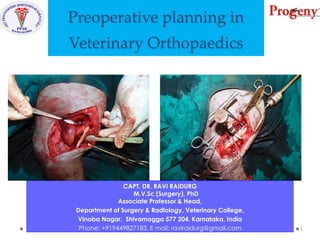
  • 1. Preoperative planning in Veterinary Orthopaedics CAPT. DR. RAVI RAIDURG M.V.Sc (Surgery), PhD Associate Professor & Head, Department of Surgery & Radiology, Veterinary College, Vinoba Nagar, Shivamogga 577 204, Karnataka, India Phone: +919449827183, E mail: raviraidurg@gmail.com 1
  • 3. S.No Webinar Topics 1 Preoperative planning in Veterinary Orthopaedics 2 Evolution of Internal Fixation in Veterinary Orthopaedics 3 Methods of fracture repair in Veterinary Orthopaedics 4 Plate Osteosynthesis – Terminologies and Instrumentation 5 Open Reduction and Internal Fixation (ORIF) for long bone fracture repair in dogs 6 Management of long bone diaphyseal fractures with minimal invasive plate osteosynthesis (MIPO)in dogs raviraidurg@gmail.com 3 PROGENY PPAK WEBINAR SERIES
  • 4. For every fracture, there are more than one method of fixation that will lead to acceptable results raviraidurg@gmail.com 4
  • 5. For every fracture, there are more than one method of fixation that will lead to acceptable results and many more that will lead to unacceptable results raviraidurg@gmail.com 5
  • 6. Preoperative planning in Veterinary Orthopaedics Aim : Introduce the concept of “ Preoperative planning in Veterinary Orthopaedics” Learning Objectives At the end of this session you should be able to: • Describe Fracture healing (Primary vs Secondary bone healing) • Describe Fracture Classification • Describe AO Fracture Classification of Long bones (eg AO 32 A3 ???) • Derive a Fracture Patient Assessment Score (FPAS) and describe factors (mechanical, biological and clinical) which support the score. • List Preoperative planning in MIO (Minimally Invasive Osteosynthesis) 6 raviraidurg@gmail.com
  • 7. S. No Topic Time (min) Slides 1 Introduction 10 01-12 2 Case presentations 15 13 - 39 3 Fracture healing 20 40 – 47 4 Fracture classification 48 – 59 5 AO Fracture classification of long bones 60 – 68 6 FPAS (Fracture Patient Assessment Score) 30 70 – 79 7 Treatment guidelines based on FPAS 80 – 140 8 Preoperative planning in MIO (Minimally Invasive Osteosynthesis) 10 141 – 158 9 Summary 05 159 – 160 raviraidurg@gmail.com 7 Preoperative planning in Veterinary Orthopaedics SESSION OUTLINE (90 Min)
  • 8. Cases presented to Veterinary College Shivamogga (2015 – 2018) Humerus raviraidurg@gmail.com 8
  • 9. Case example 14 Year old, 5 kg dog (SPITZ) ie GERIATRIC DOG Diaphyseal long oblique / Spiral fracture raviraidurg@gmail.com 9 How could I fix this 1. POP cast (Conservative method) 2. Wiring 3. External fixator (ESF) 4. Internal fixator IMP IMP + wiring IMP + Thomas splint ILN (Interlocking Nails) TENS (Titanium elastic nail system) PO (Plate osteosynthesis ie Bone plating)
  • 10. Results of the Poll raviraidurg@gmail.com 10
  • 11. Case example 14 Year old, 5 kg dog (SPITZ) ie GERIATRIC DOG Diaphyseal long oblique / Spiral fracture raviraidurg@gmail.com 11 How could I fix this Internal fixator 1. IMP 2. IMP + wiring 3. ILN (Interlocking Nails) 4. Lag Screw with Plate osteosynthesis (Bone plating)
  • 12. Results of the Poll raviraidurg@gmail.com 12
  • 14. Humerus Case No 4946_25 Nov 2015 raviraidurg@gmail.com 14 A 14 Year old, 5 kg dog ie GERIATRIC DOG Diaphyseal long oblique / Spiral fracture
  • 15. Humerus Case No 4946_25 Nov 2015 A 14 Year old, 5 kg dog ie GERIATRIC DOG Diaphyseal long oblique / Spiral fracture raviraidurg@gmail.com 15
  • 16. Humerus Case No 4946_25 Nov 2015 A 14 Year old, 5 kg dog ie GERIATRIC DOG Diaphyseal long oblique / Spiral fracture raviraidurg@gmail.com 16
  • 17. Humerus Case No 4946_25 Nov 2015 A 14 Year old, 5 kg dog ie GERIATRIC DOG Diaphyseal long oblique / Spiral fracture raviraidurg@gmail.com 17
  • 18. Humerus Case No 4946_25 Nov 2015 A 14 Year old, 5 kg dog ie GERIATRIC DOG Diaphyseal long oblique / Spiral fracture raviraidurg@gmail.com 18
  • 19. Humerus Case No 4946_25 Nov 2015 A 14 Year old, 5 kg dog ie GERIATRIC DOG Diaphyseal long oblique / Spiral fracture raviraidurg@gmail.com 19 6 th Week Post Operative
  • 20. Humerus Case No 4946_25 Nov 2015 A 14 Year old, 5 kg dog ie GERIATRIC DOG Diaphyseal long oblique / Spiral fracture raviraidurg@gmail.com 20 6 th Week Post Operative
  • 22. Humerus Case No 8698 dated 07 Dec 2015 raviraidurg@gmail.com 22 A 05 mth, 45 kg dog ie JUVENILE DOG Diaphyseal Transverse fracture with longitudinal slit in distal fragment
  • 23. Humerus_ Case No 8698 dated 07 Dec 2015 A 05 mth, 45 kg Great Dane Pup ie JUVENILE DOG Diaphyseal Transverse fracture with longitudinal slit in distal fragment raviraidurg@gmail.com 23
  • 24. raviraidurg@gmail.com 24 Humerus_ Case No 8698 dated 07 Dec 2015 A 05 mth, 45 kg Great Dane Pup ie JUVENILE DOG Diaphyseal Transverse fracture with longitudinal slit in distal fragment
  • 25. Humerus_ Case No 8698_GD_07 Dec 2015 A 05 mth, 45 kg dog ie JUVENILE DOG Diaphyseal Transverse fracture with longitudinal slit in distal fragment raviraidurg@gmail.com 25
  • 26. raviraidurg@gmail.com 26 Humerus_ Case No 8698 dated 07 Dec 2015 A 05 mth, 45 kg Great Dane Pup ie JUVENILE DOG Diaphyseal Transverse fracture with longitudinal slit in distal fragment
  • 27. raviraidurg@gmail.com 27 Humerus_ Case No 8698 dated 07 Dec 2015 A 05 mth, 45 kg Great Dane Pup ie JUVENILE DOG Diaphyseal Transverse fracture with longitudinal slit in distal fragment
  • 28. raviraidurg@gmail.com 28 Humerus_ Case No 8698 dated 07 Dec 2015 A 05 mth, 45 kg Great Dane Pup ie JUVENILE DOG Diaphyseal Transverse fracture with longitudinal slit in distal fragment
  • 29. raviraidurg@gmail.com 29 10 th Post Operative day Humerus_ Case No 8698 dated 07 Dec 2015 A 05 mth, 45 kg Great Dane Pup ie JUVENILE DOG Diaphyseal Transverse fracture with longitudinal slit in distal fragment
  • 30. raviraidurg@gmail.com 303 rd Week Post Operative day Humerus_ Case No 8698 dated 07 Dec 2015 A 05 mth, 45 kg Great Dane Pup ie JUVENILE DOG Diaphyseal Transverse fracture with longitudinal slit in distal fragment
  • 31. raviraidurg@gmail.com 31 3 rd Week Post Operative day Humerus_ Case No 8698 dated 07 Dec 2015 A 05 mth, 45 kg Great Dane Pup ie JUVENILE DOG Diaphyseal Transverse fracture with longitudinal slit in distal fragment
  • 32. raviraidurg@gmail.com 32 17 th Week Post Operative day Humerus_ Case No 8698 dated 07 Dec 2015 A 05 mth, 45 kg Great Dane Pup ie JUVENILE DOG Diaphyseal Transverse fracture with longitudinal slit in distal fragment
  • 33. Humerus_ Case No 8698_GD_07 Dec 2015 A 05 mth, 45 kg dog ie JUVENILE DOG Diaphyseal Transverse fracture with longitudinal slit in distal fragment raviraidurg@gmail.com 33 30th Month Post Operative day
  • 35. Humerus Case No 10148 dated 27 June 2016 raviraidurg@gmail.com 35 A 2 ½ Year old, 22 kg Mudhol Hound dog Transverse fracture in distal fragment
  • 40. Preoperative planning in Veterinary Orthopaedics 40 raviraidurg@gmail.com
  • 41. Preoperative planning in Veterinary Orthopaedics Learning Objectives At the end of this session you should be able to: • Describe Fracture healing (Primary vs Secondary bone healing) • Describe Fracture Classification • Describe AO Fracture Classification of Long bones (eg AO 32 A3 ???) • Derive a Fracture Patient Assessment Score (FPAS) and describe factors (mechanical, biological and clinical) which support the score. • List Preoperative planning in MIO (Minimally Invasive Osteosynthesis) 41 raviraidurg@gmail.com
  • 42. FRACTURE HEALING PRIMARY BONE HEALING SECONDARY BONE HEALING 42 raviraidurg@gmail.com
  • 43. FRACTURE HEALING PRIMARY BONE HEALING • Rigidly stabilised fracture fragments with direct contact between them heal by migration of ‘cutting cones’ (comprising osteoclasts and osteoblasts) from one fragment to the other, resulting in reformation of lamellar bone (Haversian remodelling) without any requirement for precursor tissue or callus. • The lack of a callus phase can be very important in some cases, particularly when joints are affected. 43 raviraidurg@gmail.com
  • 44. FRACTURE HEALING PRIMARY BONE HEALING • ‘Gap healing’, in which rigidly stabilised fragments are separated by a gap of up to 1 mm, is a slight variation of this mechanism. • The space is initially filled by lamellar bone orientated at 90° to the long axis of the bone, which enables the cutting cones to migrate across the gap and reorientate the lamellar bone. 44 raviraidurg@gmail.com
  • 45. FRACTURE HEALING PRIMARY BONE HEALING • Clinical union takes significantly longer to achieve with primary bone healing than secondary bone healing, and therefore relies on implants for longer. • Primary bone healing can only be obtained with the use of internal fixation due to the requirement for absolute rigidity with less than 2 per cent strain at the fracture site. 45 raviraidurg@gmail.com
  • 46. FRACTURE HEALING SECONDARY BONE HEALING • Fragments separated by a gap of more than 1 mm or with an interfragmentary strain greater than 2 per cent will undergo secondary bone healing. • This involves the formation of tissues (ie, callus) that can cope with the strain occurring at the fracture site. As increased stability is conferred by the callus, the next tissue in the sequence forms, which increases stability still further and, in turn, allows the next tissue to form, until bone tissue can once again fill the fracture gap. 46 raviraidurg@gmail.com
  • 48. Preoperative planning in Veterinary Orthopaedics Learning Objectives At the end of this section you should be able to: • Describe Fracture healing (Primary vs Secondary bone healing) • Describe Fracture Classification • Describe AO Fracture Classification of Long bones (eg AO 32 A3 ???) • Derive a Fracture Patient Assessment Score (FPAS) and describe factors (mechanical, biological and clinical) which support the score. • List Preoperative planning in MIO (Minimally Invasive Osteosynthesis) 48 raviraidurg@gmail.com
  • 49. Fracture classification ■ Cause ■ Open or closed ■ Extent of bone damage ■ Number and position of fracture lines ■ Direction ■ Location ■ Forces acting on the fracture ■ Stability ■ Degree of soft tissue damage raviraidurg@gmail.com 49
  • 50. Fracture classification CAUSE OF THE FRACTURE Intrinsic ■ MUSCULAR (eg, tibial tuberosity avulsion secondary to quadriceps muscle contraction). ■ PATHOLOGICAL (eg, secondary to a primary bone neoplasm). ■ STRESS (a multiple low-grade trauma can eventually result in a fracture). Extrinsic • Extrinsic fractures are caused by external trauma (eg, a • road traffic accident). raviraidurg@gmail.com 50
  • 51. Fracture classification OPEN/CLOSED FRACTURE • Radiographs should be examined for signs of gas shadows within the soft tissue planes that may suggest communication with the external environment. • Open fractures should be graded raviraidurg@gmail.com 51
  • 52. Fracture classification ■ OPEN VERSUS CLOSED • Open fractures are more challenging to treat due to the risks of infection and the potential for damage to the soft tissues surrounding the bone. The importance of soft tissue damage is reflected in the grading system used to describe these fractures: GRADE 1. The skin has been punctured by a sharp bone fragment that has usually retracted back beneath the surface. There is typically only mild soft tissue damage associated with the relatively small wound; GRADE 2. The skin lesion is due to extrinsic trauma leaving a variable-sized soft tissue deficit and more severe damage to the surrounding tissues. Foreign material may be present within the wound; GRADE 3. There is extensive loss of multiple tissue types due to extrinsic trauma. The fracture is usually fragmented due to the high-energy nature of the injury. Neurovascular injury is likely and could cause complications. raviraidurg@gmail.com 52
  • 53. Fracture classification EXTENT OF BONE DAMAGE Incomplete ■ GREENSTICK. Young animals can fracture one cortex, with the opposite cortex undergoing plastic deformation rather than fracture (like snapping a green stick). ■ FISSURE. Radiographs should be examined carefully for fissures leading away from a fracture line, as they can greatly increase the complexity of the repair or progress to a full fracture if not addressed. A fissure is often associated with comminuted fractures. ■ DEPRESSED. Depressed fractures usually occur in the skull. Complete • Complete fractures involve both cortices of the bone. raviraidurg@gmail.com 53
  • 54. Fracture classification NUMBER AND POSITION OF THE FRACTURE LINES ■ SIMPLE. One fracture line without small fragments of bone. ■ SEGMENTAL. Two or more fracture lines that do not communicate with each other. ■ COMMINUTED. Two or more fracture lines that do communicate with each other. This type of fracture is usually associated with high-energy trauma and therefore extensive soft tissue damage. raviraidurg@gmail.com 54
  • 55. Fracture classification raviraidurg@gmail.com 55 DIRECTION OF FRACTURE LINES ■ TRANSVERSE. The fracture line is perpendicular to the long axis of the bone. ■ OBLIQUE. Generally, a fracture that is 1·5 to two times longer than the diameter of the bone (or longer) is considered oblique. The point when a short oblique fracture is classified as a transverse fracture is a unclear. ■ SPIRAL. The fracture line curves around the bone (usually due to a twisting force) creating a complex three-dimensional fracture.
  • 56. Fracture classification raviraidurg@gmail.com 56 FRACTURE LOCATION ■ DIAPHYSEAL. Cortical bone is affected and the fracture can be described as proximal, mid or distal. ■ METAPHYSEAL. Cancellous bone situated towards the ends of the bones (metaphyses) has differing characteristics of implant holding and will often heal more quickly once stabilised than diaphyseal bone. ■ ARTICULAR. Once a joint suffers a fracture, a degree of secondary osteoarthrosis is inevitable, but the extent of the degeneration and effect on joint mobility can be minimised by appropriate and prompt fracture management. ■ CONDYLAR.
  • 57. Fracture classification raviraidurg@gmail.com 57 FRACTURE LOCATION ■ PHYSEAL. Fractures involving the physis of skeletally immature bones are classified according to the Salter-Harris system. Smooth implants that cross the physis perpendicular to the direction of growth and occupy as little of the physeal area as possible are ideal for repair in order to preserve as much growth potential as possible. Fracture location and configuration usually dictate implant orientation away from this ideal, with the result that implants are removed in very immature animals to allow continued growth. Unfortunately, despite gentle handling, growth disturbances are common following physeal injury and owners should be made aware of this.
  • 58. Forces acting on Bone Bending Torsional Axial compression Axial tension Shearing
  • 59. Preoperative planning in Veterinary Orthopaedics Learning Objectives At the end of this section you should be able to: • Describe Fracture healing (Primary vs Secondary bone healing) • Describe Fracture Classification • Describe AO Fracture Classification of Long bones (eg AO 32 A3 ???) • Derive a Fracture Patient Assessment Score (FPAS) and describe factors (mechanical, biological and clinical) which support the score. • List Preoperative planning in MIO (Minimally Invasive Osteosynthesis) 59 raviraidurg@gmail.com
  • 60. AO FRACTURE CLASSIFICATION OF LONG BONES 60 raviraidurg@gmail.com
  • 61. 1958 AO/ASIF (Arbeitsgemeinschaft fur osteosynthesefragen) Maurice Müller, Martin Allgöwer, Hans Willenegger, Robert Schneider and Robert Mathys (Switzerland) • Plate and screw osteosynthesis. • A group of Swiss orthopaedic surgeons formed the Arbeitsgemeinschaft fur osteosynthesefragen (AO), also known as the Association for the Study of Internal Fixation (ASIF). • The principles for fracture management developed by the AO group defined the standard of care for fracture 61
  • 62. 62 1958 AO founded in Biel (Bienne) November 6, 1958, Hotel Elite Biel (Bienne), Switzerland
  • 63. 63 1958 AO Founders Arbeitsgemeinschaft für Osteosynthesefragen Maurice Müller Zürich 1918−2009 Martin Allgöwer Chur 1917−2007 Walter Bandi Interlaken 1912−1997 Robert Schneider Grosshöchstetten 1912−1990 Hans Willenegger Liestal 1910−1998
  • 64. Fracture classification raviraidurg@gmail.com 64 • The first symbol of the alpha-numeric code represents the fractured bone, humerus is coded as 1, Radius – Ulna as 2, femur as 3 and tibia which was coded as 4. • The second symbol represents “location of the fracture” ie Proximal as 1, middle diaphyseal as 2 and Distal as 3 • Simple (A), wedge (B) and complex fractures (C). • A1 incomplete fractures. • A2 indicates diaphyseal simple oblique fractures, • A3 indicates diaphyseal simple transverse fractures, • B1 indicates diaphyseal one reducible wedge, • B2 indicates diaphyseal several reducible wedge • B3 indicated diaphyseal non-reducible wedge. • C1 indicates Complex reducible wedge • C 2 indicates Complex segmental wedge, • C3 indicates Complex non reducible wedge, AO 32 A3
  • 69. Preoperative planning in Veterinary Orthopaedics Learning Objectives At the end of this section you should be able to: • Describe Fracture healing (Primary vs Secondary bone healing) • Describe Fracture Classification • Describe AO Fracture Classification of Long bones (eg AO 32 A3 ???) • Derive a Fracture Patient Assessment Score (FPAS) and describe factors (mechanical, biological and clinical) which support the score. • List Preoperative planning in MIO (Minimally Invasive Osteosynthesis) 69 raviraidurg@gmail.com
  • 70. FPAS (Fracture Patient Assessment Score) 70 raviraidurg@gmail.com
  • 71. FPAS • The FPAS is a simple 1 -10 scoring system that is determined by evaluating pertinent mechanical, biological, and clinical factors to determine the relative needs of the fracture case. • The “10” end of the scale indicates ideal conditions (i.e. good news) and the “1” end represents the most challenging conditions (i.e. bad news) . • Acknowledging that each and every orthopedic patient is different, it is advisable to derive a FPAS for each patient prior to treatment. raviraidurg@gmail.com 71
  • 72. FPAS raviraidurg@gmail.com 72 1 ☺10 Marked instability Moderate instability Mild instability MECHANICAL Non-Load-Sharing Partial Load-Sharing Ideal Load-Sharing Large Patient Size Medium Patient Small Patient Size FACTORS Multi-limb dysfunction Moderate multi-limb Single limb dysfunction CLINICAL FACTORS Unwilling to follow-up Questionable followup Willing/able to follow-up Owner Unable to restrict activity Willing to try Able to restrict activity Intolerant of activity restrict Somewhat tolerant Tolerant of activ. restr.. Patient Rambunctious Active, but responsive Calm Intolerant of treatments Difficult to treat Easy to treat BIOLOGICAL FACTORS Severe tissue injury Moderate tissue injury Minimal tissue injuryLocal Factors Comminuted Transverse/Oblique Spiral Greenstick Open Fx – Type III Type II Type I Closed Fracture Marked instability Moderate instability Mild instability Previous radiation Tx Systemic Geriatric Mature Adult Immature Patient Factors Debilitated/ill Compensated illness Healthy Second-hand smoke?
  • 73. FPAS (Fracture Patient Assessment Score) MECHANICAL FACTORS 73 raviraidurg@gmail.com
  • 74. FPAS MECHANICAL FACTORS raviraidurg@gmail.com 74 The amount of loading that a given fixation device must withstand is influenced by the following factors: • palpable stability of the fracture zone, • • the number of dysfunctional limbs, • • the patient’s coordination and agility • • the patient’s weight,/size, and • • the degree of “load-sharing” anticipated between the fixation device and the bony column. 1 ☺10 Marked instability Moderate instability Mild instability MECHANICAL Non-Load-Sharing Partial Load-Sharing Ideal Load-Sharing Large Patient Size Medium Patient Small Patient Size FACTORS Multi-limb dysfunction Moderate multi-limb Single limb dysfunction CLINICAL FACTORS Unwilling to follow-up Questionable followup Willing/able to follow-up Owner Unable to restrict activity Willing to try Able to restrict activity Intolerant of activity restrict Somewhat tolerant Tolerant of activ. restr.. Patient Rambunctious Active, but responsive Calm Intolerant of treatments Difficult to treat Easy to treat BIOLOGICAL FACTORS Severe tissue injury Moderate tissue injury Minimal tissue injuryLocal Factors Comminuted Transverse/Oblique Spiral Greenstick Open Fx – Type III Type II Type I Closed Fracture Marked instability Moderate instability Mild instability Previous radiation Tx Systemic Geriatric Mature Adult Immature Patient Factors Debilitated/ill Compensated illness Healthy Second-hand smoke?
  • 75. raviraidurg@gmail.com 75 Fig 1: Ideal load-sharing Fig 2: Non-load-sharing Fig 3: Partial load-sharing The mechanical FPAS shifts downward (toward 1) when there is multi-limb dysfunction, a large, clumsy patient, and/or the anticipation of a non-load-sharing osteosynthesis.
  • 76. FPAS (Fracture Patient Assessment Score) CLINICAL FACTORS 76 raviraidurg@gmail.com
  • 77. FPAS CLINICAL FACTORS raviraidurg@gmail.com 77 • Poor patient/owner compliance leads to excessive patient activity and/or insufficient physical therapy. • Fracture treatment plans should be made with possibility of poor exercise restriction kept in mind. • Poor anticipated compliance shifts the FPAS downward. Since poor patient/owner compliance invariably increases the mechanical demand upon the fixation, the clinical FCAS is averaged with the mechanical FPAS to derive a total mechanical score. • Sometimes owner compliance concerns cause us to avoid the use of External Skeletal Fixation. • In attempt to shift the FPAS toward 10, I spend extra time with pet owners explaining the importance of exercise restriction as well as the potential consequences of failing to do so (especially in challenging fracture cases). 1 ☺10 Marked instability Moderate instability Mild instability MECHANICAL Non-Load-Sharing Partial Load-Sharing Ideal Load-Sharing Large Patient Size Medium Patient Small Patient Size FACTORS Multi-limb dysfunction Moderate multi-limb Single limb dysfunction CLINICAL FACTORS Unwilling to follow-up Questionable followup Willing/able to follow-up Owner Unable to restrict activity Willing to try Able to restrict activity Intolerant of activity restrict Somewhat tolerant Tolerant of activ. restr.. Patient Rambunctious Active, but responsive Calm Intolerant of treatments Difficult to treat Easy to treat BIOLOGICAL FACTORS Severe tissue injury Moderate tissue injury Minimal tissue injuryLocal Factors Comminuted Transverse/Oblique Spiral Greenstick Open Fx – Type III Type II Type I Closed Fracture Marked instability Moderate instability Mild instability Previous radiation Tx Systemic Geriatric Mature Adult Immature Patient Factors Debilitated/ill Compensated illness Healthy Second-hand smoke?
  • 78. FPAS (Fracture Patient Assessment Score) BIOLOGICAL FACTORS 78 raviraidurg@gmail.com
  • 79. FPAS BIOLOGICAL FACTORS raviraidurg@gmail.com 79 • Bone healing is influenced by • local factors(fracture zone): open fractures, severe soft tissue disruption, and previous local radiation fields. • Systemic factors: old age, cachexia, and systemic illness. • Each of these local and systemic factors will shift the biological FPAS toward 1. Conversely, bone healing is more rapid in closed fractures with minimal soft tissue injury, and in young, healthy, well-nourished patients; each of which shifts the FPAS toward 10. 1 ☺10 Marked instability Moderate instability Mild instability MECHANICAL Non-Load-Sharing Partial Load-Sharing Ideal Load-Sharing Large Patient Size Medium Patient Small Patient Size FACTORS Multi-limb dysfunction Moderate multi-limb Single limb dysfunction CLINICAL FACTORS Unwilling to follow-up Questionable followup Willing/able to follow-up Owner Unable to restrict activity Willing to try Able to restrict activity Intolerant of activity restrict Somewhat tolerant Tolerant of activ. restr.. Patient Rambunctious Active, but responsive Calm Intolerant of treatments Difficult to treat Easy to treat BIOLOGICAL FACTORS Severe tissue injury Moderate tissue injury Minimal tissue injuryLocal Factors Comminuted Transverse/Oblique Spiral Greenstick Open Fx – Type III Type II Type I Closed Fracture Marked instability Moderate instability Mild instability Previous radiation Tx Systemic Geriatric Mature Adult Immature Patient Factors Debilitated/ill Compensated illness Healthy Second-hand smoke?
  • 80. TREATMENT GUIDELINES BASED ON PRE-OPERATIVE FPAS 80 raviraidurg@gmail.com
  • 82. raviraidurg@gmail.com 82 FPAS 1 – 3 1 ☺10 Marked instability Moderate instability Mild instability MECHANICAL Non-Load-Sharing Partial Load-Sharing Ideal Load-Sharing Large Patient Size Medium Patient Small Patient Size FACTORS Multi-limb dysfunction Moderate multi-limb Single limb dysfunction CLINICAL FACTORS Unwilling to follow-up Questionable followup Willing/able to follow-up Owner Unable to restrict activity Willing to try Able to restrict activity Intolerant of activity restrict Somewhat tolerant Tolerant of activ. restr.. Patient Rambunctious Active, but responsive Calm Intolerant of treatments Difficult to treat Easy to treat BIOLOGICAL FACTORS Severe tissue injury Moderate tissue injury Minimal tissue injuryLocal Factors Comminuted Transverse/Oblique Spiral Greenstick Open Fx – Type III Type II Type I Closed Fracture Marked instability Moderate instability Mild instability Previous radiation Tx Systemic Geriatric Mature Adult Immature Patient Factors Debilitated/ill Compensated illness Healthy Second-hand smoke? AO 32 C1
  • 83. • Cases in which the overall FCAS =1-3 require an orthopedist who is an expert (5-10 years of frequent use) with the chosen fixation system. • Fixation systems considered are bone plates, interlocking nails (IN), or plate + IM pin combinations, ESF +/- IM pin. Percutaneous application of an internal fixator is a consideration. • There is no room for technical or judgment error or excessive soft tissue manipulation. • A careful balance between mechanical stability and preservation of soft tissue integrity is vital as dictated by the mechanical and biological FPAS. • There is significant risk of complications requiring surgical revision even with logical use of the FPAS, error-free application of the fixation, and attentive postoperative care. raviraidurg@gmail.com 83 FPAS 1 - 3
  • 84. • When mechanical FCAS = 1-3, strict attention must be paid to maximizing fixation strength. The fixation device must be modern and its application must use flawless technique. • When biologic FCAS = 1 -3 or preoperative radiographs suggest that anatomic fracture reconstruction is unlikely or extremely cumbersome (non-reconstructable), the surgical priorities shift from anatomic reconstruction to spatial alignment, preservation of soft tissue viability and rigid fixation capable of withstanding the stresses of non-load-sharing fixation for the estimated healing period. • Closed (percutaneous) approaches are desired and can often be applied to the radius and tibia utilizing a hanging limb position. • Such applications to the humerus and femur are challenging; Open But Do Not Touch (OBDNT) applications are used if needed. Cancellous bone grafting is employed in open approaches. • Attentive convalescent care and monitoring is critical. raviraidurg@gmail.com 84 FPAS 1 - 3
  • 85. FPAS 1 - 3 Case no 10295 Femur_26 July 2016 AO 32C1 raviraidurg@gmail.com 85
  • 86. FPAS 1 - 3 Case no 10295, 26 July 2016, Femur raviraidurg@gmail.com 86 Preoperative Planning
  • 87. FPAS 1 - 3 Case no 10295, 26 July 2016, Femur Pre Op Post Op raviraidurg@gmail.com 87
  • 88. Post Op 03 Weeks Post Op raviraidurg@gmail.com 88 FPAS 1 - 3 Case no 10295, 26 July 2016, Femur
  • 90. raviraidurg@gmail.com 90 FPAS 4– 6 1 ☺10 Marked instability Moderate instability Mild instability MECHANICAL Non-Load-Sharing Partial Load-Sharing Ideal Load-Sharing Large Patient Size Medium Patient Small Patient Size FACTORS Multi-limb dysfunction Moderate multi-limb Single limb dysfunction CLINICAL FACTORS Unwilling to follow-up Questionable followup Willing/able to follow-up Owner Unable to restrict activity Willing to try Able to restrict activity Intolerant of activity restrict Somewhat tolerant Tolerant of activ. restr.. Patient Rambunctious Active, but responsive Calm Intolerant of treatments Difficult to treat Easy to treat BIOLOGICAL FACTORS Severe tissue injury Moderate tissue injury Minimal tissue injuryLocal Factors Comminuted Transverse/Oblique Spiral Greenstick Open Fx – Type III Type II Type I Closed Fracture Marked instability Moderate instability Mild instability Previous radiation Tx Systemic Geriatric Mature Adult Immature Patient Factors Debilitated/ill Compensated illness Healthy Second-hand smoke? AO 13 A1
  • 91. • Cases in which the overall FPAS = 4-6 require an orthopedist with considerable experience (3-5 years of frequent use) with the chosen fixation system. • Fixation systems considered are bone plates, interlocking nails (IN), plate + IM pin combinations and ESF +/- IM pin fixation. Minor technical error or slightly excessive soft tissue manipulation often causes failure. • The balance between mechanical stability and preservation of soft tissue integrity is important and is not forgiving in these patients. Attentive postoperative care is important. There is risk of implant complications, but revision surgeries are uncommon if logical application of the FPAS, error-free application, and thoughtful convalescent care are employed. raviraidurg@gmail.com 91 FPAS 4 - 6
  • 92. • When mechanical FPAS = 4-6, the need for fixation strength cannot be ignored. • When biological FPAS = 4-6, preservation of fracture zone viability remains a priority. • Surgical approaches may be percutaneous or OBDNT if anatomic reconstruction is not a goal. • Open approaches can be used if it will aid anatomic reconstruction (and thereby load-sharing) of reconstructable fractures. Cancellous grafting is employed whenever open approaches are used. raviraidurg@gmail.com 92 FPAS 4 - 6
  • 93. FPAS 4 - 6 Case no 15503_21 Apr 2018 AO 13 A1 raviraidurg@gmail.com 93
  • 94. FPAS 4 – 6 Case no 15503_21 Apr 2018_Humerus Case no 15503_21 Apr 2018 AO 13 A1 raviraidurg@gmail.com 94
  • 95. FPAS 4 – 6 Case no 15503_21 Apr 2018_Humerus Craniolateral approach Fractured ends raviraidurg@gmail.com 95
  • 96. FPAS 4 – 6 Case no 15503_21 Apr 2018_Humerus IMP in situ LCP on caudal aspect raviraidurg@gmail.com 96
  • 97. FPAS 4 – 6 Case no 15503_21 Apr 2018_Humerus raviraidurg@gmail.com 97
  • 98. FPAS 4 – 6 Case no 15503_21 Apr 2018_Humerus Post Op_18 hrs_22.04.2018 4th Month Post Op_21.08.2018 raviraidurg@gmail.com 98
  • 99. FPAS 4 – 6 Case no 15503_21 Apr 2018_Humerus AO 13 A1 raviraidurg@gmail.com 99
  • 101. raviraidurg@gmail.com 101 FPAS 7– 9 1 ☺10 Marked instability Moderate instability Mild instability MECHANICAL Non-Load-Sharing Partial Load-Sharing Ideal Load-Sharing Large Patient Size Medium Patient Small Patient Size FACTORS Multi-limb dysfunction Moderate multi-limb Single limb dysfunction CLINICAL FACTORS Unwilling to follow-up Questionable followup Willing/able to follow-up Owner Unable to restrict activity Willing to try Able to restrict activity Intolerant of activity restrict Somewhat tolerant Tolerant of activ. restr.. Patient Rambunctious Active, but responsive Calm Intolerant of treatments Difficult to treat Easy to treat BIOLOGICAL FACTORS Severe tissue injury Moderate tissue injury Minimal tissue injuryLocal Factors Comminuted Transverse/Oblique Spiral Greenstick Open Fx – Type III Type II Type I Closed Fracture Marked instability Moderate instability Mild instability Previous radiation Tx Systemic Geriatric Mature Adult Immature Patient Factors Debilitated/ill Compensated illness Healthy Second-hand smoke?
  • 102. • Cases in which the overall FPAS = 7-9 require an orthopedist with a thorough academic training and some clinical experience (1-2 years) with the chosen fixation system. • Fixation systems considered are bone plates, interlocking nails (IN), intramedullary pin + full cerclage wire, plate + IM pin combinations and ESF +/- IM pin fixation. • Minor technical errors or slightly excessive soft tissue manipulation are usually tolerated. • The balance between mechanical stability and preservation of soft tissue integrity is important, but somewhat forgiving. raviraidurg@gmail.com 102 FPAS 7 - 9
  • 103. • When mechanical FPAS = 7-9, fixation strength is often not the great priority. • When biological FPAS = 7-9, a full open approach is used when it will aid anatomic reconstruction of reconstructable fractures (to achieve load-sharing); otherwise, closed or OBDNT approaches are used with non-reconstructable fractures. • Cancellous grafting is often employed with many open surgical approaches, but may be less critical in these cases. raviraidurg@gmail.com 103 FPAS 7 - 9
  • 104. FPAS 7 - 9 Case no 17633_11.01.2019 raviraidurg@gmail.com 104
  • 105. FPAS 7 - 9 Case no 17633 dt 11.01.2019 Preoperative Plan raviraidurg@gmail.com 105
  • 106. FPAS 7 – 9 Case no 17633_11.01.2019_Mandible 3.5 mm, 06 hole Recon LCP raviraidurg@gmail.com 106
  • 107. FPAS 7 – 9 Case no 17633_11.01.2019_Mandible WIRING raviraidurg@gmail.com 107
  • 108. FPAS 7 – 9 Case no 17633_11.01.2019_Mandible 3.5 mm, 06 hole Recon LCP raviraidurg@gmail.com 108
  • 109. Ravi Raidurg., Chandrashekharappa M and Naveen M (2020): Plate osteosynthesis for mandibular body fracture repair in a Mudhol Hound using Locking Reconstruction Plate. Ind J Vet Surg: 40 (2): 144. raviraidurg@gmail.com 109
  • 111. raviraidurg@gmail.com 111 FPAS 10 1 ☺10 Marked instability Moderate instability Mild instability MECHANICAL Non-Load-Sharing Partial Load-Sharing Ideal Load-Sharing Large Patient Size Medium Patient Small Patient Size FACTORS Multi-limb dysfunction Moderate multi-limb Single limb dysfunction CLINICAL FACTORS Unwilling to follow-up Questionable followup Willing/able to follow-up Owner Unable to restrict activity Willing to try Able to restrict activity Intolerant of activity restrict Somewhat tolerant Tolerant of activ. restr.. Patient Rambunctious Active, but responsive Calm Intolerant of treatments Difficult to treat Easy to treat BIOLOGICAL FACTORS Severe tissue injury Moderate tissue injury Minimal tissue injuryLocal Factors Comminuted Transverse/Oblique Spiral Greenstick Open Fx – Type III Type II Type I Closed Fracture Marked instability Moderate instability Mild instability Previous radiation Tx Systemic Geriatric Mature Adult Immature Patient Factors Debilitated/ill Compensated illness Healthy Second-hand smoke? AO 32 A2
  • 112. • Cases in which the overall FPAS = 10 are ideal for building experience in fracture treatment or in getting comfortable with a new fixation system. • Fixation systems considered are bone plates, interlocking nails (IN), ESF or intramedullary pin + full cerclage wire combinations. • An academic training background is presumed, but little previous clinical experience is required because minor technical error or excessive soft tissue manipulations are almost uniformly tolerated. The balance between mechanical stability and preservation of soft tissue integrity is not particularly critical and novice surgeons may opt for relatively “mechanical” or “biological” fixations with little fear of failure. raviraidurg@gmail.com 112 FPAS 10
  • 113. • When mechanical FPAS = 10, success can achieved even with less than ideal fixation devices and systems. • When biological FPAS = 10, a full open approach can safely be used for anatomic reduction of reconstructable fractures (which is the case for most of these patients). • Extensive surgical time and tissue manipulations, which commonly occur with less experienced surgeons, are tolerated for these patients. • Bone grafting is not necessary for these cases. Because many of these patients are skeletally immature, care should be taken to preserve physeal function and to avoid manipulations that may risk soft tissue contractures (ie, quadriceps contracture). Additionally, early restoration of limb function is vital for normal skeletal development in dogs < 6 months of age. raviraidurg@gmail.com 113 FPAS 10
  • 114. FPAS 10 Case no 16059_04 July 2018 AO 32 A2 raviraidurg@gmail.com 114
  • 115. FPAS 10 Case no 16059_04 July 2018 AO 32 A2 raviraidurg@gmail.com 115
  • 116. FPAS 10 Case no 16059_04 July 2018 AO 32 A2 raviraidurg@gmail.com 116
  • 117. FPAS 10 Case no 16059_04 July 2018 AO 32 A2 raviraidurg@gmail.com 117
  • 118. • Apparently the TITANIUM nail came out from proximal end 03 days after surgery (Informed telephonically). • Case was not presented for re evaluation as it was presented from small town THIRTHAHALLI (70 km away). • Post operative video showed animal to be highly AGILE. • 11th week Post Op radiograph showed “Formation of good callus” raviraidurg@gmail.com 118 FPAS 10
  • 119. FPAS 10 Case no 16059_04 July 2018 11th week Post Op raviraidurg@gmail.com 119
  • 121. • The FPAS will help you know when to treat a fracture yourself and when to recommend referral to a surgical specialist. • What should be the “Fixation system” based on the surgeons experience with a chosen fixation system. • You can also use the FPAS as a tool by which you can monitor your progress (or the progress of those you are mentoring) as an orthopaedist over time. raviraidurg@gmail.com 121 CONCLUSION FPAS
  • 122. Case example 1 ½ Year old, 18 kg dog (Dobermann) Distal third oblique fracture Duration = 02 days old # raviraidurg@gmail.com 122 How could I fix this 1. POP cast (Conservative method) 2. Wiring 3. External fixator (ESF) 4. Internal fixator IMP IMP + wiring IMP + Thomas splint ILN (Interlocking Nails) TENS (Titanium elastic nail system) PO (Plate osteosynthesis ie Bone plating)
  • 123. Results of the Poll raviraidurg@gmail.com 123
  • 125. Humerus Case No 11506 dated 17 Jan 2017 raviraidurg@gmail.com 125 1 ½ Year old, 18 kg dog (Dobermann) Distal third oblique fracture Duration = 02 days old #
  • 126. Case example 1 ½ Year old, 18 kg dog (Dobermann) Distal third oblique fracture Duration = 02 days old # raviraidurg@gmail.com 126 How could I fix this Internal fixatIon (ORIF) 1. PLAN A - PO ( Lag screw + 3.5 mm LCP) 2. PLAN B - PO ( Wiring + 3.5 mm LCP) 3. PLAN C - PO PLATE ROD TECHNIQUE ( IMP + Wiring + 3.5 mm LCP)
  • 127. Case example 1 ½ Year old, 18 kg dog (Dobermann) Distal third oblique fracture Duration = 02 days old # raviraidurg@gmail.com 127 How could I fix this Internal fixatIon (ORIF) 1. PLAN A - PO ( Lag screw + 3.5 mm LCP) 2. PLAN B - PO ( Wiring + 3.5 mm LCP) 3. PLAN C - PO PLATE ROD TECHNIQUE ( IMP + Wiring + 3.5 mm LCP) Surgical approach to the bone • Medial approach • Lateral approach
  • 128. Case example 1 ½ Year old, 18 kg dog (Dobermann) Distal third oblique fracture Duration = 02 days old # raviraidurg@gmail.com 128 How could I fix this Internal fixatIon (ORIF) 1. PLAN A - PO ( Lag screw + 3.5 mm LCP) 2. PLAN B - PO ( Wiring + 3.5 mm LCP) 3. PLAN C - PO PLATE ROD TECHNIQUE ( IMP + Wiring + 3.5 mm LCP) Surgical approach to the bone • Medial approach • Lateral approach Cranial / Caudal / Medial / Lateral PLATING
  • 129. Case example 1 ½ Year old, 18 kg dog (Dobermann) Distal third oblique fracture Duration = 02 days old # raviraidurg@gmail.com 129 How could I fix this Internal fixatIon (ORIF) 1. PLAN A - PO ( Lag screw + 3.5 mm LCP) 2. PLAN B - PO ( Wiring + 3.5 mm LCP) 3. PLAN C - PO PLATE ROD TECHNIQUE ( IMP + Wiring + 3.5 mm LCP) Surgical approach to the bone • Medial approach • Cranio - Lateral approach Cranial / Caudal / Medial / Lateral PLATING
  • 130. Humerus Case No 11506 dated 17 Jan 2017 A 1 ½ Year old, 18 kg dog (Dobermann) Distal third oblique fracture Duration = 02 days old # raviraidurg@gmail.com 130
  • 131. Humerus Case No 11506 dated 17 Jan 2017 A 1 ½ Year old, 18 kg dog (Dobermann) Distal third oblique fracture Duration = 02 days old # raviraidurg@gmail.com 131
  • 132. Humerus Case No 11506 dated 17 Jan 2017 A 1 ½ Year old, 18 kg dog (Dobermann) Distal third oblique fracture Duration = 02 days old # raviraidurg@gmail.com 132
  • 133. Humerus Case No 11506 dated 17 Jan 2017 A 1 ½ Year old, 18 kg dog (Dobermann) Distal third oblique fracture Duration = 02 days old # raviraidurg@gmail.com 133
  • 134. Humerus Case No 11506 dated 17 Jan 2017 A 1 ½ Year old, 18 kg dog (Dobermann) Distal third oblique fracture Duration = 02 days old # raviraidurg@gmail.com 134
  • 135. raviraidurg@gmail.com 135 Humerus Case No 11506 dated 17 Jan 2017 A 1 ½ Year old, 18 kg dog (Dobermann) Distal third oblique fracture Duration = 02 days old #
  • 136. raviraidurg@gmail.com 136 Humerus Case No 11506 dated 17 Jan 2017 A 1 ½ Year old, 18 kg dog (Dobermann) Distal third oblique fracture Duration = 02 days old # 6 th Week Post Operative
  • 137. OPEN REDUCTION & INTERNAL FIXATION (ORIF) OF RIGHT HUMERAL DISTAL THIRD SPIRAL FRACTURE IN A DOBERMANN BY PLATE ROD TECHNIQUE Case no 11506 dated 17 Jan 2017 Department Of Surgery and Radiology, Veterinary College, Vinoba Nagar, Shivamogga 577204 0 day Post Op 6th Week Post Op 9th Month Post Op
  • 138. raviraidurg@gmail.com 138 Humerus Case No 11506 dated 17 Jan 2017 A 1 ½ Year old, 18 kg dog (Dobermann) Distal third oblique fracture Duration = 02 days old # 01 YEAR Post Operative
  • 139. Lesson learnt • 1. ORTHOGONAL Radiographic views ie TWO views at 90 ° angle. • 2. Decide “Method of fracture repair” based on “FPAS” and “Availability of Instrumentation” and “Availability of Expertise w.r.t particular method of fracture repair system”. • 3. Have PLAN “A’, PLAN “B’ & PLAN “C” for a fracture repair. • 4. Execute your Plan accordingly raviraidurg@gmail.com 139
  • 140. Preoperative planning in Veterinary Orthopaedics Learning Objectives At the end of this section you should be able to: • Describe Fracture healing (Primary vs Secondary bone healing) • Describe Fracture Classification • Describe AO Fracture Classification of Long bones (eg AO 32 A3 ???) • Derive a Fracture Patient Assessment Score (FPAS) and describe factors (mechanical, biological and clinical) which support the score. • List Preoperative planning in MIO (Minimally Invasive Osteosynthesis) 140 raviraidurg@gmail.com
  • 141. PREOPERATIVE PLANNING IN MIO (Minimally Invasive Osteosynthesis) 141 raviraidurg@gmail.com
  • 142. • KEY POINTS • High quality radiographs of both affected and normal contralateral limb segments are critical to adequate surgical planning. • Additional imaging modalities, including CT and intra-operative fluoroscopy (C arm image intensifier), may be necessary to identify the full extent of lesions and to assist with reduction and fixation. • Loco-regional anatomy thorough knowledge is a prerequisite in MIO. • Fracture specific patient positioning is necessary to guarantee continuous visualization of adjacent joints and to allow unhindered procedures throughout surgery. raviraidurg@gmail.com 142 PREOPERATIVE PLANNING IN MIO (Minimally Invasive Osteosynthesis)
  • 144. PREOPERATIVE IMAGING • High quality radiographs of both affected and normal contralateral limb segments are critical to adequate surgical planning ie (ORTHOGONAL VIEWS) • Image quality greatly impacts diagnostic accuracy and therefore operative planning and execution. • To enhance patient comfort and improve quality of care, avoid motion artifacts, and optimize image quality, appropriate pain management, up to general anesthesia, should be provided during diagnostic imaging. raviraidurg@gmail.com 144 PREOPERATIVE PLANNING IN MIO (Minimally Invasive Osteosynthesis)
  • 145. Importance of Orthogonal views in Orthopaedics raviraidurg@gmail.com 145
  • 146. PREOPERATIVE IMAGING • Imaging of the contralateral segment is highly recommended in MIO for several reasons: 1) it optimizes preoperative implant selection, particularly with regard to length and type (e.g. plate vs. interlocking nail) 2) it allows comparison with the fractured segment to identify normal vs. abnormal structures and 3) it allows accurate comparative evaluation of postoperative alignment. raviraidurg@gmail.com 146 PREOPERATIVE PLANNING IN MIO (Minimally Invasive Osteosynthesis)
  • 147. ADDITIONAL IMAGING MODALITIES • CT scan: The major benefit of computed tomography is its ability to generate sectional images and allows a 3-D reconstruction of the fractured bone(s). It also provides detailed images of the fracture configuration and fragment displacement (Figure 2). • The use of 3-D imaging helps in understanding complex intra-articular fractures and improves preoperative planning. Furthermore, 3-D reconstruction is beneficial in evaluating fragment distribution and can be used to guide reduction maneuvers. • The main shortcoming associated with CT imaging is the cost. However, because the benefits of accurate planning and subsequent avoidance of intra-operative complications likely overcomes this limitation, CT imaging should be considered an integral part of pre-operative planning when using MIO.raviraidurg@gmail.com 147 PREOPERATIVE PLANNING IN MIO (Minimally Invasive Osteosynthesis)
  • 148. ADDITIONAL IMAGING MODALITIES • MRI is useful in the diagnosis of central nervous system lesions, its application in appendicular traumatology is limited. • Ultrasound is not readily used in the diagnosis of appendicular musculoskeletal trauma. • C arm imaging is of great help in MIO raviraidurg@gmail.com 148 PREOPERATIVE PLANNING IN MIO (Minimally Invasive Osteosynthesis)
  • 149. SURGICALAPPROACHES • Loco-regional anatomy thorough knowledge is a prerequisite in MIO as direct visualization of the fracture site is not available to the surgeon. • Surgeon relies exclusively on percutaneous landmarks and on intraoperative fluoroscopy to achieve fracture reduction, to restore alignment and to complete fixation. • Because a comprehensive 3-D understanding of the bone’s anatomy is necessary to enable adequate implant contouring and fixation, using dry specimens, during pre- operative planning and in the operating room in addition to CT reconstruction, is highly recommended. raviraidurg@gmail.com 149 PREOPERATIVE PLANNING IN MIO (Minimally Invasive Osteosynthesis)
  • 150. IMPLANT SELECTION • The choice of a specific implant is made based upon fracture configuration, patient signalment and the surgeon’s preferences. • Once a fixation system is selected, specific implant dimensions must be determined. Appropriate templating is mainly based on the preoperative radiographs of the contralateral intact side, corrected for magnification. In contrast, implant position is based on a detailed evaluation of the fractured bone. The fracture pattern, including the presence and extent of fissures, as well as the spread of the fragments, should be carefully evaluated as it may influence the choice and/or position of an implant. raviraidurg@gmail.com 150 PREOPERATIVE PLANNING IN MIO (Minimally Invasive Osteosynthesis)
  • 151. raviraidurg@gmail.com 151 A 05 mth, 45 kg GREAT DANE PUP Diaphyseal Transverse fracture with longitudinal slit in distal fragment Humerus_ Case No 8698 dated 07 Dec 2015 A 05 mth, 45 kg Great Dane Pup Diaphyseal Transverse fracture with longitudinal slit in distal fragment
  • 152. raviraidurg@gmail.com 152 IMPLANT SELECTION • As an example, the choice of a bone plate may be ill advised for the repair of a comminuted tibial fracture with medial longitudinal fissures extending in the proximal and/or distal metaphysis. • In such a case, the choice of an interlocking nail (ILN)with locking bolts oriented in the sagittal plane may be more appropriate. Humerus_ Case No 8698 dated 07 Dec 2015 A 05 mth, 45 kg Great Dane Pup ie JUVENILE DOG Diaphyseal Transverse fracture with longitudinal slit in distal fragment
  • 153. PATIENT POSITIONING • Preoperative planning includes identification of optimal patient positioning on the surgery table. • The importance of this step is often underestimated and it should be emphasized that adequate positioning is essential to the smooth execution of the surgical procedure from fracture reduction to restoration of alignment to proper implant position. • Ultimately, patient positioning may also affect the post-operative outcome. raviraidurg@gmail.com 153 PREOPERATIVE PLANNING IN MIO (Minimally Invasive Osteosynthesis)
  • 154. PATIENT POSITIONING • From an anesthetic standpoint, the final patient position should allow easy access to airways, prevent ventilation compromise, permit adequate monitoring and allow the utilization of an extra-corporal warming apparatus. raviraidurg@gmail.com 154 PREOPERATIVE PLANNING IN MIO (Minimally Invasive Osteosynthesis)
  • 155. raviraidurg@gmail.com 155 PATIENT POSITIONING • From a surgical standpoint, the patient should be positioned so as to facilitate all surgical phases including approach, reduction maneuvers as well as implant insertion and fixation. It must also permit unrestricted C-arm mobility around the patient so that intra-operative views of adjacent joints in both sagittal and frontal planes can be easily obtained throughout the surgical procedure PREOPERATIVE PLANNING IN MIO (Minimally Invasive Osteosynthesis) Figure: Pre-operative view of the operating room set up illustrating proper positioning of the patient for the treatment of a humeral fracture. The chest has been elevated to allow unrestricted C-arm motion around the limb. In turn, this provides intra-operative views of the joints adjacent to the fracture in 2 orthogonal planes and facilitaes restoration of alignment.
  • 157. Importance of preoperative planning • Tibial fractures have the highest rate of non-union after those of the radius (25% and 60% of all non-unions, respectively). • The majority of fracture complications come as a result of poor decision-making by, rather than poor technical expertise of, the attending veterinary surgeon. • Pre-operative assessment of the fracture and planning the repair helps to limit complication rates of tibial fractures. ( Glyde Mark and Arnett Richard 2006 : Tibial fractures in dogs and cat: options for management. Irish Veterinary Journal., 59 (5), 290 – 295.) raviraidurg@gmail.com 157
  • 158. Summary / Take Home Points • 1. ORTHOGONAL Radiographic views are MUST ie TWO views at 90 ° angle. • 2. Decide “Method of fracture repair” based on “FPAS” and “Availability of Instrumentation” and “Availability of Expertise w.r.t particular method of fracture repair system”. • 3. Always have PLAN “A’, PLAN “B’ & PLAN “C” for a fracture repair. raviraidurg@gmail.com 158
  • 160. THANK YOU SINGLE Take Home Point Always have PLAN “A’, PLAN “B’ & PLAN “C” for a FRACTURE REPAIR 160 raviraidurg@gmail.com DOI: 10.13140/RG.2.2.14258.63683
  • 161. Summary / Take Home Points • 1. Always perform complete and thorough physical examination of the animal presented with history of “vehicular accident”. • 2. ORTHOGONAL Radiographic views are MUST ie TWO views at 90 ° angle. • 3. Not all fractures need to be “OPERATED” on the same day of presentation • 4. When in doubt about handling the orthopaedic case, then “STABILISE” the animal by “FIRST AID” and “ROBERT JONES BANDAGE” and REFER to immediate nearest “Veterinary orthopaedic surgeon” after duly consulting him / her. (Remember he/she too is another human being like you and NOT a MAGICIAN) raviraidurg@gmail.com 161
  • 162. Summary / Take Home Points • 5. Decide “Method of fracture repair” based on “FPAS” and “Availability of Instrumentation” and “Availability of Expertise w.r.t particular method of fracture repair system”. • 6. Perform a “ASEPTIC SURGERY”. • 7. Always have PLAN “A’, PLAN “B’ & PLAN “C”. raviraidurg@gmail.com 162
  • 163. Summary / Take Home Points • Bone healing occurs even during INFECTION, provided the Bone fragments are in apposition. • Preoperative ANTIBIOTICS • Premptive ANALGESIA raviraidurg@gmail.com 163
  • 164. Cases presented to Veterinary College Shivamogga (2015 – 2018) • 14 Year old, 5 kg dog (SPITZ) ie GERIATRIC DOG Diaphyseal long oblique / Spiral fracture • 05 mth, 45 kg dog (GREAT DANE) ie JUVENILE DOG Diaphyseal Transverse fracture with longitudinal slit in distal fragment • 2 ½ Year old, 22 kg dog DOCILE DOG (Mudhol Hound) Transverse fracture in distal fragment • 03 Year old, 28 kg dog (Dobermann), HYPERACTIVE BREED Oblique / Spiral distal third fracture • 03 Year old, 38 kg dog (Rottweiller), FEROCIOUS BREED Oblique / Spiral distal third fracture raviraidurg@gmail.com 164
  • 165. • Conventional method (POP cast / Thomas splint / Velpeau sling) • IMP & wiring • IMP and Lag screw • ESF (Type ?) • TIE – In Configuration (IMP + ESF) • ILN (Size ?) • Plate Osteosynthesis o Size of Plate o (ORIF / MIPO) o Plate – rod technique For every fracture, there are more than one method of fixation that will lead to acceptable results and many more that will lead to unacceptable results raviraidurg@gmail.com 165 Pre – Operative planning
  • 166. Triage of the fracture patient 166 raviraidurg@gmail.com
  • 167. TRIAGE OF THE FRACTURE PATIENT ■ Stabilisation and triage should take priority over investigation of potential fractures, even though orthopaedic injuries are often obvious when a trauma patient is presented for treatment. ■ Open fractures should be covered immediately with a sterile dressing while basic first aid is being performed. ■ Blunt trauma patients may have significant internal injuries such as pneumothorax, diaphragm rupture, pulmonary contusions or urinary tract rupture, which require investigation and treatment before the more obvious fractures are managed. ■ Triage should follow the ABC mnemonic: • – Airway • – Breathing • – Circulation ■ Once the patient is stabilised, fractures below the stifle and elbow can be stabilised with splints or modified Robert–Jones dressings pending radiographic assessment. ■ POCUS (Point of care Us ie Bedside Us) raviraidurg@gmail.com 167