SlideShare a Scribd company logo
1 of 37
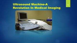
Ultrasound Machine-A
Revolution In Medical Imaging
1
Contents:-
1. What is medical imaging?
2. Why ultrasound imaging is required?
3. History of ultrasound
4. What is ultrasound
A. Physical definition
B. Medical definition
5. Ultrasound production
6. The Returning echo
7. Doppler effect
8. What is Doppler ultrasound
9. Principles of instrumentation in ultrasonography
10. Transmitter and receiver circuits of ultrasound
11. Mechanical assembly of ultrasound machine
12. Manufacturing companies of USG
13. Sonoscape S40 color Doppler ultrasound system
14. Clinical applications of ultrasound
15. Future of ultrasound
16. Conclusion
2
What is Medical Imaging?
It is a technique and process used to create
image of the internal as well as external human
body parts for clinical purpose .
3
Why ULTRASOUND Imaging Is
Required?
• Ultrasound imaging provides a pictorial
status of particular organ which is to be
treated
• Ultrasound makes a surgical targets more
clear and precise
• Ultrasound provides a pictorial status of fetus
development right from 4th week to 36th- 38th
week
• Ultrasound make therapeutic targets easy to
detect and treat
4
HISTORY OF
ULTRASOUND……………..
•In 1880 brothers Jacques et Pierre Curie noted
that the electricity is created in the crystal of
quartz under mechanical vibration. This
phenomenon was described as
the piezoelectric effect.
•Diagnosis medical application in use since
late 1950’s like visualizing cerebral chamber.
5
What is Ultrasound :Physical
Definition
• Ultrasound is a mechanical, longitudinal wave with a
frequency exceeding the upper limit of human
hearing, which is 20,000 Hz or 20 kHz.
6
What is Ultrasound : Medical
Definition
• Diagnostic Medical Ultrasound is the use of
high frequency sound to aid in diagnosis
and treatment of patient.
• Frequency Ranges Used In Medical
Ultrasound are 2.5-40MHZ
7
Ultrasound Production:
 Transducer contains piezoelectric
elements/crystals which produce the
ultrasound pulses (transmit 1% of the time)
 These elements convert electrical energy into
a mechanical ultrasound wave
8
 Reflected echoes return to the scan head
where the piezoelectric elements convert the
ultrasound wave back into an electrical signal
 The electrical signal is then processed by the
ultrasound system
9
Doppler Effect
10
• The Doppler effect is the apparent change in frequency
detected when the sound is moving relative to the
hearer. It is important to note that the effect does not
result because of an actual change in the frequency of
the source.
Modes of ultrasound
 A-mode (A=amplitude)
The amplitude of reflected ultrasound is displayed on an oscilloscope
screen
 M-mode (M=motion) It reflects a motion of the heart structures over time
Due to its excellent temporal resolution (high sampling rate), M-mode is
extremely valuable for accurate evaluation of rapid movements.
 B-mode (B=brightness) (2D in echocardiography) This is now the
essential imaging modality in the diagnostic ultrasound. An amplitude of
the reflected ultrasound signals is converted into a gray scale image
 D-mode (D=Doppler)
This imaging mode is based on the Doppler effect ie. change in frequency
(Doppler shift) caused by the reciprocal movement of the sound generator
and the observer.
11
What is Doppler Ultrasound?
• A Doppler ultrasound is a noninvasive test that can be used to
estimate our blood flow through blood vessels by bouncing
high-frequency sound waves (ultrasound) off circulating red
blood cells.
APPLICATIONS:
• Blood clots
• Poorly functioning valves in leg veins (venous insufficiency)
• Heart valve defects and congenital heart disease
• A blocked artery (arterial occlusion)
• Decreased blood circulation into legs (peripheral artery disease)
• Bulging arteries (aneurysms)
• Narrowing of an artery, such as in our neck (carotid artery stenosis)
12
Principles of Instrumentation In
Ultrasonography
13
• All ultrasound scanners consist of similar
components that perform the same key
functions.
• One of these is a transmitter that sends pulses
to the transducer, a receiver and a processor
that detects and amplifies the backscattered
energy.
Transmitter circuit of ultrasound
Around transistor T1 (BC549) generates an 8MHz signal, which serves as input to
the first decade counter built around IC1. The decade counter divides the oscillator
frequency to 800 kHz. The output of IC1 is fed to the second CD4017 decade
counter (IC2), which further divides the frequency to 80 kHz.
The flip-flop (IC3) divides 80kHz signal by 2 to give 40kHz signal, which is transmitted
by ultrasonic transducer TX.
14
Receiver circuit of ultrasound
It is necessary to down-convert the 40kHz signal into 4kHz to bring it
in the audible range. The receiver’s transducer unit (RX) detects the
transmitted 40kHz signal, which is amplified by the amplifier built
around transistor BC549 (T2). The amplified signal is fed to decade
counter IC4, which divides the frequency to 4 kHz.
Transistor T3 (SL100) amplifies the 4kHz signal to drive the speaker.
15
Mechanical assembly Of Ultrasound
Machine
1.Transducer:
Probe
Pulse Control
2.CPU (Central Processing Unit)
3.Key Board
4.Display
5.Storage device
6.Printer
16
Images 17
Different components of US
Machine
18
The following parts:
1. Transducer (probe)
• The probe is the mouth and ears of the ultrasound
machine.
• In the probe, there are one or more quartz crystals
called piezoelectric crystals.
• When an electric current is applied to these
crystals, they change shape rapidly.
Different components of US
Machine
19
Transducer (pulse controls)
• The operator, called the ultrasonographer,
changes the amplitude, frequency and duration
of the pulses emitted from the transducer probe
Different components of US
Machine
20
2. Central Processing Unit (CPU)
• The CPU is the brain of an ultrasound machine.
• The CPU is a computer that contains the
microprocessor, memory, amplifiers and power
supplies for the microprocessor and transducer
probe.
• The transducer receives electrical currents from the
CPU and sends electrical pulses that are created by
returning echoes.
Different components of US
Machine
21
3.Keyboard/Cursor
• Ultrasound machines have a keyboard and a
cursor.
• The keyboard allows the operator to add notes
and to take measurements of the image.
Different components of US
Machine
22
5 Display
• Displays the image from the ultrasound data
processed by the CPU.
• This image can be either in black-and-white or color,
depending upon the model of the ultrasound
machine
Different components of US
Machine
23
5. Disk Storage
• The processed data and/or images can be stored
on disks.
• These disks can be hard disks, floppy disks,
compact disks (CDs), or digital video disks
(DVDs).
• Most of the time, ultrasound scans are filled on
floppy disks and stored with the patient's medical
records.
Different components of US
Machine
24
6. Printers
Most ultrasound machines have printers which are
thermal . These can be used to capture a printed
picture of the image from the monitor.
Working of the USG machine: 25
MANUFACTURING COMPANIES
OF USG 26
MANUFACTURER MODEL COST (Rs)
1.PHILIPS HEALTHCARE EPIQ 7 243754.00
2.GE HEALTHCARE LOGIQ E9 225443.00
3.SIEMENS ACUSON P500 211553.00
4.CARESTREAM HEALTH CARESTREAM
TOUCH
ULTRASOUND
149239.00
5.TOSHIBA MEDICAL
SYSTEMS CORPORATION
EB1970UK
endobronchial
148255.00
6.HITACHI MEDICAL
CORPORATION
Aplio™ 500/400/300 175358.00
:
SONOSCAPE S40 COLOR DOPPLER ULTRASOUND
SYSTEM
TECHNICAL SPECIFICATIONS:
1. Full Digital Super-wide Band Beam Former,
2. Digital Dynamic Focusing,
3. Variable Aperture and Dynamic Tracing,
4. Wide Band Dynamic Range,
5. Multi-Beam Parallel Processing designed to
comply with applicable international standards and
regulations, ensuring the safety and availability of
this product.
6. Based on the computer technology and Linux
operation system, which make the system more
flexible and stable.
7. User Friendly.
27
Different ultrasound probes
 C362, convex high-density probe for adults' examinations
Field of use: Abdominal examinations, obstetrics,
 L742, linear high-density probe for examinations of vessels
Field of use: Abdominal examinations, pediatry,
 5P1, sectorial phased probe for pediatric examinations
Field of use: Cardiology, pediatry
 МРТЕЕ, transesophageal probe
Field of use: Cardiology, surgery.

28
FURTHER MORE DETAILS…
ADVANTAGES:
• New generation digital front-end
technology
• Spatial compound imaging
• Post-processing technology
• Tissue harmonic imaging
• High pulse repetition frequency
• Panoramic imaging
• 4D imaging
• Graphic diagnosis icon
• Touch screen with human-computer
interaction technology
• Keyboard lifting system
DISADVANTAGES:
• Bulky
• Heat development and cavity
formation
• Sensors having minimum sensing
distance
• Targets of low density like foam and
cloth tend to absorb sound energy,
these materials may be difficult to
sense at long range
• Temperature, pressure, humidity, air
turbulence affect the ultrasonic
response
29
Clinical application of ultrasound
• Anaesthesiology-
Ultrasound is commonly used by
anesthesiologists to guide injecting
needles when placing local anaesthetic
solutions near nerves
• Cardiology-
Echocardiography is an essential tool in
cardiology, to diagnose e.g. dilatation of
parts of the heart and function of heart
ventricles and valves
• Gastroenterology-
In abdominal sonography, the solid organs
of the abdomen the pancreas, aorta, inferior
vena cava, liver, gall bladder, bile ducts,
kidneys, and spleen are imaged.
• Obstetrics-
Obstetrical is commonly used during
pregnancy to check on the development of the
fetus.
30
Clinical application of
ultrasound
• Urology-
To determine, for example, the amount of
fluid retained in a patient's bladder.
In a pelvic sonogram, organs of the
pelvic region are imaged.
This includes the uterus and ovaries or
urinary bladder.
Males are sometimes given a pelvic
sonogram to check on the health of their
bladder, the prostate, or their testicles
(for example to distinguish epididymitis
from testicular torsion).
31
•Gynecology-
Gynecologic sonography is used
extensively:
To assess pelvic organs,
To diagnose and manage gynecologic
problems including, leiomyoma, ovarian
cysts and lesions,
To Identify ectopic Pregnancy,
To Diagnose Gynecologic Cancer
The Future OF Ultrasound:
HIGH POWER ULTRASOUND TECHNOLOGY:
• New powerful, technology that is not only safe and environmentally friendly in
its application but is also efficient and economical.
• Reduce or eliminate the need for chemicals or heat application in a variety of
industrial processes.
•This innovative new technology, of low frequency, high-power ultrasound (20kHz
- 1MHz), can be applied to a large number of industry processing applications
including food safety related areas.
•Innovative Ultrasonics is a company specializing in process development,
engineering design, installation and equipment in the area of high-powered
ultrasonics for new and existing industrial applications.
• The use of high-power ultrasonics in industry is rapidly expanding throughout
Europe and North America.
32
CONCLUSION:
• Sonography is effective for soft tissues imaging of
many different systems
• Ultrasound machine is rapid ,easy to use .
• Ultrasound machine is free from ionizing radiations.
• Ultrasound has not only been used to made or
exclude diagnoses, but it has also become the
modality of choice in the imaging of both the stable
and unstable pediatric patients.
33
REFERENCES
http://www3.gehealthcare.in/en/products/categories/ultrasound
http://gpsmedical.typepad.com/
http://dinesh-projects.blogspot.in/2012/04/ultrasonic-transmitter-and-
receiver.html
http://www.sciencedirect.com/science/article/pii/S1875389210000064
http://www.meditech.cn/meditech-edu/ultrasound-scanner-2.asp
http://dothealth.com/Content/Uploads/live/preview/20131220121521-Color-
Doppler-Ultrasound-System-S11.jpg
http://www.solutionmedical.gr/default.aspx?pageid=9413&type=product
34
35
36
37

More Related Content

What's hot

Digital Fluoroscopy Imaging System
Digital Fluoroscopy Imaging SystemDigital Fluoroscopy Imaging System
Digital Fluoroscopy Imaging SystemMuhammad Arif Afridi
 
Interactions of ultrasound with matter
Interactions of ultrasound with matterInteractions of ultrasound with matter
Interactions of ultrasound with matterSwapnil Shetty
 
Mammographic equipment
Mammographic equipmentMammographic equipment
Mammographic equipmentSharmaRajan4
 
Encoding and image formation
Encoding and image formationEncoding and image formation
Encoding and image formationAnkit Mishra
 
"high tension generator", "high tension","tension generator", "generator", ...
"high tension generator",  "high tension","tension generator",  "generator", ..."high tension generator",  "high tension","tension generator",  "generator", ...
"high tension generator", "high tension","tension generator", "generator", ...Prem Murti
 
Ultrasound & properties
Ultrasound &  propertiesUltrasound &  properties
Ultrasound & propertiesMahesh Kumar
 
Ultrasound Physics Made easy - By Dr Chandni Wadhwani
Ultrasound Physics Made easy - By Dr Chandni WadhwaniUltrasound Physics Made easy - By Dr Chandni Wadhwani
Ultrasound Physics Made easy - By Dr Chandni WadhwaniChandni Wadhwani
 
Advances in ct technology
Advances in ct technologyAdvances in ct technology
Advances in ct technologyMitusha Verma
 
Basic physics of ultrasound.JH
Basic physics of ultrasound.JHBasic physics of ultrasound.JH
Basic physics of ultrasound.JHhari baskar
 
Introduction to ultarsound machine and physics
Introduction to ultarsound machine and physicsIntroduction to ultarsound machine and physics
Introduction to ultarsound machine and physicsmanishyadav513
 
Post processing of computed tomography
Post processing of computed tomographyPost processing of computed tomography
Post processing of computed tomographyBeuniquewithNehaSing
 
Types of Ultrasound Probes
Types of Ultrasound ProbesTypes of Ultrasound Probes
Types of Ultrasound Probeshafsatalat1
 
Basics of radiation and production of x rays
Basics of radiation and production of x raysBasics of radiation and production of x rays
Basics of radiation and production of x raysdbc9427
 
Production of xray
Production of xrayProduction of xray
Production of xrayPrajwith Rai
 
Mobile & portable radiography
Mobile & portable radiographyMobile & portable radiography
Mobile & portable radiographyPriyanka Parimala
 
CT image quality and image resolution.pptx
CT image quality and image resolution.pptxCT image quality and image resolution.pptx
CT image quality and image resolution.pptxRohit Bansal
 
Ct quality control
Ct quality controlCt quality control
Ct quality controlSAJIDEJAZ1
 

What's hot (20)

Digital Fluoroscopy Imaging System
Digital Fluoroscopy Imaging SystemDigital Fluoroscopy Imaging System
Digital Fluoroscopy Imaging System
 
Interactions of ultrasound with matter
Interactions of ultrasound with matterInteractions of ultrasound with matter
Interactions of ultrasound with matter
 
Mammographic equipment
Mammographic equipmentMammographic equipment
Mammographic equipment
 
Encoding and image formation
Encoding and image formationEncoding and image formation
Encoding and image formation
 
"high tension generator", "high tension","tension generator", "generator", ...
"high tension generator",  "high tension","tension generator",  "generator", ..."high tension generator",  "high tension","tension generator",  "generator", ...
"high tension generator", "high tension","tension generator", "generator", ...
 
Ultrasound & properties
Ultrasound &  propertiesUltrasound &  properties
Ultrasound & properties
 
Ultrasound Physics Made easy - By Dr Chandni Wadhwani
Ultrasound Physics Made easy - By Dr Chandni WadhwaniUltrasound Physics Made easy - By Dr Chandni Wadhwani
Ultrasound Physics Made easy - By Dr Chandni Wadhwani
 
Advances in ct technology
Advances in ct technologyAdvances in ct technology
Advances in ct technology
 
Basic physics of ultrasound.JH
Basic physics of ultrasound.JHBasic physics of ultrasound.JH
Basic physics of ultrasound.JH
 
Introduction to ultarsound machine and physics
Introduction to ultarsound machine and physicsIntroduction to ultarsound machine and physics
Introduction to ultarsound machine and physics
 
Post processing of computed tomography
Post processing of computed tomographyPost processing of computed tomography
Post processing of computed tomography
 
Basic principles of CT scanning
Basic principles of CT scanningBasic principles of CT scanning
Basic principles of CT scanning
 
Types of Ultrasound Probes
Types of Ultrasound ProbesTypes of Ultrasound Probes
Types of Ultrasound Probes
 
Basics of radiation and production of x rays
Basics of radiation and production of x raysBasics of radiation and production of x rays
Basics of radiation and production of x rays
 
Fluoroscopy
 Fluoroscopy  Fluoroscopy
Fluoroscopy
 
Production of xray
Production of xrayProduction of xray
Production of xray
 
Mobile & portable radiography
Mobile & portable radiographyMobile & portable radiography
Mobile & portable radiography
 
CT image quality and image resolution.pptx
CT image quality and image resolution.pptxCT image quality and image resolution.pptx
CT image quality and image resolution.pptx
 
Slip ring
Slip ringSlip ring
Slip ring
 
Ct quality control
Ct quality controlCt quality control
Ct quality control
 

Viewers also liked

Ultrasound probe diagnostic results
Ultrasound probe diagnostic resultsUltrasound probe diagnostic results
Ultrasound probe diagnostic resultsProbelogic Pty Ltd
 
Image Characteristic & Interpretation
Image Characteristic & InterpretationImage Characteristic & Interpretation
Image Characteristic & InterpretationWAlid Salem
 
Radiographic interpretation
Radiographic interpretationRadiographic interpretation
Radiographic interpretationmoix rafiq
 

Viewers also liked (6)

Image Contrast, Noise, Resolution
Image Contrast, Noise, ResolutionImage Contrast, Noise, Resolution
Image Contrast, Noise, Resolution
 
Film interpretation
Film interpretationFilm interpretation
Film interpretation
 
Ultrasound probe diagnostic results
Ultrasound probe diagnostic resultsUltrasound probe diagnostic results
Ultrasound probe diagnostic results
 
Image Characteristic & Interpretation
Image Characteristic & InterpretationImage Characteristic & Interpretation
Image Characteristic & Interpretation
 
MRI sequences
MRI sequencesMRI sequences
MRI sequences
 
Radiographic interpretation
Radiographic interpretationRadiographic interpretation
Radiographic interpretation
 

Similar to Ultrasound Machine-A Revolution In Medical Imaging

Ultrasound imaging
Ultrasound imagingUltrasound imaging
Ultrasound imagingAbid Hussain
 
Ultrasonography
UltrasonographyUltrasonography
UltrasonographySai Ram
 
Computed Tomography.pptx
Computed Tomography.pptxComputed Tomography.pptx
Computed Tomography.pptxjerelubarco1
 
Ultrasound Imaging_2023.pdf
Ultrasound Imaging_2023.pdfUltrasound Imaging_2023.pdf
Ultrasound Imaging_2023.pdfghadaElbanby1
 
Radiology_Equipment_Lec-1_Dr. Emad Taleb.pdf
Radiology_Equipment_Lec-1_Dr. Emad Taleb.pdfRadiology_Equipment_Lec-1_Dr. Emad Taleb.pdf
Radiology_Equipment_Lec-1_Dr. Emad Taleb.pdfEmadTaleb1
 
Use of computers in veterinary surgery
Use of computers in veterinary surgeryUse of computers in veterinary surgery
Use of computers in veterinary surgeryAjith Y
 
Telemedicine System For Cardiac Patients
Telemedicine System For Cardiac PatientsTelemedicine System For Cardiac Patients
Telemedicine System For Cardiac PatientsSharad Karwa
 
Basics of Diagnostic Ultrasound
Basics of Diagnostic UltrasoundBasics of Diagnostic Ultrasound
Basics of Diagnostic UltrasoundIbrahim Tayyan
 
ULTRASOUND MACHINE
ULTRASOUND  MACHINEULTRASOUND  MACHINE
ULTRASOUND MACHINEkaushikbaro
 
Ultrasonography - History, evolution and principles
Ultrasonography - History, evolution and principlesUltrasonography - History, evolution and principles
Ultrasonography - History, evolution and principlesaparna666
 
Image Processing in Measurement guided Radiotherapy and Geometric accuracy
Image Processing in Measurement guided Radiotherapy and Geometric accuracyImage Processing in Measurement guided Radiotherapy and Geometric accuracy
Image Processing in Measurement guided Radiotherapy and Geometric accuracyajayhakkumar
 
Role Of Computers IN VETERINARY SURGERY,DRF.MUDASIR BASHIR
Role Of Computers IN VETERINARY SURGERY,DRF.MUDASIR BASHIRRole Of Computers IN VETERINARY SURGERY,DRF.MUDASIR BASHIR
Role Of Computers IN VETERINARY SURGERY,DRF.MUDASIR BASHIRguestafb98a0
 
Ct computed tomography
Ct computed tomographyCt computed tomography
Ct computed tomographySaadNasser6
 
APPLICATION OF DSP IN BIOMEDICAL ENGINEERING
APPLICATION OF DSP IN BIOMEDICAL ENGINEERINGAPPLICATION OF DSP IN BIOMEDICAL ENGINEERING
APPLICATION OF DSP IN BIOMEDICAL ENGINEERINGpirh khan
 

Similar to Ultrasound Machine-A Revolution In Medical Imaging (20)

Ultrasound imaging
Ultrasound imagingUltrasound imaging
Ultrasound imaging
 
Ultrasonography
UltrasonographyUltrasonography
Ultrasonography
 
Computed Tomography.pptx
Computed Tomography.pptxComputed Tomography.pptx
Computed Tomography.pptx
 
Ct scan
Ct scanCt scan
Ct scan
 
Ultrasound Imaging_2023.pdf
Ultrasound Imaging_2023.pdfUltrasound Imaging_2023.pdf
Ultrasound Imaging_2023.pdf
 
Ultrasonography ppt[1]
Ultrasonography ppt[1]Ultrasonography ppt[1]
Ultrasonography ppt[1]
 
Radiology_Equipment_Lec-1_Dr. Emad Taleb.pdf
Radiology_Equipment_Lec-1_Dr. Emad Taleb.pdfRadiology_Equipment_Lec-1_Dr. Emad Taleb.pdf
Radiology_Equipment_Lec-1_Dr. Emad Taleb.pdf
 
Use of computers in veterinary surgery
Use of computers in veterinary surgeryUse of computers in veterinary surgery
Use of computers in veterinary surgery
 
Telemedicine System For Cardiac Patients
Telemedicine System For Cardiac PatientsTelemedicine System For Cardiac Patients
Telemedicine System For Cardiac Patients
 
Usg
UsgUsg
Usg
 
Basics of Diagnostic Ultrasound
Basics of Diagnostic UltrasoundBasics of Diagnostic Ultrasound
Basics of Diagnostic Ultrasound
 
ultrasonography.pptx
ultrasonography.pptxultrasonography.pptx
ultrasonography.pptx
 
ULTRASOUND MACHINE
ULTRASOUND  MACHINEULTRASOUND  MACHINE
ULTRASOUND MACHINE
 
Ultrasonography - History, evolution and principles
Ultrasonography - History, evolution and principlesUltrasonography - History, evolution and principles
Ultrasonography - History, evolution and principles
 
Image Processing in Measurement guided Radiotherapy and Geometric accuracy
Image Processing in Measurement guided Radiotherapy and Geometric accuracyImage Processing in Measurement guided Radiotherapy and Geometric accuracy
Image Processing in Measurement guided Radiotherapy and Geometric accuracy
 
Role Of Computers IN VETERINARY SURGERY,DRF.MUDASIR BASHIR
Role Of Computers IN VETERINARY SURGERY,DRF.MUDASIR BASHIRRole Of Computers IN VETERINARY SURGERY,DRF.MUDASIR BASHIR
Role Of Computers IN VETERINARY SURGERY,DRF.MUDASIR BASHIR
 
Perineal Hernia Animals
Perineal Hernia  AnimalsPerineal Hernia  Animals
Perineal Hernia Animals
 
Ct computed tomography
Ct computed tomographyCt computed tomography
Ct computed tomography
 
Ultrasonic_Based_Security_System
Ultrasonic_Based_Security_SystemUltrasonic_Based_Security_System
Ultrasonic_Based_Security_System
 
APPLICATION OF DSP IN BIOMEDICAL ENGINEERING
APPLICATION OF DSP IN BIOMEDICAL ENGINEERINGAPPLICATION OF DSP IN BIOMEDICAL ENGINEERING
APPLICATION OF DSP IN BIOMEDICAL ENGINEERING
 

Recently uploaded

Oxy acetylene welding presentation note.
Oxy acetylene welding presentation note.Oxy acetylene welding presentation note.
Oxy acetylene welding presentation note.eptoze12
 
Arduino_CSE ece ppt for working and principal of arduino.ppt
Arduino_CSE ece ppt for working and principal of arduino.pptArduino_CSE ece ppt for working and principal of arduino.ppt
Arduino_CSE ece ppt for working and principal of arduino.pptSAURABHKUMAR892774
 
INFLUENCE OF NANOSILICA ON THE PROPERTIES OF CONCRETE
INFLUENCE OF NANOSILICA ON THE PROPERTIES OF CONCRETEINFLUENCE OF NANOSILICA ON THE PROPERTIES OF CONCRETE
INFLUENCE OF NANOSILICA ON THE PROPERTIES OF CONCRETEroselinkalist12
 
complete construction, environmental and economics information of biomass com...
complete construction, environmental and economics information of biomass com...complete construction, environmental and economics information of biomass com...
complete construction, environmental and economics information of biomass com...asadnawaz62
 
UNIT III ANALOG ELECTRONICS (BASIC ELECTRONICS)
UNIT III ANALOG ELECTRONICS (BASIC ELECTRONICS)UNIT III ANALOG ELECTRONICS (BASIC ELECTRONICS)
UNIT III ANALOG ELECTRONICS (BASIC ELECTRONICS)Dr SOUNDIRARAJ N
 
Architect Hassan Khalil Portfolio for 2024
Architect Hassan Khalil Portfolio for 2024Architect Hassan Khalil Portfolio for 2024
Architect Hassan Khalil Portfolio for 2024hassan khalil
 
Gurgaon ✡️9711147426✨Call In girls Gurgaon Sector 51 escort service
Gurgaon ✡️9711147426✨Call In girls Gurgaon Sector 51 escort serviceGurgaon ✡️9711147426✨Call In girls Gurgaon Sector 51 escort service
Gurgaon ✡️9711147426✨Call In girls Gurgaon Sector 51 escort servicejennyeacort
 
Decoding Kotlin - Your guide to solving the mysterious in Kotlin.pptx
Decoding Kotlin - Your guide to solving the mysterious in Kotlin.pptxDecoding Kotlin - Your guide to solving the mysterious in Kotlin.pptx
Decoding Kotlin - Your guide to solving the mysterious in Kotlin.pptxJoão Esperancinha
 
Churning of Butter, Factors affecting .
Churning of Butter, Factors affecting  .Churning of Butter, Factors affecting  .
Churning of Butter, Factors affecting .Satyam Kumar
 
Call Us ≽ 8377877756 ≼ Call Girls In Shastri Nagar (Delhi)
Call Us ≽ 8377877756 ≼ Call Girls In Shastri Nagar (Delhi)Call Us ≽ 8377877756 ≼ Call Girls In Shastri Nagar (Delhi)
Call Us ≽ 8377877756 ≼ Call Girls In Shastri Nagar (Delhi)dollysharma2066
 
Gfe Mayur Vihar Call Girls Service WhatsApp -> 9999965857 Available 24x7 ^ De...
Gfe Mayur Vihar Call Girls Service WhatsApp -> 9999965857 Available 24x7 ^ De...Gfe Mayur Vihar Call Girls Service WhatsApp -> 9999965857 Available 24x7 ^ De...
Gfe Mayur Vihar Call Girls Service WhatsApp -> 9999965857 Available 24x7 ^ De...srsj9000
 
Past, Present and Future of Generative AI
Past, Present and Future of Generative AIPast, Present and Future of Generative AI
Past, Present and Future of Generative AIabhishek36461
 
Introduction-To-Agricultural-Surveillance-Rover.pptx
Introduction-To-Agricultural-Surveillance-Rover.pptxIntroduction-To-Agricultural-Surveillance-Rover.pptx
Introduction-To-Agricultural-Surveillance-Rover.pptxk795866
 
Why does (not) Kafka need fsync: Eliminating tail latency spikes caused by fsync
Why does (not) Kafka need fsync: Eliminating tail latency spikes caused by fsyncWhy does (not) Kafka need fsync: Eliminating tail latency spikes caused by fsync
Why does (not) Kafka need fsync: Eliminating tail latency spikes caused by fsyncssuser2ae721
 
Application of Residue Theorem to evaluate real integrations.pptx
Application of Residue Theorem to evaluate real integrations.pptxApplication of Residue Theorem to evaluate real integrations.pptx
Application of Residue Theorem to evaluate real integrations.pptx959SahilShah
 
CCS355 Neural Network & Deep Learning Unit II Notes with Question bank .pdf
CCS355 Neural Network & Deep Learning Unit II Notes with Question bank .pdfCCS355 Neural Network & Deep Learning Unit II Notes with Question bank .pdf
CCS355 Neural Network & Deep Learning Unit II Notes with Question bank .pdfAsst.prof M.Gokilavani
 
Electronically Controlled suspensions system .pdf
Electronically Controlled suspensions system .pdfElectronically Controlled suspensions system .pdf
Electronically Controlled suspensions system .pdfme23b1001
 
Artificial-Intelligence-in-Electronics (K).pptx
Artificial-Intelligence-in-Electronics (K).pptxArtificial-Intelligence-in-Electronics (K).pptx
Artificial-Intelligence-in-Electronics (K).pptxbritheesh05
 

Recently uploaded (20)

Oxy acetylene welding presentation note.
Oxy acetylene welding presentation note.Oxy acetylene welding presentation note.
Oxy acetylene welding presentation note.
 
Call Us -/9953056974- Call Girls In Vikaspuri-/- Delhi NCR
Call Us -/9953056974- Call Girls In Vikaspuri-/- Delhi NCRCall Us -/9953056974- Call Girls In Vikaspuri-/- Delhi NCR
Call Us -/9953056974- Call Girls In Vikaspuri-/- Delhi NCR
 
Arduino_CSE ece ppt for working and principal of arduino.ppt
Arduino_CSE ece ppt for working and principal of arduino.pptArduino_CSE ece ppt for working and principal of arduino.ppt
Arduino_CSE ece ppt for working and principal of arduino.ppt
 
INFLUENCE OF NANOSILICA ON THE PROPERTIES OF CONCRETE
INFLUENCE OF NANOSILICA ON THE PROPERTIES OF CONCRETEINFLUENCE OF NANOSILICA ON THE PROPERTIES OF CONCRETE
INFLUENCE OF NANOSILICA ON THE PROPERTIES OF CONCRETE
 
complete construction, environmental and economics information of biomass com...
complete construction, environmental and economics information of biomass com...complete construction, environmental and economics information of biomass com...
complete construction, environmental and economics information of biomass com...
 
UNIT III ANALOG ELECTRONICS (BASIC ELECTRONICS)
UNIT III ANALOG ELECTRONICS (BASIC ELECTRONICS)UNIT III ANALOG ELECTRONICS (BASIC ELECTRONICS)
UNIT III ANALOG ELECTRONICS (BASIC ELECTRONICS)
 
Architect Hassan Khalil Portfolio for 2024
Architect Hassan Khalil Portfolio for 2024Architect Hassan Khalil Portfolio for 2024
Architect Hassan Khalil Portfolio for 2024
 
Gurgaon ✡️9711147426✨Call In girls Gurgaon Sector 51 escort service
Gurgaon ✡️9711147426✨Call In girls Gurgaon Sector 51 escort serviceGurgaon ✡️9711147426✨Call In girls Gurgaon Sector 51 escort service
Gurgaon ✡️9711147426✨Call In girls Gurgaon Sector 51 escort service
 
Decoding Kotlin - Your guide to solving the mysterious in Kotlin.pptx
Decoding Kotlin - Your guide to solving the mysterious in Kotlin.pptxDecoding Kotlin - Your guide to solving the mysterious in Kotlin.pptx
Decoding Kotlin - Your guide to solving the mysterious in Kotlin.pptx
 
Churning of Butter, Factors affecting .
Churning of Butter, Factors affecting  .Churning of Butter, Factors affecting  .
Churning of Butter, Factors affecting .
 
Call Us ≽ 8377877756 ≼ Call Girls In Shastri Nagar (Delhi)
Call Us ≽ 8377877756 ≼ Call Girls In Shastri Nagar (Delhi)Call Us ≽ 8377877756 ≼ Call Girls In Shastri Nagar (Delhi)
Call Us ≽ 8377877756 ≼ Call Girls In Shastri Nagar (Delhi)
 
Gfe Mayur Vihar Call Girls Service WhatsApp -> 9999965857 Available 24x7 ^ De...
Gfe Mayur Vihar Call Girls Service WhatsApp -> 9999965857 Available 24x7 ^ De...Gfe Mayur Vihar Call Girls Service WhatsApp -> 9999965857 Available 24x7 ^ De...
Gfe Mayur Vihar Call Girls Service WhatsApp -> 9999965857 Available 24x7 ^ De...
 
Past, Present and Future of Generative AI
Past, Present and Future of Generative AIPast, Present and Future of Generative AI
Past, Present and Future of Generative AI
 
Introduction-To-Agricultural-Surveillance-Rover.pptx
Introduction-To-Agricultural-Surveillance-Rover.pptxIntroduction-To-Agricultural-Surveillance-Rover.pptx
Introduction-To-Agricultural-Surveillance-Rover.pptx
 
Why does (not) Kafka need fsync: Eliminating tail latency spikes caused by fsync
Why does (not) Kafka need fsync: Eliminating tail latency spikes caused by fsyncWhy does (not) Kafka need fsync: Eliminating tail latency spikes caused by fsync
Why does (not) Kafka need fsync: Eliminating tail latency spikes caused by fsync
 
young call girls in Green Park🔝 9953056974 🔝 escort Service
young call girls in Green Park🔝 9953056974 🔝 escort Serviceyoung call girls in Green Park🔝 9953056974 🔝 escort Service
young call girls in Green Park🔝 9953056974 🔝 escort Service
 
Application of Residue Theorem to evaluate real integrations.pptx
Application of Residue Theorem to evaluate real integrations.pptxApplication of Residue Theorem to evaluate real integrations.pptx
Application of Residue Theorem to evaluate real integrations.pptx
 
CCS355 Neural Network & Deep Learning Unit II Notes with Question bank .pdf
CCS355 Neural Network & Deep Learning Unit II Notes with Question bank .pdfCCS355 Neural Network & Deep Learning Unit II Notes with Question bank .pdf
CCS355 Neural Network & Deep Learning Unit II Notes with Question bank .pdf
 
Electronically Controlled suspensions system .pdf
Electronically Controlled suspensions system .pdfElectronically Controlled suspensions system .pdf
Electronically Controlled suspensions system .pdf
 
Artificial-Intelligence-in-Electronics (K).pptx
Artificial-Intelligence-in-Electronics (K).pptxArtificial-Intelligence-in-Electronics (K).pptx
Artificial-Intelligence-in-Electronics (K).pptx
 

Ultrasound Machine-A Revolution In Medical Imaging

  • 2. Contents:- 1. What is medical imaging? 2. Why ultrasound imaging is required? 3. History of ultrasound 4. What is ultrasound A. Physical definition B. Medical definition 5. Ultrasound production 6. The Returning echo 7. Doppler effect 8. What is Doppler ultrasound 9. Principles of instrumentation in ultrasonography 10. Transmitter and receiver circuits of ultrasound 11. Mechanical assembly of ultrasound machine 12. Manufacturing companies of USG 13. Sonoscape S40 color Doppler ultrasound system 14. Clinical applications of ultrasound 15. Future of ultrasound 16. Conclusion 2
  • 3. What is Medical Imaging? It is a technique and process used to create image of the internal as well as external human body parts for clinical purpose . 3
  • 4. Why ULTRASOUND Imaging Is Required? • Ultrasound imaging provides a pictorial status of particular organ which is to be treated • Ultrasound makes a surgical targets more clear and precise • Ultrasound provides a pictorial status of fetus development right from 4th week to 36th- 38th week • Ultrasound make therapeutic targets easy to detect and treat 4
  • 5. HISTORY OF ULTRASOUND…………….. •In 1880 brothers Jacques et Pierre Curie noted that the electricity is created in the crystal of quartz under mechanical vibration. This phenomenon was described as the piezoelectric effect. •Diagnosis medical application in use since late 1950’s like visualizing cerebral chamber. 5
  • 6. What is Ultrasound :Physical Definition • Ultrasound is a mechanical, longitudinal wave with a frequency exceeding the upper limit of human hearing, which is 20,000 Hz or 20 kHz. 6
  • 7. What is Ultrasound : Medical Definition • Diagnostic Medical Ultrasound is the use of high frequency sound to aid in diagnosis and treatment of patient. • Frequency Ranges Used In Medical Ultrasound are 2.5-40MHZ 7
  • 8. Ultrasound Production:  Transducer contains piezoelectric elements/crystals which produce the ultrasound pulses (transmit 1% of the time)  These elements convert electrical energy into a mechanical ultrasound wave 8
  • 9.  Reflected echoes return to the scan head where the piezoelectric elements convert the ultrasound wave back into an electrical signal  The electrical signal is then processed by the ultrasound system 9
  • 10. Doppler Effect 10 • The Doppler effect is the apparent change in frequency detected when the sound is moving relative to the hearer. It is important to note that the effect does not result because of an actual change in the frequency of the source.
  • 11. Modes of ultrasound  A-mode (A=amplitude) The amplitude of reflected ultrasound is displayed on an oscilloscope screen  M-mode (M=motion) It reflects a motion of the heart structures over time Due to its excellent temporal resolution (high sampling rate), M-mode is extremely valuable for accurate evaluation of rapid movements.  B-mode (B=brightness) (2D in echocardiography) This is now the essential imaging modality in the diagnostic ultrasound. An amplitude of the reflected ultrasound signals is converted into a gray scale image  D-mode (D=Doppler) This imaging mode is based on the Doppler effect ie. change in frequency (Doppler shift) caused by the reciprocal movement of the sound generator and the observer. 11
  • 12. What is Doppler Ultrasound? • A Doppler ultrasound is a noninvasive test that can be used to estimate our blood flow through blood vessels by bouncing high-frequency sound waves (ultrasound) off circulating red blood cells. APPLICATIONS: • Blood clots • Poorly functioning valves in leg veins (venous insufficiency) • Heart valve defects and congenital heart disease • A blocked artery (arterial occlusion) • Decreased blood circulation into legs (peripheral artery disease) • Bulging arteries (aneurysms) • Narrowing of an artery, such as in our neck (carotid artery stenosis) 12
  • 13. Principles of Instrumentation In Ultrasonography 13 • All ultrasound scanners consist of similar components that perform the same key functions. • One of these is a transmitter that sends pulses to the transducer, a receiver and a processor that detects and amplifies the backscattered energy.
  • 14. Transmitter circuit of ultrasound Around transistor T1 (BC549) generates an 8MHz signal, which serves as input to the first decade counter built around IC1. The decade counter divides the oscillator frequency to 800 kHz. The output of IC1 is fed to the second CD4017 decade counter (IC2), which further divides the frequency to 80 kHz. The flip-flop (IC3) divides 80kHz signal by 2 to give 40kHz signal, which is transmitted by ultrasonic transducer TX. 14
  • 15. Receiver circuit of ultrasound It is necessary to down-convert the 40kHz signal into 4kHz to bring it in the audible range. The receiver’s transducer unit (RX) detects the transmitted 40kHz signal, which is amplified by the amplifier built around transistor BC549 (T2). The amplified signal is fed to decade counter IC4, which divides the frequency to 4 kHz. Transistor T3 (SL100) amplifies the 4kHz signal to drive the speaker. 15
  • 16. Mechanical assembly Of Ultrasound Machine 1.Transducer: Probe Pulse Control 2.CPU (Central Processing Unit) 3.Key Board 4.Display 5.Storage device 6.Printer 16
  • 18. Different components of US Machine 18 The following parts: 1. Transducer (probe) • The probe is the mouth and ears of the ultrasound machine. • In the probe, there are one or more quartz crystals called piezoelectric crystals. • When an electric current is applied to these crystals, they change shape rapidly.
  • 19. Different components of US Machine 19 Transducer (pulse controls) • The operator, called the ultrasonographer, changes the amplitude, frequency and duration of the pulses emitted from the transducer probe
  • 20. Different components of US Machine 20 2. Central Processing Unit (CPU) • The CPU is the brain of an ultrasound machine. • The CPU is a computer that contains the microprocessor, memory, amplifiers and power supplies for the microprocessor and transducer probe. • The transducer receives electrical currents from the CPU and sends electrical pulses that are created by returning echoes.
  • 21. Different components of US Machine 21 3.Keyboard/Cursor • Ultrasound machines have a keyboard and a cursor. • The keyboard allows the operator to add notes and to take measurements of the image.
  • 22. Different components of US Machine 22 5 Display • Displays the image from the ultrasound data processed by the CPU. • This image can be either in black-and-white or color, depending upon the model of the ultrasound machine
  • 23. Different components of US Machine 23 5. Disk Storage • The processed data and/or images can be stored on disks. • These disks can be hard disks, floppy disks, compact disks (CDs), or digital video disks (DVDs). • Most of the time, ultrasound scans are filled on floppy disks and stored with the patient's medical records.
  • 24. Different components of US Machine 24 6. Printers Most ultrasound machines have printers which are thermal . These can be used to capture a printed picture of the image from the monitor.
  • 25. Working of the USG machine: 25
  • 26. MANUFACTURING COMPANIES OF USG 26 MANUFACTURER MODEL COST (Rs) 1.PHILIPS HEALTHCARE EPIQ 7 243754.00 2.GE HEALTHCARE LOGIQ E9 225443.00 3.SIEMENS ACUSON P500 211553.00 4.CARESTREAM HEALTH CARESTREAM TOUCH ULTRASOUND 149239.00 5.TOSHIBA MEDICAL SYSTEMS CORPORATION EB1970UK endobronchial 148255.00 6.HITACHI MEDICAL CORPORATION Aplio™ 500/400/300 175358.00 :
  • 27. SONOSCAPE S40 COLOR DOPPLER ULTRASOUND SYSTEM TECHNICAL SPECIFICATIONS: 1. Full Digital Super-wide Band Beam Former, 2. Digital Dynamic Focusing, 3. Variable Aperture and Dynamic Tracing, 4. Wide Band Dynamic Range, 5. Multi-Beam Parallel Processing designed to comply with applicable international standards and regulations, ensuring the safety and availability of this product. 6. Based on the computer technology and Linux operation system, which make the system more flexible and stable. 7. User Friendly. 27
  • 28. Different ultrasound probes  C362, convex high-density probe for adults' examinations Field of use: Abdominal examinations, obstetrics,  L742, linear high-density probe for examinations of vessels Field of use: Abdominal examinations, pediatry,  5P1, sectorial phased probe for pediatric examinations Field of use: Cardiology, pediatry  МРТЕЕ, transesophageal probe Field of use: Cardiology, surgery.  28
  • 29. FURTHER MORE DETAILS… ADVANTAGES: • New generation digital front-end technology • Spatial compound imaging • Post-processing technology • Tissue harmonic imaging • High pulse repetition frequency • Panoramic imaging • 4D imaging • Graphic diagnosis icon • Touch screen with human-computer interaction technology • Keyboard lifting system DISADVANTAGES: • Bulky • Heat development and cavity formation • Sensors having minimum sensing distance • Targets of low density like foam and cloth tend to absorb sound energy, these materials may be difficult to sense at long range • Temperature, pressure, humidity, air turbulence affect the ultrasonic response 29
  • 30. Clinical application of ultrasound • Anaesthesiology- Ultrasound is commonly used by anesthesiologists to guide injecting needles when placing local anaesthetic solutions near nerves • Cardiology- Echocardiography is an essential tool in cardiology, to diagnose e.g. dilatation of parts of the heart and function of heart ventricles and valves • Gastroenterology- In abdominal sonography, the solid organs of the abdomen the pancreas, aorta, inferior vena cava, liver, gall bladder, bile ducts, kidneys, and spleen are imaged. • Obstetrics- Obstetrical is commonly used during pregnancy to check on the development of the fetus. 30
  • 31. Clinical application of ultrasound • Urology- To determine, for example, the amount of fluid retained in a patient's bladder. In a pelvic sonogram, organs of the pelvic region are imaged. This includes the uterus and ovaries or urinary bladder. Males are sometimes given a pelvic sonogram to check on the health of their bladder, the prostate, or their testicles (for example to distinguish epididymitis from testicular torsion). 31 •Gynecology- Gynecologic sonography is used extensively: To assess pelvic organs, To diagnose and manage gynecologic problems including, leiomyoma, ovarian cysts and lesions, To Identify ectopic Pregnancy, To Diagnose Gynecologic Cancer
  • 32. The Future OF Ultrasound: HIGH POWER ULTRASOUND TECHNOLOGY: • New powerful, technology that is not only safe and environmentally friendly in its application but is also efficient and economical. • Reduce or eliminate the need for chemicals or heat application in a variety of industrial processes. •This innovative new technology, of low frequency, high-power ultrasound (20kHz - 1MHz), can be applied to a large number of industry processing applications including food safety related areas. •Innovative Ultrasonics is a company specializing in process development, engineering design, installation and equipment in the area of high-powered ultrasonics for new and existing industrial applications. • The use of high-power ultrasonics in industry is rapidly expanding throughout Europe and North America. 32
  • 33. CONCLUSION: • Sonography is effective for soft tissues imaging of many different systems • Ultrasound machine is rapid ,easy to use . • Ultrasound machine is free from ionizing radiations. • Ultrasound has not only been used to made or exclude diagnoses, but it has also become the modality of choice in the imaging of both the stable and unstable pediatric patients. 33
  • 35. 35
  • 36. 36
  • 37. 37