Western blotting is a technique used to detect specific proteins in a tissue sample. It involves three main steps - separating proteins by size through gel electrophoresis, transferring them to a membrane, and detecting the target protein using primary and secondary antibodies. The target protein band can then be visualized, allowing identification and quantification of proteins in a complex mixture. Western blotting is commonly used to identify proteins, estimate their size and amount, and diagnose conditions by detecting antibodies against specific antigens.
Unit-IV; Professional Sales Representative (PSR).pptx
Western Blotting.pdf
1. B.Sc.III (Sem-VI), Unit-II, Paper-II:Biotechnology
Notes on Western Blotting Techniques
Authored by: Dr. Rajendra Chavhan, RMG College Nagbhid
WESTERN BLOTTING
Introduction
Western blot is the analytical technique used in molecular biology,
immunogenetics, and other molecular biology to detect specific proteins in a
sample of tissue homogenate or extract. Western blotting is called so as the
procedure is similar to Southern blotting. While Southern blotting is done to
detect DNA, Western blotting is done for the detection of proteins. Western
blotting is also called protein immunoblotting because an antibody is used to
specifically detect its antigen.
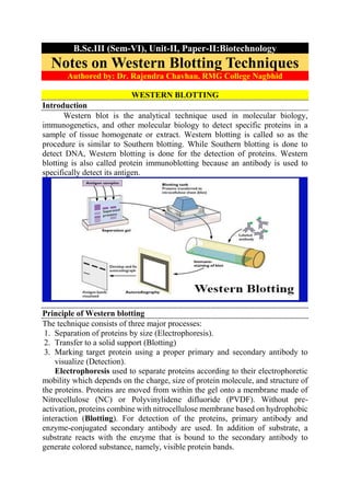
Principle of Western blotting
The technique consists of three major processes:
1. Separation of proteins by size (Electrophoresis).
2. Transfer to a solid support (Blotting)
3. Marking target protein using a proper primary and secondary antibody to
visualize (Detection).
Electrophoresis used to separate proteins according to their electrophoretic
mobility which depends on the charge, size of protein molecule, and structure of
the proteins. Proteins are moved from within the gel onto a membrane made of
Nitrocellulose (NC) or Polyvinylidene difluoride (PVDF). Without pre-
activation, proteins combine with nitrocellulose membrane based on hydrophobic
interaction (Blotting). For detection of the proteins, primary antibody and
enzyme-conjugated secondary antibody are used. In addition of substrate, a
substrate reacts with the enzyme that is bound to the secondary antibody to
generate colored substance, namely, visible protein bands.
2. In this technique, a mixture of proteins is separated based on molecular
weight, and thus by type, through gel electrophoresis. These results are then
transferred to a membrane producing a band for each protein. The membrane is
then incubated with labels antibodies specific to the protein of interest. The
unbound antibody is washed off leaving only the bound antibody to the protein
of interest. The bound antibodies are then detected by developing the film. As the
antibodies only bind to the protein of interest, only one band should be visible.
The thickness of the band corresponds to the amount of protein present; thus
doing a standard can indicate the amount of protein present.
Western blotting is usually done on a tissue homogenate or extract. It
utilizes SDS-PAGE (Sodium dodecyl sulfate polyacrylamide gel
electrophoresis), a type of gel electrophoresis to first separate various proteins
in a mixture on the basis of their shape and size. The protein bands thus obtained
are transferred onto a nitrocellulose or nylon membrane where they are
“probed” with antibodies specific to the protein to be detected. The antigen-
antibody complexes that form on the band containing the protein recognized by
the antibody can be visualized in a variety of ways.
If the protein of interest is bound by a radioactive antibody, its position on
the blot can be determined by exposing the membrane to a sheet of X-ray film, a
procedure called autoradiography. However, the most generally used detection
procedures employ enzyme-linked antibodies against the protein. After binding
of the enzyme–antibody conjugate, the addition of a chromogenic substrate that
produces a highly colored and insoluble product causes the appearance of a
colored band at the site of the target antigen. The site of the protein of interest can
be determined with a much higher sensitivity if a chemiluminescent compound
along with suitable enhancing agents is used to produce light at the antigen site.
Applications of Western blotting
1. Identification of a specific protein in a complex mixture of proteins. In this
method, known antigens of well-defined molecular weight are separated by
SDS-PAGE and blotted onto nitrocellulose. The separated bands of known
antigens are then probed with the sample suspected of containing antibodies
specific to one or more of these antigens. The reaction of an antibody with a
band is detected by using either a radiolabeled or enzyme-linked secondary
antibody that is specific for the species of the antibodies in the test sample.
2. Estimation of the size of the protein as well as the amount of protein present
in the mixture.
3. It is most widely used as a confirmatory test for the diagnosis of HIV, where
this procedure is used to determine whether the patient has antibodies that
react with one or more viral proteins or not.
4. Demonstration of specific antibodies in the serum for diagnosis of
neurocysticercosis and tubercular meningitis.