Magnetic Resonance Perfusion
•Download as PPTX, PDF•
17 likes•4,854 views

MR Perfusion Weighted Imaging Basic Principles and Physics Diagnostic values of MRP

Recommended
More Related Content
What's hot
What's hot (20)
Similar to Magnetic Resonance Perfusion
Similar to Magnetic Resonance Perfusion (20)
More from Rahman Ud Din
More from Rahman Ud Din (11)
Recently uploaded
Recently uploaded (20)
Magnetic Resonance Perfusion
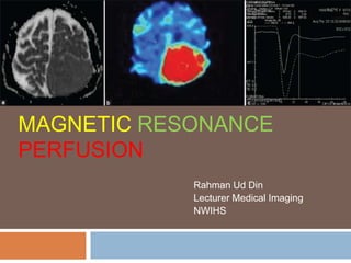
- 1. MAGNETIC RESONANCE PERFUSION Rahman Ud Din Lecturer Medical Imaging NWIHS
- 2. Introduction Blood flow and metabolism of human tissues Studied in nuclear medicine with tracer Now it is still studied with PET But PET lack spatial and temporal resolution Also lack in specificity Recent methods introduced like CT & MRI perfusion MR perfusion performed with Exogenous contrast agent (Gd) Endogenous contrast agent (no contrast administration)
- 3. Principles of MRP Imaging Perfusion refers to passage of blood from arterial supply to venous drainage through microcirculation Perfusion is vital for nutritive supply to tissues For clearance of products as well Perfusion can be affect by various disease processes Hence to detect such changes in perfusion can help in diagnosis of certain diseases, monitoring and assessing the treatment response
- 4. Gd (paramagnetic agents) cause shortening of both T1 and T2 of the tissue or region in which they go Decrease T1 relaxation time on T1-w images results into increased signals or brightening Decrease in the T2 relaxation time on T2 or T2*-w images results into signal drop or blackening As Gd passes through the microvasculature in high concentration there is decrease in signal in surrounding tissues Signal drop (shows disease)
- 5. Before and after Gd adminitration in T1-w Sequence
- 6. Technique of MRP with Exogenous Contrast A dose of 0.1mmol/kg of Gd injected IV Using power injector at rate of 5ml/s Fast T2*-w EPI seq is run to catch first pass of the contrast through microcirculation This seq: takes 15-20 slices from entire brain 1-2 s About 60 such run acquired before, during and after dynamic injection of the contrast media Raw data images converted into various color coded maps using software rCBV=relative Cerebral Blood Volume CBF= Cerebral blood flow TTP=Time to Peak MTT =Mean Transit Time
- 8. Permeability or Leakiness Areas of severe blood-brain barrier break-down are seen in necrotic tumor and irradiated tumor beds Increase in leakiness bcoz break in BBB results in accumulation of Gd- based contrast in extravascular space T1-enhancing effects of extravascular Gd may predominate to counteract the T2 signal
- 9. Clinical Applications of MRP MRP performed in various clinical conditions Stroke Brain tumors Dementia Psychiatric illnesses Migraine Headaches Trauma Epilepsy Multiple sclerosis
- 10. MRP in Stroke Stroke is brain attack To detect brain ischemia & salvageable tissue in early window period of 3-6 hours DWI and PWI or MRP effective for early ischemia PWI more sensitive than DWI detecting ischemia in early period of after onset of arterial occlusion
- 12. MRP in Brain Tumours Glioma Glioblastoma
- 13. Other Clinical Conditions MRP used to assess ischemic or under perfused areas Like moyamoya disease and CNS vasculities CNS vasculities