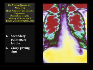
Secondary pulmonary lobule crazy paving sign
•Als PPTX, PDF herunterladen•
11 gefällt mir•3,818 views

Secondary pulmonary lobule crazy paving sign

Empfohlen
Weitere ähnliche Inhalte
Was ist angesagt?
Was ist angesagt? (20)
Ähnlich wie Secondary pulmonary lobule crazy paving sign
Ähnlich wie Secondary pulmonary lobule crazy paving sign (20)
Mehr von Minstry of health ,Ibn alnafis hoapital, Damascus
Mehr von Minstry of health ,Ibn alnafis hoapital, Damascus (10)
Kürzlich hochgeladen
Kürzlich hochgeladen (20)
Secondary pulmonary lobule crazy paving sign
- 1. Dr MazenQusaibatyMD, DISHead Pulmonary and Internist Department Ibnalnafisse HospitalMinistry of Syrian healthEmail: Qusaibaty@gmail.com Secondary pulmonary lobule Crazy paving sign
- 2. Topic Outline Secondary pulmonary lobule Crazy paving sign 2
- 4. 4
- 5. Secondary pulmonary lobule Unit of lung (0.5-3 cm) Irregularly polyhedral 5
- 6. Secondary pulmonary lobule A group of terminal bronchioles 6
- 7. Secondary pulmonary lobule Accompanyingpulmonary arterioles 7
- 8. Secondary pulmonary lobule Surrounded by lymph vessels 8
- 9. Secondary pulmonary lobule Demarcated by “interlobular septa” 9
- 10. Secondary pulmonary lobule Pulmonary veins 10
- 11. Secondary pulmonary lobule Pulmonary lymphatics 11
- 12. 12 Connective Tissue Stroma
- 13. 13 Quiz
- 14. 14 Quiz
- 15. 15 Quiz
- 16. 16 Quiz
- 17. 17 Quiz
- 18. 18 Quiz
- 19. 19 Quiz
- 21. Crazy paving sign Refers to the combination of ground glass attenuation superimposed on a network of interlobular septal thickening giving it an appearance of a surface paved with slabs of differing shapes 21
- 22. 22 Crazy paving sign Crazy Paving is a combination of ground glass opacity with superimposed net of septal thickening
- 23. Crazy paving sign Alveolar proteinosis 23
- 24. Crazy paving sign Thin-section CT scan targeted to the left upper lobe in a 41-year-old man with alveolar proteinosis 24
- 25. 25 Crazy Paving sign
- 26. 26 Crazy Paving
- 27. 27 Crazy Paving sign
- 28. 28 Crazy Paving sign
- 29. 29 Crazy Paving sign
- 30. 30 Crazy Paving sign
- 31. 31 Crazy Paving sign
- 32. 32 Crazy Paving sign
- 33. Causes of the crazy-paving pattern 33 http://radiographics.rsna.org/content/23/6/1509/F2.expansion.html
- 34. Case 01 /Crazy Paving sign Thin-section CT scan of right upper lobe 34
- 35. Case 01 / Crazy Paving sign A fine reticular pattern superimposed on a background of ground-glass opacity—that is, the crazy-paving pattern. 35
- 36. Case 01 / Crazy Paving sign Fluid within the major fissure(arrowheads) 36
- 37. Case 01 / Crazy Paving sign A 32-year-old man with ARDS 37
- 38. Case 02 / Thin-section CT scan of right lower lobe The crazy-paving pattern is present in the right lower lobe, particularly in the region circumscribed by the arrowheads 38
- 39. Case 02 / Crazy Paving sign The minimum and maximum sizes of each frame of the network are 2 and 6 mm, respectively. 39
- 40. Case 02 / Crazy Paving sign A 65-year-old woman with chronic eosinophilic pneumonia 40
- 41. Case 03 / Thin-section CT scan of the left lower lobe Crazy-paving appearance, which is best seen in the region circumscribed by the arrows. 41
- 42. Case 03 / Postmortem findings in a 41-year-old woman Alveolar Hemorrhage 42
- 43. Case 04 / High-resolution CT scan Areas of ground-glass attenuation with intralobular lines 43 http://radiographics.rsna.org/content/23/6/1509/F3.expansion.html
- 44. P carinii pneumonia in a 32-year-old man with acquired immunodeficiency syndrome. 44 http://radiographics.rsna.org/content/23/6/1509/F3.expansion.html
- 45. Case 04 / Photomicrograph (original magnification, ×400; Grocott stain) of a specimen obtained with bronchoalveolar lavage shows alveolar exudates that contain cystic forms of P carinii (arrows) 45 http://radiographics.rsna.org/content/23/6/1509/F4.expansion.html
- 46. Case 05 / Diffuse mucinous bronchioloalveolar carcinoma in a 78-year-old man / High-resolution CT scan shows a bilateral crazy-paving pattern and centrilobular nodules. 46 http://radiographics.rsna.org/content/23/6/1509/F5.expansion.html
- 47. Case 06 / Sarcoidosis in a 25-year-old asymptomatic man High-resolution CT scan Shows scattered bilateral areas of ground-glass attenuation associated with inter- and intralobular lines. 47 http://radiographics.rsna.org/content/23/6/1509/F11.expansion.html
- 48. Case 07 Methotrexate-induced NSIP in a 41-year-old woman with rheumatoid arthritis who presented with dyspnea and decreased diffusing capacity of the lungs for carbon monoxide (DLCO). 48 http://radiographics.rsna.org/content/23/6/1509/F12.expansion.html
- 50. Reactive hyperplastic type II pneumonocytes(arrow)49 http://radiographics.rsna.org/content/23/6/1509/F13.expansion.html
- 51. Case 07 / Photomicrograph (original magnification, ×400; hematoxylin-eosin stain) of a specimen from lung biopsy Findings consistent with NSIP induced by the pulmonary toxic effects of Methotrexate. 50 http://radiographics.rsna.org/content/23/6/1509/F13.expansion.html
- 52. Case 08 / Amiodarone-induced NSIP in an 88-year-old man with severe dyspnea High-resolution CT scan shows bilateral diffuse ground-glass attenuation and inter- and intralobular lines. Note the traction bronchiectasis. 51 http://radiographics.rsna.org/content/23/6/1509/F14.expansion.html
- 53. Case 09 Bleomycin induced organizing pneumonia in a 44-year-old woman with Hodgkin lymphoma who presented with: A nonproductive cough Dyspnea Decreased DLCO 52
- 54. Case 09 / High-resolution CT/ Bleomycin inducedBOOP Crazy-paving sign 53 http://radiographics.rsna.org/content/23/6/1509/F17.expansion.html
- 55. Case 09 / High-resolution CT/ Bleomycin inducedBOOP The diagnosis of was confirmed with transthoracic biopsy 54 http://radiographics.rsna.org/content/23/6/1509/F17.expansion.html
- 57. Initiation of corticosteroid therapy55 http://radiographics.rsna.org/content/23/6/1509/F17.expansion.html
- 58. Case 10 Lipoid pneumonia in a 64-year-old woman with a 20-year history of scleroderma who presented: Progressive dyspnea Dry cough 56
- 59. Case 10 /High-resolution CT scan/ Lipoid pneumonia Shows Crazy-paving sign(arrow) 57 http://radiographics.rsna.org/content/23/6/1509/F19.expansion.html
- 60. Case 10 /High-resolution CT scan/ Lipoid pneumonia The results of bronchoalveolar lavage and transbronchial biopsy were nondiagnostic 58 http://radiographics.rsna.org/content/23/6/1509/F19.expansion.html
- 61. Case 10 / Photomicrograph (original magnification, ×250; hematoxylin-eosin stain) of a specimen from open lung biopsy shows numerous lipid-laden macrophages that fill and distend the alveoli (arrow) and interstitium. 59 http://radiographics.rsna.org/content/23/6/1509/F20.expansion.html
- 62. Case 11 / Posteroanterior chest radiograph Shows a centrally located mass adjacent to an area of diffuse ground-glass opacity in the right upper lobe. Note the air trapping in the lung base. 60 http://radiographics.rsna.org/content/23/6/1509/F24.expansion.html
- 63. Case 11 / Posteroanterior chest radiograph CT scan shows typical crazy-paving surrounding the mass, which is perihilar. 61 http://radiographics.rsna.org/content/23/6/1509/F24.expansion.html
- 64. Case 11 / What diagnosis that do you expect? Adenocarcinoma BOOP. Lipoidpneumonia Diffuse pulmonary hemorrhage P carinii pneumonia 62 http://radiographics.rsna.org/content/23/6/1509/F24.expansion.html
- 65. Case 11 / What diagnosis that do you expect? Diffuse pulmonary hemorrhage BOOP. Lipoidpneumonia Adenocarcinoma P carinii pneumonia 63 http://radiographics.rsna.org/content/23/6/1509/F24.expansion.html
- 66. Case 11 / How do you explain the ground glass? Lymphangitic carcinomatosis surrounding the central mass Adenocarcinoma with surrounding pulmonary hemorrhage 64 http://radiographics.rsna.org/content/23/6/1509/F24.expansion.html
- 67. Case 11 / How do you explain the ground glass? Lymphangitic carcinomatosis surrounding the central mass Adenocarcinoma with surrounding pulmonary hemorrhage 65 http://radiographics.rsna.org/content/23/6/1509/F24.expansion.html