SlideShare ist ein Scribd-Unternehmen logo
1 von 34
Cell signalling due to
change in cytosolic Ca2+
concentration.
Protein Kinase C
PRADEEP SINGH
M.Sc. MED. BIOCHEMISTRY
HIMSR, JAMIA HAMDARD
Content
 Introduction
 Calcium Signalling
1. Ca2+ signalling by Voltage Operated Channels
2. Ca2+ signalling by Receptor Operated Channels
3. Ca2+ signalling by Hormones (Activation of Phospholipase)
A. Phospholipase C (IP3 & DAG)
 Activation of Protein Kinase C
B. Phospholipase D
Introduction
 Ca2+ is an essential element which regulate large number of
physiological processes such as proliferation, neural signaling,
learning, contraction, secretion, and fertilization.
 Concentration of calcium varies greatly in intracellular and
extracellular environment.
 Extracellular concentration of calcium is 10-3 M/L while the
intracellular concentration 10-7 M/L [10000 times lower than ECF].
So, regulation of Ca2+ levels in the cell is very important.
 In mitochondria, increase in the concentration of free Ca2+ in the mitochondrial
matrix accelerate pyruvate oxidation and ATP production.
 In muscle, increases in the concentration of Ca2+ are used both to induce
contraction and to coordinately increase mitochondrial ATP synthesis to provide
the energy for contraction.
Calcium Signalling
 Calcium signalling pathway fall into two main groups
depending on how they are activated:
1. External Stimuli
2. Internal Stimuli
Calcium signalling by external stimuli takes place by
following pathways:
1. Calcium signalling by VOC (Voltage operated calcium)channels
2. Calcium signalling by ROC (Receptor operated calcium) channels
3. Calcium signalling by PLC (Phospholipase C)
4. Calcium signalling by PLD (Phospholipase D)
1. Ca2+ Signalling by VOC
 Secretory vesicles wait near the plasma Membrane (neurolemma)
until signaled to release their contents.
 Membrane depolarization in the presynaptic membrane activate a
specific isoform of VOC.
 VOC in the presynaptic endings are associated with the synaptic
vesicles, thus producing a highly localized puff of Ca2+ to trigger
exocytosis.
Exocytosis of Synaptic vesicles
 Exocytosis of synaptic vesicles involve 3 steps:
1. TEHTERING
2. DOCKING
3. FUSION
 Exocytosis of vesicles require 3 major proteins:
1. SNARE Proteins [t-SNARE, v-SNARE &
Synaptotagmin]
2. Rab-GTPase
3. Rab Effector Proteins
 Exocytosis of synaptic
vesicles involve 3 steps:
1. TEHTERING
2. DOCKING
3. FUSION
2. Calcium signalling by ROC
 Receptor-operated channels (ROCs)
1. Nicotinic acetylcholine receptors
2. 5-HT5 [5-Hydroxytryptamine receptor or Serotonin Receptor]
3. AMPA receptors [α-Amino-3-hydroxy-5-methylisoxazole-4-
propionic acid receptor]
4. NMDA receptors [N-methyl-D-aspartate receptor]
5. P2X receptors
Mechanism of
Nicotinic
Acetylcholine
Receptors (Skeletal
Muscle
Contraction)
Steps of skeletal muscle contraction
1. Release of ACh at the neuromuscular junction
2. Generation of Motor End Plate potential
3. Opening of Dihydropyridine Receptors (Influx of Extracellular Ca2+)
4. Release of Calcium from Sarcoplasmic Reticulum by ryanodine
receptors (via physical coupling to the dihydropyridine receptors)
5. Ca2+ binds to troponin; blocking action of tropomyosin released
6. Contraction via crossbridge formation; ATP hydrolysis
7. Removal of Ca2+ by active transport
8. Tropomyosin bloackage restored; contraction ends
3. Calcium Signalling by Phospholipase C
 PLCs are a family of enzymes that hydrolyze a phosphoester bond in
certain phospholipids (Phosphatidylinositol & Phosphatidylcholine).
 Breakdown of phospholipids yields two second messengers DAG & IP3.
 DAG & IP3 function in elevating both the cytosolic and mitochondrial-
matrix Ca2+ levels.
 Elevated Cytosolic Ca2+ levels and activate a family of cytosolic kinases
known as protein kinases (PKC & PKD)
 Protein kinases in turn affect many important cellular processes such as
growth and differentiation as well as altering the activity of many
proteins.
ISOTYPE TYPES
Beta (β) PLC-β1, PLC-β2, PLC-β3, PLC-β4
Gamma (γ) PLC-γ1, PLC-γ2
Delta (δ) PLC-δ1, PLC-δ3, PLC-δ4
Epsilon (ε) PLC-ε1
Eta (ζ) PLC-ζ1, PLC-ζ2
Zeta (η) PLC-η1
Breakdown of:
Phosphatidylinositol – Activate Phospholipase C
Phosphatidylcholine – Activate Phospholipase D
 Isotypes of Phospholipase C:
 PLCβ-Isotype: Act
through G-Protein
coupled receptors
 PLCγ-Isotype: Act
through tyrosine
kinase receptors
Synthesis of DAG
& IP3 from
Phosphoinositol
Phospholipase C
Phosphoinositol
pyrophosphate
(PIP2)
Inositol 1,4,5-
triphosphate
(IP3)
IP3 gated calcium
release from ER
Activation of
calcium sensitive
intracellular
proteins
Diacylglycerol
(DAG)
Remains
embedded in the
plasma
membrane
Activation of
Protein Kinase C
A) IP3 Induced formation of calcium
calmodulin complex
 IP3 induced release of calcium from the ER leads to 10-20 fold increase in
cytosolic calcium concentration.
 Various Ca2+ binding protein acts as calcium buffers and restrict the diffusion
of increased cytosolic calcium to ECF.
 Calcium binding proteins includes troponin, calbindin, calmodulin and
calcineurin.
 The calcium ion oscillations occur in the pituitary gland cells that secrete luteinizing
hormone (LH), which plays an important role in controlling ovulation and thus female
fertility.
 LH secretion is induced by the binding of luteinizing hormone-releasing hormone
(LHRH) to its G protein–coupled receptors on the surfaces of pituitary cells.
 The signal often remains localized to the site
where the Ca2+ enters the cytosol.
 Spikes of calcium concentration controls the gene
expression i.e., one frequency of Ca2+ spikes
activates the transcription of one set of genes,
while a higher frequency activates the
transcription of a different set of genes.
Ca2+/Calmodulin
Complex
 Calmodulin consist of a highly
conserved single polypeptide
chain having two globular ends
which resembles dumbbell
shape.
 Each globular head has 2 Ca2+
binding sites.
 Binding of Ca2+ with calmodulin
induces conformational change
in the calmodulin which leads to
formation of Ca2+/Calmodulin
complex.
Functions of Ca2+/Calmodulin
complex
Ca2+/Calmodulin
complex
Ca2+/Calmodulin
complex bind to various
target proteins in the
cell to alter their activity
Ca2+/Calmodulin complex
activates the plasma membrane
Ca2+ pump that uses ATP to
pump Ca2+ out of the cells.
1.Ca2+/Calmodulin
complex activates
Ca2+/Calmodulin-
dependent kinases such
as CaM-kinase II
Ca2+/Calmodulin-Dependent Kinases (CaM-Kinase II)
 CaM-kinase II is one of the most studied Ca2+/Calmodulin-dependent
Kinase.
 CaM-kinase II plays an important role in learning and memory.
 CaM-kinase II protein has two major domains: an amino terminal kinase
domain and a carboxy-terminal hub domain, linked by regulatory
protein.
 The complete enzyme contains two stacked rings around the central hub, for a
total of 12 kinase proteins (one ring of 6 kinase proteins on both side of the hub
domain).
 In inactive state, the regulatory subunit is buried in the active site of the kinase
thereby blocking its catalytic activity.
 When a kinase domain has popped out from the central hub domain, the
regulatory subunit is now accessible to the Ca2+/Calmodulin complex.
 Ca2+/Calmodulin complex (if present) will bind the regulatory
segment and prevent it from inhibiting the kinase thereby activates
the kinase activity.
 If the adjacent kinase subunit also pops out from the hub, it will
also be activated by Ca2+/Calmodulin complex and the two kinases
will then phosphorylate each other on their regulatory segment.
 It converts the enzyme to Ca2+ independent form which activates
other kinases, so that the kinase remain active even after
dissociation of Ca2+/Calmodulin complex.
B) DAG based activation of Protein Kinase C
 After the formation by phospholipase C, the hydrophobic DAG remains
associated with the plasma membrane.
 The principle function of DAG is to activate Protein Kinase C (PKC).
 In the absence of hormone stimulation, protein kinase C is present as a
soluble cytosolic protein that is catalytically inactive.
 Increase in cytosolic Ca2+ levels causes protein kinase C to translocate to
cytosolic leaflet of plasma membrane, where it can interact with membrane
associated DAG.
 The activation of PKC in different cells plays an important role in
many aspects of cellular growth and metabolism.
 PKC phosphorylates transcription factors that are localized in the
cytosol, triggering their movement into the nucleus, where they
activate genes necessary for cell division.
 In liver cells, PKC helps regulate glycogen metabolism by
phosphorylating and so inhibiting glycogen synthase.
Functions of Protein Kinase C
Cell Responses in which GPCRs
Activate PLCβ
Target tissue Signal molecule Major response
Liver Vasopressin Glycogen
breakdown
Pancreas Acetylcholine Amylase secretion
Smooth muscle Acetylcholine Muscle contraction
Blood platelets Thrombin Platelet aggregation
Hormone-induced Cell Responses
Mediated by Cyclic-AMP
Target tissue Hormone Major Response
Thyroid gland Thyroid-stimulating hormone (TSH) Thyroid hormone synthesis and
secretion
Adrenal cortex Adrenocorticotrophic hormone (ACTH) Cortisol secretion
Ovary Luteinzing hormone (LH) Progesterone secretion
Muscle Adrenaline Glycogen breakdown
Bone Parathormone Bone resorption
Heart Adrenaline Increase in heart rate and force
of contraction
Liver Glucagon Glycogen breakdown
Kidney Vasopressin Water resorption
Fat Adrenaline, ACTH, Glucagon, TSH Triglyceride breakdown
4. Calcium Signalling By Phospholipase D
Summary
Thank you !!!

Weitere ähnliche Inhalte

Was ist angesagt?

Receptors and signal transduction
Receptors and signal transductionReceptors and signal transduction
Receptors and signal transductionaljeirou
 
Serine threonine kinase rceptors
Serine threonine kinase rceptorsSerine threonine kinase rceptors
Serine threonine kinase rceptorsSupriyaSDS
 
Calmodulin and its regulatory role
Calmodulin and its regulatory roleCalmodulin and its regulatory role
Calmodulin and its regulatory rolenelson dsouza
 
Signal tranduction
Signal tranductionSignal tranduction
Signal tranductionhamza15688
 
Receptor tyrosine kinase
Receptor tyrosine kinaseReceptor tyrosine kinase
Receptor tyrosine kinaseSobia
 
Secondary messengers system
Secondary messengers systemSecondary messengers system
Secondary messengers systemFoziyaKhan
 
Cell signaling -_introduction[1]
Cell signaling -_introduction[1]Cell signaling -_introduction[1]
Cell signaling -_introduction[1]Dilip Pandya
 
Chemical Messengers cAMP and cGMP
Chemical Messengers cAMP and cGMPChemical Messengers cAMP and cGMP
Chemical Messengers cAMP and cGMPOrchid Lozano
 
Cell Adhesion Molecules(cams) and its types, Cadherins and Integrins and inte...
Cell Adhesion Molecules(cams) and its types, Cadherins and Integrins and inte...Cell Adhesion Molecules(cams) and its types, Cadherins and Integrins and inte...
Cell Adhesion Molecules(cams) and its types, Cadherins and Integrins and inte...Tahir Ali,Punjab University Lahore
 
Protein phosphorylation, kinases and phosphatases
Protein phosphorylation, kinases and phosphatasesProtein phosphorylation, kinases and phosphatases
Protein phosphorylation, kinases and phosphatasesJeju National University
 
Recent role of calcium in cell signaling
Recent role of calcium in cell signalingRecent role of calcium in cell signaling
Recent role of calcium in cell signalingDr Muhammad Mustansar
 
Map kinase and jak stat pathway
Map kinase and jak stat pathway Map kinase and jak stat pathway
Map kinase and jak stat pathway DHINESHKUMAR V
 
Diacylglycerol pathway
Diacylglycerol pathwayDiacylglycerol pathway
Diacylglycerol pathwaySohil Shah
 
G protein coupled receptor and pharmacotherapeutics
G protein coupled receptor and pharmacotherapeuticsG protein coupled receptor and pharmacotherapeutics
G protein coupled receptor and pharmacotherapeuticspriyanka527
 

Was ist angesagt? (20)

Receptors and signal transduction
Receptors and signal transductionReceptors and signal transduction
Receptors and signal transduction
 
Serine threonine kinase rceptors
Serine threonine kinase rceptorsSerine threonine kinase rceptors
Serine threonine kinase rceptors
 
Calmodulin and its regulatory role
Calmodulin and its regulatory roleCalmodulin and its regulatory role
Calmodulin and its regulatory role
 
Signal tranduction
Signal tranductionSignal tranduction
Signal tranduction
 
Cell signaling
Cell signaling Cell signaling
Cell signaling
 
Cell surface receptors and signalling molecules
Cell surface receptors and signalling moleculesCell surface receptors and signalling molecules
Cell surface receptors and signalling molecules
 
Convergence, divergence and crosstalk
Convergence, divergence and crosstalkConvergence, divergence and crosstalk
Convergence, divergence and crosstalk
 
Receptor tyrosine kinase
Receptor tyrosine kinaseReceptor tyrosine kinase
Receptor tyrosine kinase
 
Cell Signaling
Cell SignalingCell Signaling
Cell Signaling
 
Secondary messengers system
Secondary messengers systemSecondary messengers system
Secondary messengers system
 
Cell signaling -_introduction[1]
Cell signaling -_introduction[1]Cell signaling -_introduction[1]
Cell signaling -_introduction[1]
 
Signal transduction
Signal transductionSignal transduction
Signal transduction
 
Chemical Messengers cAMP and cGMP
Chemical Messengers cAMP and cGMPChemical Messengers cAMP and cGMP
Chemical Messengers cAMP and cGMP
 
G Proteins
G ProteinsG Proteins
G Proteins
 
Cell Adhesion Molecules(cams) and its types, Cadherins and Integrins and inte...
Cell Adhesion Molecules(cams) and its types, Cadherins and Integrins and inte...Cell Adhesion Molecules(cams) and its types, Cadherins and Integrins and inte...
Cell Adhesion Molecules(cams) and its types, Cadherins and Integrins and inte...
 
Protein phosphorylation, kinases and phosphatases
Protein phosphorylation, kinases and phosphatasesProtein phosphorylation, kinases and phosphatases
Protein phosphorylation, kinases and phosphatases
 
Recent role of calcium in cell signaling
Recent role of calcium in cell signalingRecent role of calcium in cell signaling
Recent role of calcium in cell signaling
 
Map kinase and jak stat pathway
Map kinase and jak stat pathway Map kinase and jak stat pathway
Map kinase and jak stat pathway
 
Diacylglycerol pathway
Diacylglycerol pathwayDiacylglycerol pathway
Diacylglycerol pathway
 
G protein coupled receptor and pharmacotherapeutics
G protein coupled receptor and pharmacotherapeuticsG protein coupled receptor and pharmacotherapeutics
G protein coupled receptor and pharmacotherapeutics
 

Ähnlich wie Signal transduction processes connected to the changes in cytosolic calcium concentration (calcium signaling)

Secondary messengers.pptx
Secondary messengers.pptxSecondary messengers.pptx
Secondary messengers.pptxrubinawatangi1
 
Secondary messengers.pdf
Secondary messengers.pdfSecondary messengers.pdf
Secondary messengers.pdfAyush Roy
 
Slideshare second messengers aj
Slideshare second messengers ajSlideshare second messengers aj
Slideshare second messengers ajAnu Priya
 
Cell Signalling and Secondary messengers
Cell Signalling and Secondary messengers Cell Signalling and Secondary messengers
Cell Signalling and Secondary messengers VenkatramanAnanthakr
 
Signal transduction presentation
Signal transduction presentationSignal transduction presentation
Signal transduction presentationManish Kumar
 
Cell signaling
Cell signalingCell signaling
Cell signalingPavana K A
 
Second Messenger(F-28).pptx
Second Messenger(F-28).pptxSecond Messenger(F-28).pptx
Second Messenger(F-28).pptxZubair Manzoor
 
Pathways of intracellular signal transduction
Pathways of intracellular signal transductionPathways of intracellular signal transduction
Pathways of intracellular signal transductionMohamedEramHosen
 
ROLE OF SECOND MESSENGERS IN RECEPTOR SIGNAL TRABSDUCTION
ROLE OF SECOND MESSENGERS IN RECEPTOR SIGNAL TRABSDUCTION ROLE OF SECOND MESSENGERS IN RECEPTOR SIGNAL TRABSDUCTION
ROLE OF SECOND MESSENGERS IN RECEPTOR SIGNAL TRABSDUCTION Priyansha Singh
 
Second messenger system
Second messenger systemSecond messenger system
Second messenger systemdamarisb
 
Drug transducing machinery
Drug transducing machineryDrug transducing machinery
Drug transducing machinerySahilIndora
 
Cell signaling
Cell signalingCell signaling
Cell signalingJayaBellad
 
Protein kinases
Protein kinasesProtein kinases
Protein kinasesMukulTambe
 
Hormones and signal transductionfhtfyrdty1.pdf
Hormones and signal transductionfhtfyrdty1.pdfHormones and signal transductionfhtfyrdty1.pdf
Hormones and signal transductionfhtfyrdty1.pdfSriRam071
 
Carbohydrate metabolism, part 3
Carbohydrate metabolism, part 3Carbohydrate metabolism, part 3
Carbohydrate metabolism, part 3enamifat
 
Carbohydrate & lipid Metabolism_food Sci.pdf
Carbohydrate & lipid Metabolism_food Sci.pdfCarbohydrate & lipid Metabolism_food Sci.pdf
Carbohydrate & lipid Metabolism_food Sci.pdfEmmanuelSimonMarino
 
Cell Signaling through GCPRs and intracellular receptors.pptx
Cell Signaling through GCPRs and intracellular receptors.pptxCell Signaling through GCPRs and intracellular receptors.pptx
Cell Signaling through GCPRs and intracellular receptors.pptxZeleke Endalew
 

Ähnlich wie Signal transduction processes connected to the changes in cytosolic calcium concentration (calcium signaling) (20)

Secondary messengers.pptx
Secondary messengers.pptxSecondary messengers.pptx
Secondary messengers.pptx
 
Secondary messengers.pdf
Secondary messengers.pdfSecondary messengers.pdf
Secondary messengers.pdf
 
Slideshare second messengers aj
Slideshare second messengers ajSlideshare second messengers aj
Slideshare second messengers aj
 
Cell Signalling and Secondary messengers
Cell Signalling and Secondary messengers Cell Signalling and Secondary messengers
Cell Signalling and Secondary messengers
 
Signal transduction presentation
Signal transduction presentationSignal transduction presentation
Signal transduction presentation
 
Second Messenger Systems
Second Messenger SystemsSecond Messenger Systems
Second Messenger Systems
 
Cell signaling
Cell signalingCell signaling
Cell signaling
 
Second Messenger(F-28).pptx
Second Messenger(F-28).pptxSecond Messenger(F-28).pptx
Second Messenger(F-28).pptx
 
Pathways of intracellular signal transduction
Pathways of intracellular signal transductionPathways of intracellular signal transduction
Pathways of intracellular signal transduction
 
ROLE OF SECOND MESSENGERS IN RECEPTOR SIGNAL TRABSDUCTION
ROLE OF SECOND MESSENGERS IN RECEPTOR SIGNAL TRABSDUCTION ROLE OF SECOND MESSENGERS IN RECEPTOR SIGNAL TRABSDUCTION
ROLE OF SECOND MESSENGERS IN RECEPTOR SIGNAL TRABSDUCTION
 
Second messenger system
Second messenger systemSecond messenger system
Second messenger system
 
Drug transducing machinery
Drug transducing machineryDrug transducing machinery
Drug transducing machinery
 
Cell signaling
Cell signalingCell signaling
Cell signaling
 
Protein kinases
Protein kinasesProtein kinases
Protein kinases
 
corrections.pptx
corrections.pptxcorrections.pptx
corrections.pptx
 
Sukhjinder singh
Sukhjinder singhSukhjinder singh
Sukhjinder singh
 
Hormones and signal transductionfhtfyrdty1.pdf
Hormones and signal transductionfhtfyrdty1.pdfHormones and signal transductionfhtfyrdty1.pdf
Hormones and signal transductionfhtfyrdty1.pdf
 
Carbohydrate metabolism, part 3
Carbohydrate metabolism, part 3Carbohydrate metabolism, part 3
Carbohydrate metabolism, part 3
 
Carbohydrate & lipid Metabolism_food Sci.pdf
Carbohydrate & lipid Metabolism_food Sci.pdfCarbohydrate & lipid Metabolism_food Sci.pdf
Carbohydrate & lipid Metabolism_food Sci.pdf
 
Cell Signaling through GCPRs and intracellular receptors.pptx
Cell Signaling through GCPRs and intracellular receptors.pptxCell Signaling through GCPRs and intracellular receptors.pptx
Cell Signaling through GCPRs and intracellular receptors.pptx
 

Mehr von Pradeep Singh Narwat

Comparative biology of rat and human
Comparative biology of rat and humanComparative biology of rat and human
Comparative biology of rat and humanPradeep Singh Narwat
 
Applications of Isotopes in Medicine
Applications of Isotopes in MedicineApplications of Isotopes in Medicine
Applications of Isotopes in MedicinePradeep Singh Narwat
 
Automation in Biochemistry (Autoanalyzers)
Automation in Biochemistry (Autoanalyzers)Automation in Biochemistry (Autoanalyzers)
Automation in Biochemistry (Autoanalyzers)Pradeep Singh Narwat
 
Major histocompatility complex (Antigen Presentation to T cells, Autoimmunity...
Major histocompatility complex (Antigen Presentation to T cells, Autoimmunity...Major histocompatility complex (Antigen Presentation to T cells, Autoimmunity...
Major histocompatility complex (Antigen Presentation to T cells, Autoimmunity...Pradeep Singh Narwat
 
Cell membrane (Plasma Membrane) & Cellular Junctions
Cell membrane (Plasma Membrane) & Cellular JunctionsCell membrane (Plasma Membrane) & Cellular Junctions
Cell membrane (Plasma Membrane) & Cellular JunctionsPradeep Singh Narwat
 
Junk DNA/ Non-coding DNA and its Importance (Regulatory RNAs, RNA interferen...
Junk DNA/ Non-coding DNA and its Importance  (Regulatory RNAs, RNA interferen...Junk DNA/ Non-coding DNA and its Importance  (Regulatory RNAs, RNA interferen...
Junk DNA/ Non-coding DNA and its Importance (Regulatory RNAs, RNA interferen...Pradeep Singh Narwat
 
Hemostatsis (Fibrinolysis - Plasminogen)
Hemostatsis (Fibrinolysis - Plasminogen)Hemostatsis (Fibrinolysis - Plasminogen)
Hemostatsis (Fibrinolysis - Plasminogen)Pradeep Singh Narwat
 
G protein coupled receptors (Type II Hormone Signaling)
G protein coupled receptors (Type II Hormone Signaling)G protein coupled receptors (Type II Hormone Signaling)
G protein coupled receptors (Type II Hormone Signaling)Pradeep Singh Narwat
 
Purification of proteins (purification of enzymes)
Purification of proteins (purification of enzymes)Purification of proteins (purification of enzymes)
Purification of proteins (purification of enzymes)Pradeep Singh Narwat
 
Role of Nitric oxide & Nitric oxide synthases (NOS)
Role of Nitric oxide & Nitric oxide synthases (NOS)Role of Nitric oxide & Nitric oxide synthases (NOS)
Role of Nitric oxide & Nitric oxide synthases (NOS)Pradeep Singh Narwat
 
Metabolic syndrome & its complications
Metabolic syndrome & its complicationsMetabolic syndrome & its complications
Metabolic syndrome & its complicationsPradeep Singh Narwat
 
Determination of primary structure of proteins
Determination of primary structure of proteinsDetermination of primary structure of proteins
Determination of primary structure of proteinsPradeep Singh Narwat
 
Cytoskeleton & Extracellular matrix
Cytoskeleton & Extracellular matrixCytoskeleton & Extracellular matrix
Cytoskeleton & Extracellular matrixPradeep Singh Narwat
 

Mehr von Pradeep Singh Narwat (20)

Intellectual property rights
Intellectual property rightsIntellectual property rights
Intellectual property rights
 
Comparative biology of rat and human
Comparative biology of rat and humanComparative biology of rat and human
Comparative biology of rat and human
 
Applications of Isotopes in Medicine
Applications of Isotopes in MedicineApplications of Isotopes in Medicine
Applications of Isotopes in Medicine
 
Human gene therapy
Human gene therapyHuman gene therapy
Human gene therapy
 
Chromatography
ChromatographyChromatography
Chromatography
 
Automation in Biochemistry (Autoanalyzers)
Automation in Biochemistry (Autoanalyzers)Automation in Biochemistry (Autoanalyzers)
Automation in Biochemistry (Autoanalyzers)
 
Major histocompatility complex (Antigen Presentation to T cells, Autoimmunity...
Major histocompatility complex (Antigen Presentation to T cells, Autoimmunity...Major histocompatility complex (Antigen Presentation to T cells, Autoimmunity...
Major histocompatility complex (Antigen Presentation to T cells, Autoimmunity...
 
Cell membrane (Plasma Membrane) & Cellular Junctions
Cell membrane (Plasma Membrane) & Cellular JunctionsCell membrane (Plasma Membrane) & Cellular Junctions
Cell membrane (Plasma Membrane) & Cellular Junctions
 
Junk DNA/ Non-coding DNA and its Importance (Regulatory RNAs, RNA interferen...
Junk DNA/ Non-coding DNA and its Importance  (Regulatory RNAs, RNA interferen...Junk DNA/ Non-coding DNA and its Importance  (Regulatory RNAs, RNA interferen...
Junk DNA/ Non-coding DNA and its Importance (Regulatory RNAs, RNA interferen...
 
Hemostatsis (Fibrinolysis - Plasminogen)
Hemostatsis (Fibrinolysis - Plasminogen)Hemostatsis (Fibrinolysis - Plasminogen)
Hemostatsis (Fibrinolysis - Plasminogen)
 
G protein coupled receptors (Type II Hormone Signaling)
G protein coupled receptors (Type II Hormone Signaling)G protein coupled receptors (Type II Hormone Signaling)
G protein coupled receptors (Type II Hormone Signaling)
 
Purification of proteins (purification of enzymes)
Purification of proteins (purification of enzymes)Purification of proteins (purification of enzymes)
Purification of proteins (purification of enzymes)
 
Role of Nitric oxide & Nitric oxide synthases (NOS)
Role of Nitric oxide & Nitric oxide synthases (NOS)Role of Nitric oxide & Nitric oxide synthases (NOS)
Role of Nitric oxide & Nitric oxide synthases (NOS)
 
Sodium - Potassium Pump
Sodium - Potassium Pump Sodium - Potassium Pump
Sodium - Potassium Pump
 
Metabolic syndrome & its complications
Metabolic syndrome & its complicationsMetabolic syndrome & its complications
Metabolic syndrome & its complications
 
Transport across Plasma Membrane
Transport across Plasma Membrane Transport across Plasma Membrane
Transport across Plasma Membrane
 
Flow cytometry
Flow cytometryFlow cytometry
Flow cytometry
 
Electrophoresis
ElectrophoresisElectrophoresis
Electrophoresis
 
Determination of primary structure of proteins
Determination of primary structure of proteinsDetermination of primary structure of proteins
Determination of primary structure of proteins
 
Cytoskeleton & Extracellular matrix
Cytoskeleton & Extracellular matrixCytoskeleton & Extracellular matrix
Cytoskeleton & Extracellular matrix
 

Kürzlich hochgeladen

97111 47426 Call Girls In Delhi MUNIRKAA
97111 47426 Call Girls In Delhi MUNIRKAA97111 47426 Call Girls In Delhi MUNIRKAA
97111 47426 Call Girls In Delhi MUNIRKAAjennyeacort
 
epilepsy and status epilepticus for undergraduate.pptx
epilepsy and status epilepticus  for undergraduate.pptxepilepsy and status epilepticus  for undergraduate.pptx
epilepsy and status epilepticus for undergraduate.pptxMohamed Rizk Khodair
 
PNEUMOTHORAX AND ITS MANAGEMENTS.pdf
PNEUMOTHORAX   AND  ITS  MANAGEMENTS.pdfPNEUMOTHORAX   AND  ITS  MANAGEMENTS.pdf
PNEUMOTHORAX AND ITS MANAGEMENTS.pdfDolisha Warbi
 
Hematology and Immunology - Leukocytes Functions
Hematology and Immunology - Leukocytes FunctionsHematology and Immunology - Leukocytes Functions
Hematology and Immunology - Leukocytes FunctionsMedicoseAcademics
 
maternal mortality and its causes and how to reduce maternal mortality
maternal mortality and its causes and how to reduce maternal mortalitymaternal mortality and its causes and how to reduce maternal mortality
maternal mortality and its causes and how to reduce maternal mortalityhardikdabas3
 
MedDRA-A-Comprehensive-Guide-to-Standardized-Medical-Terminology.pdf
MedDRA-A-Comprehensive-Guide-to-Standardized-Medical-Terminology.pdfMedDRA-A-Comprehensive-Guide-to-Standardized-Medical-Terminology.pdf
MedDRA-A-Comprehensive-Guide-to-Standardized-Medical-Terminology.pdfSasikiranMarri
 
Introduction to Sports Injuries by- Dr. Anjali Rai
Introduction to Sports Injuries by- Dr. Anjali RaiIntroduction to Sports Injuries by- Dr. Anjali Rai
Introduction to Sports Injuries by- Dr. Anjali RaiGoogle
 
SYNDESMOTIC INJURY- ANATOMICAL REPAIR.pptx
SYNDESMOTIC INJURY- ANATOMICAL REPAIR.pptxSYNDESMOTIC INJURY- ANATOMICAL REPAIR.pptx
SYNDESMOTIC INJURY- ANATOMICAL REPAIR.pptxdrashraf369
 
Nutrition of OCD for my Nutritional Neuroscience Class
Nutrition of OCD for my Nutritional Neuroscience ClassNutrition of OCD for my Nutritional Neuroscience Class
Nutrition of OCD for my Nutritional Neuroscience Classmanuelazg2001
 
ANTI-DIABETICS DRUGS - PTEROCARPUS AND GYMNEMA
ANTI-DIABETICS DRUGS - PTEROCARPUS AND GYMNEMAANTI-DIABETICS DRUGS - PTEROCARPUS AND GYMNEMA
ANTI-DIABETICS DRUGS - PTEROCARPUS AND GYMNEMADivya Kanojiya
 
Presentation on General Anesthetics pdf.
Presentation on General Anesthetics pdf.Presentation on General Anesthetics pdf.
Presentation on General Anesthetics pdf.Prerana Jadhav
 
PULMONARY EMBOLISM AND ITS MANAGEMENTS.pdf
PULMONARY EMBOLISM AND ITS MANAGEMENTS.pdfPULMONARY EMBOLISM AND ITS MANAGEMENTS.pdf
PULMONARY EMBOLISM AND ITS MANAGEMENTS.pdfDolisha Warbi
 
Biomechanics- Shoulder Joint!!!!!!!!!!!!
Biomechanics- Shoulder Joint!!!!!!!!!!!!Biomechanics- Shoulder Joint!!!!!!!!!!!!
Biomechanics- Shoulder Joint!!!!!!!!!!!!ibtesaam huma
 
SWD (Short wave diathermy)- Physiotherapy.ppt
SWD (Short wave diathermy)- Physiotherapy.pptSWD (Short wave diathermy)- Physiotherapy.ppt
SWD (Short wave diathermy)- Physiotherapy.pptMumux Mirani
 
Study on the Impact of FOCUS-PDCA Management Model on the Disinfection Qualit...
Study on the Impact of FOCUS-PDCA Management Model on the Disinfection Qualit...Study on the Impact of FOCUS-PDCA Management Model on the Disinfection Qualit...
Study on the Impact of FOCUS-PDCA Management Model on the Disinfection Qualit...MehranMouzam
 
LUNG TUMORS AND ITS CLASSIFICATIONS.pdf
LUNG TUMORS AND ITS  CLASSIFICATIONS.pdfLUNG TUMORS AND ITS  CLASSIFICATIONS.pdf
LUNG TUMORS AND ITS CLASSIFICATIONS.pdfDolisha Warbi
 
Wessex Health Partners Wessex Integrated Care, Population Health, Research & ...
Wessex Health Partners Wessex Integrated Care, Population Health, Research & ...Wessex Health Partners Wessex Integrated Care, Population Health, Research & ...
Wessex Health Partners Wessex Integrated Care, Population Health, Research & ...Wessex Health Partners
 
Statistical modeling in pharmaceutical research and development.
Statistical modeling in pharmaceutical research and development.Statistical modeling in pharmaceutical research and development.
Statistical modeling in pharmaceutical research and development.ANJALI
 
VarSeq 2.6.0: Advancing Pharmacogenomics and Genomic Analysis
VarSeq 2.6.0: Advancing Pharmacogenomics and Genomic AnalysisVarSeq 2.6.0: Advancing Pharmacogenomics and Genomic Analysis
VarSeq 2.6.0: Advancing Pharmacogenomics and Genomic AnalysisGolden Helix
 
COVID-19 (NOVEL CORONA VIRUS DISEASE PANDEMIC ).pptx
COVID-19  (NOVEL CORONA  VIRUS DISEASE PANDEMIC ).pptxCOVID-19  (NOVEL CORONA  VIRUS DISEASE PANDEMIC ).pptx
COVID-19 (NOVEL CORONA VIRUS DISEASE PANDEMIC ).pptxBibekananda shah
 

Kürzlich hochgeladen (20)

97111 47426 Call Girls In Delhi MUNIRKAA
97111 47426 Call Girls In Delhi MUNIRKAA97111 47426 Call Girls In Delhi MUNIRKAA
97111 47426 Call Girls In Delhi MUNIRKAA
 
epilepsy and status epilepticus for undergraduate.pptx
epilepsy and status epilepticus  for undergraduate.pptxepilepsy and status epilepticus  for undergraduate.pptx
epilepsy and status epilepticus for undergraduate.pptx
 
PNEUMOTHORAX AND ITS MANAGEMENTS.pdf
PNEUMOTHORAX   AND  ITS  MANAGEMENTS.pdfPNEUMOTHORAX   AND  ITS  MANAGEMENTS.pdf
PNEUMOTHORAX AND ITS MANAGEMENTS.pdf
 
Hematology and Immunology - Leukocytes Functions
Hematology and Immunology - Leukocytes FunctionsHematology and Immunology - Leukocytes Functions
Hematology and Immunology - Leukocytes Functions
 
maternal mortality and its causes and how to reduce maternal mortality
maternal mortality and its causes and how to reduce maternal mortalitymaternal mortality and its causes and how to reduce maternal mortality
maternal mortality and its causes and how to reduce maternal mortality
 
MedDRA-A-Comprehensive-Guide-to-Standardized-Medical-Terminology.pdf
MedDRA-A-Comprehensive-Guide-to-Standardized-Medical-Terminology.pdfMedDRA-A-Comprehensive-Guide-to-Standardized-Medical-Terminology.pdf
MedDRA-A-Comprehensive-Guide-to-Standardized-Medical-Terminology.pdf
 
Introduction to Sports Injuries by- Dr. Anjali Rai
Introduction to Sports Injuries by- Dr. Anjali RaiIntroduction to Sports Injuries by- Dr. Anjali Rai
Introduction to Sports Injuries by- Dr. Anjali Rai
 
SYNDESMOTIC INJURY- ANATOMICAL REPAIR.pptx
SYNDESMOTIC INJURY- ANATOMICAL REPAIR.pptxSYNDESMOTIC INJURY- ANATOMICAL REPAIR.pptx
SYNDESMOTIC INJURY- ANATOMICAL REPAIR.pptx
 
Nutrition of OCD for my Nutritional Neuroscience Class
Nutrition of OCD for my Nutritional Neuroscience ClassNutrition of OCD for my Nutritional Neuroscience Class
Nutrition of OCD for my Nutritional Neuroscience Class
 
ANTI-DIABETICS DRUGS - PTEROCARPUS AND GYMNEMA
ANTI-DIABETICS DRUGS - PTEROCARPUS AND GYMNEMAANTI-DIABETICS DRUGS - PTEROCARPUS AND GYMNEMA
ANTI-DIABETICS DRUGS - PTEROCARPUS AND GYMNEMA
 
Presentation on General Anesthetics pdf.
Presentation on General Anesthetics pdf.Presentation on General Anesthetics pdf.
Presentation on General Anesthetics pdf.
 
PULMONARY EMBOLISM AND ITS MANAGEMENTS.pdf
PULMONARY EMBOLISM AND ITS MANAGEMENTS.pdfPULMONARY EMBOLISM AND ITS MANAGEMENTS.pdf
PULMONARY EMBOLISM AND ITS MANAGEMENTS.pdf
 
Biomechanics- Shoulder Joint!!!!!!!!!!!!
Biomechanics- Shoulder Joint!!!!!!!!!!!!Biomechanics- Shoulder Joint!!!!!!!!!!!!
Biomechanics- Shoulder Joint!!!!!!!!!!!!
 
SWD (Short wave diathermy)- Physiotherapy.ppt
SWD (Short wave diathermy)- Physiotherapy.pptSWD (Short wave diathermy)- Physiotherapy.ppt
SWD (Short wave diathermy)- Physiotherapy.ppt
 
Study on the Impact of FOCUS-PDCA Management Model on the Disinfection Qualit...
Study on the Impact of FOCUS-PDCA Management Model on the Disinfection Qualit...Study on the Impact of FOCUS-PDCA Management Model on the Disinfection Qualit...
Study on the Impact of FOCUS-PDCA Management Model on the Disinfection Qualit...
 
LUNG TUMORS AND ITS CLASSIFICATIONS.pdf
LUNG TUMORS AND ITS  CLASSIFICATIONS.pdfLUNG TUMORS AND ITS  CLASSIFICATIONS.pdf
LUNG TUMORS AND ITS CLASSIFICATIONS.pdf
 
Wessex Health Partners Wessex Integrated Care, Population Health, Research & ...
Wessex Health Partners Wessex Integrated Care, Population Health, Research & ...Wessex Health Partners Wessex Integrated Care, Population Health, Research & ...
Wessex Health Partners Wessex Integrated Care, Population Health, Research & ...
 
Statistical modeling in pharmaceutical research and development.
Statistical modeling in pharmaceutical research and development.Statistical modeling in pharmaceutical research and development.
Statistical modeling in pharmaceutical research and development.
 
VarSeq 2.6.0: Advancing Pharmacogenomics and Genomic Analysis
VarSeq 2.6.0: Advancing Pharmacogenomics and Genomic AnalysisVarSeq 2.6.0: Advancing Pharmacogenomics and Genomic Analysis
VarSeq 2.6.0: Advancing Pharmacogenomics and Genomic Analysis
 
COVID-19 (NOVEL CORONA VIRUS DISEASE PANDEMIC ).pptx
COVID-19  (NOVEL CORONA  VIRUS DISEASE PANDEMIC ).pptxCOVID-19  (NOVEL CORONA  VIRUS DISEASE PANDEMIC ).pptx
COVID-19 (NOVEL CORONA VIRUS DISEASE PANDEMIC ).pptx
 

Signal transduction processes connected to the changes in cytosolic calcium concentration (calcium signaling)

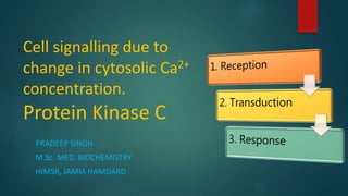
  • 1. Cell signalling due to change in cytosolic Ca2+ concentration. Protein Kinase C PRADEEP SINGH M.Sc. MED. BIOCHEMISTRY HIMSR, JAMIA HAMDARD
  • 2. Content  Introduction  Calcium Signalling 1. Ca2+ signalling by Voltage Operated Channels 2. Ca2+ signalling by Receptor Operated Channels 3. Ca2+ signalling by Hormones (Activation of Phospholipase) A. Phospholipase C (IP3 & DAG)  Activation of Protein Kinase C B. Phospholipase D
  • 3. Introduction  Ca2+ is an essential element which regulate large number of physiological processes such as proliferation, neural signaling, learning, contraction, secretion, and fertilization.  Concentration of calcium varies greatly in intracellular and extracellular environment.  Extracellular concentration of calcium is 10-3 M/L while the intracellular concentration 10-7 M/L [10000 times lower than ECF]. So, regulation of Ca2+ levels in the cell is very important.
  • 4.  In mitochondria, increase in the concentration of free Ca2+ in the mitochondrial matrix accelerate pyruvate oxidation and ATP production.  In muscle, increases in the concentration of Ca2+ are used both to induce contraction and to coordinately increase mitochondrial ATP synthesis to provide the energy for contraction.
  • 5. Calcium Signalling  Calcium signalling pathway fall into two main groups depending on how they are activated: 1. External Stimuli 2. Internal Stimuli
  • 6. Calcium signalling by external stimuli takes place by following pathways: 1. Calcium signalling by VOC (Voltage operated calcium)channels 2. Calcium signalling by ROC (Receptor operated calcium) channels 3. Calcium signalling by PLC (Phospholipase C) 4. Calcium signalling by PLD (Phospholipase D)
  • 7. 1. Ca2+ Signalling by VOC  Secretory vesicles wait near the plasma Membrane (neurolemma) until signaled to release their contents.  Membrane depolarization in the presynaptic membrane activate a specific isoform of VOC.  VOC in the presynaptic endings are associated with the synaptic vesicles, thus producing a highly localized puff of Ca2+ to trigger exocytosis.
  • 8. Exocytosis of Synaptic vesicles  Exocytosis of synaptic vesicles involve 3 steps: 1. TEHTERING 2. DOCKING 3. FUSION  Exocytosis of vesicles require 3 major proteins: 1. SNARE Proteins [t-SNARE, v-SNARE & Synaptotagmin] 2. Rab-GTPase 3. Rab Effector Proteins
  • 9.  Exocytosis of synaptic vesicles involve 3 steps: 1. TEHTERING 2. DOCKING 3. FUSION
  • 10. 2. Calcium signalling by ROC  Receptor-operated channels (ROCs) 1. Nicotinic acetylcholine receptors 2. 5-HT5 [5-Hydroxytryptamine receptor or Serotonin Receptor] 3. AMPA receptors [α-Amino-3-hydroxy-5-methylisoxazole-4- propionic acid receptor] 4. NMDA receptors [N-methyl-D-aspartate receptor] 5. P2X receptors
  • 12. Steps of skeletal muscle contraction 1. Release of ACh at the neuromuscular junction 2. Generation of Motor End Plate potential 3. Opening of Dihydropyridine Receptors (Influx of Extracellular Ca2+) 4. Release of Calcium from Sarcoplasmic Reticulum by ryanodine receptors (via physical coupling to the dihydropyridine receptors) 5. Ca2+ binds to troponin; blocking action of tropomyosin released 6. Contraction via crossbridge formation; ATP hydrolysis 7. Removal of Ca2+ by active transport 8. Tropomyosin bloackage restored; contraction ends
  • 13. 3. Calcium Signalling by Phospholipase C  PLCs are a family of enzymes that hydrolyze a phosphoester bond in certain phospholipids (Phosphatidylinositol & Phosphatidylcholine).  Breakdown of phospholipids yields two second messengers DAG & IP3.  DAG & IP3 function in elevating both the cytosolic and mitochondrial- matrix Ca2+ levels.  Elevated Cytosolic Ca2+ levels and activate a family of cytosolic kinases known as protein kinases (PKC & PKD)  Protein kinases in turn affect many important cellular processes such as growth and differentiation as well as altering the activity of many proteins.
  • 14. ISOTYPE TYPES Beta (β) PLC-β1, PLC-β2, PLC-β3, PLC-β4 Gamma (γ) PLC-γ1, PLC-γ2 Delta (δ) PLC-δ1, PLC-δ3, PLC-δ4 Epsilon (ε) PLC-ε1 Eta (ζ) PLC-ζ1, PLC-ζ2 Zeta (η) PLC-η1 Breakdown of: Phosphatidylinositol – Activate Phospholipase C Phosphatidylcholine – Activate Phospholipase D  Isotypes of Phospholipase C:
  • 15.  PLCβ-Isotype: Act through G-Protein coupled receptors  PLCγ-Isotype: Act through tyrosine kinase receptors
  • 16. Synthesis of DAG & IP3 from Phosphoinositol
  • 17. Phospholipase C Phosphoinositol pyrophosphate (PIP2) Inositol 1,4,5- triphosphate (IP3) IP3 gated calcium release from ER Activation of calcium sensitive intracellular proteins Diacylglycerol (DAG) Remains embedded in the plasma membrane Activation of Protein Kinase C
  • 18. A) IP3 Induced formation of calcium calmodulin complex  IP3 induced release of calcium from the ER leads to 10-20 fold increase in cytosolic calcium concentration.  Various Ca2+ binding protein acts as calcium buffers and restrict the diffusion of increased cytosolic calcium to ECF.  Calcium binding proteins includes troponin, calbindin, calmodulin and calcineurin.
  • 19.  The calcium ion oscillations occur in the pituitary gland cells that secrete luteinizing hormone (LH), which plays an important role in controlling ovulation and thus female fertility.  LH secretion is induced by the binding of luteinizing hormone-releasing hormone (LHRH) to its G protein–coupled receptors on the surfaces of pituitary cells.  The signal often remains localized to the site where the Ca2+ enters the cytosol.  Spikes of calcium concentration controls the gene expression i.e., one frequency of Ca2+ spikes activates the transcription of one set of genes, while a higher frequency activates the transcription of a different set of genes.
  • 20. Ca2+/Calmodulin Complex  Calmodulin consist of a highly conserved single polypeptide chain having two globular ends which resembles dumbbell shape.  Each globular head has 2 Ca2+ binding sites.  Binding of Ca2+ with calmodulin induces conformational change in the calmodulin which leads to formation of Ca2+/Calmodulin complex.
  • 21. Functions of Ca2+/Calmodulin complex Ca2+/Calmodulin complex Ca2+/Calmodulin complex bind to various target proteins in the cell to alter their activity Ca2+/Calmodulin complex activates the plasma membrane Ca2+ pump that uses ATP to pump Ca2+ out of the cells. 1.Ca2+/Calmodulin complex activates Ca2+/Calmodulin- dependent kinases such as CaM-kinase II
  • 22. Ca2+/Calmodulin-Dependent Kinases (CaM-Kinase II)  CaM-kinase II is one of the most studied Ca2+/Calmodulin-dependent Kinase.  CaM-kinase II plays an important role in learning and memory.  CaM-kinase II protein has two major domains: an amino terminal kinase domain and a carboxy-terminal hub domain, linked by regulatory protein.
  • 23.
  • 24.  The complete enzyme contains two stacked rings around the central hub, for a total of 12 kinase proteins (one ring of 6 kinase proteins on both side of the hub domain).  In inactive state, the regulatory subunit is buried in the active site of the kinase thereby blocking its catalytic activity.  When a kinase domain has popped out from the central hub domain, the regulatory subunit is now accessible to the Ca2+/Calmodulin complex.
  • 25.  Ca2+/Calmodulin complex (if present) will bind the regulatory segment and prevent it from inhibiting the kinase thereby activates the kinase activity.  If the adjacent kinase subunit also pops out from the hub, it will also be activated by Ca2+/Calmodulin complex and the two kinases will then phosphorylate each other on their regulatory segment.  It converts the enzyme to Ca2+ independent form which activates other kinases, so that the kinase remain active even after dissociation of Ca2+/Calmodulin complex.
  • 26. B) DAG based activation of Protein Kinase C  After the formation by phospholipase C, the hydrophobic DAG remains associated with the plasma membrane.  The principle function of DAG is to activate Protein Kinase C (PKC).  In the absence of hormone stimulation, protein kinase C is present as a soluble cytosolic protein that is catalytically inactive.  Increase in cytosolic Ca2+ levels causes protein kinase C to translocate to cytosolic leaflet of plasma membrane, where it can interact with membrane associated DAG.
  • 27.
  • 28.  The activation of PKC in different cells plays an important role in many aspects of cellular growth and metabolism.  PKC phosphorylates transcription factors that are localized in the cytosol, triggering their movement into the nucleus, where they activate genes necessary for cell division.  In liver cells, PKC helps regulate glycogen metabolism by phosphorylating and so inhibiting glycogen synthase.
  • 30. Cell Responses in which GPCRs Activate PLCβ Target tissue Signal molecule Major response Liver Vasopressin Glycogen breakdown Pancreas Acetylcholine Amylase secretion Smooth muscle Acetylcholine Muscle contraction Blood platelets Thrombin Platelet aggregation
  • 31. Hormone-induced Cell Responses Mediated by Cyclic-AMP Target tissue Hormone Major Response Thyroid gland Thyroid-stimulating hormone (TSH) Thyroid hormone synthesis and secretion Adrenal cortex Adrenocorticotrophic hormone (ACTH) Cortisol secretion Ovary Luteinzing hormone (LH) Progesterone secretion Muscle Adrenaline Glycogen breakdown Bone Parathormone Bone resorption Heart Adrenaline Increase in heart rate and force of contraction Liver Glucagon Glycogen breakdown Kidney Vasopressin Water resorption Fat Adrenaline, ACTH, Glucagon, TSH Triglyceride breakdown
  • 32. 4. Calcium Signalling By Phospholipase D

Hinweis der Redaktion

  1. Ca2+ increased the conductance of Complex IV (2.3-fold), Complexes I and III (2.2-fold), ATP production/transport (2.4-fold), and fuel transport/dehydrogenases (1.7-fold).
  2. External Stimuli (function to transfer information from the cell surface to internal effector system) Internal Stimuli [Respond to information generated within the cell]
  3. Cyclic ADP-Ribose/NAADP
  4. Example – exocytosis of vesicles from presynaptic membrane
  5. ‘t’ means target membrane ‘v’ means vesicle membrane
  6. Three types of proteins are involved in synaptic docking: SNARE Protein (t-SNARE & v-SNARE) “t” snare means snare protein which is present at target membrane & “v” snare means snare protein which is present at the vesicle membrane. 2. RAB-GTPase – Present on vesicle membrane. This protein helps in the recognition of the target membrane. 3. RAB Effector Protein Rab
  7. Arrival of the nerve impulse at the neuromuscular junction.
  8. Calcium induced calcium release from the ER in skeletal muscle cells
  9. G protein called Gaq activates the inositol phospholipid signalling pathway. The activated phospholipase ten cleaves the Phosphatidylinositol 4,5-bisphosphate [PI(4,5)P2] to generate two products
  10. Pi-4 kinase introduces a phosphate group at the 4th position.
  11. Protein Kinase C is activated by combination of DAG, Ca2+ & negatively charged membrane phospholipid phosphatidylserine. Protein Kinase C in turn phosphorylates target proteins that vary depending on the cell type.
  12. Ca2+/Calmodulin has no enzymatic activity itself but it acts by binding to and activating other proteins.
  13. Ca2+/Calmodulin complex itself has no enzyme activity.
  14. Front Back
  15. Complete enzyme contains two stacked rings of 12 CaM-kinase II proteins. Each ring contain 6 CaM-kinase II proteins on both sides of the hub domain. In inactive state, the regulatory subunit is buried in the active site of the kinase thereby blocks the kinase activity. When kinase domain has popped out and linked to the central hub by regulatory subunit. If present
  16. Ca2+/Calmodulin complex binds the regulatory subunit of CaM-Kinase II
  17. Activation of PKC thus depends on an increase of both Ca2+ ions and DAG.
  18. Phosphatidylcholine breaks into Phosphatidic Acid & Choline. Lyso PC = Lysophospholipids