The human face is a fascinating study of physiology and psychology. Face is the mirror of one’s personality. It is our most useful and most underestimated tool for communication.
Face is the most beautiful and attractive part of the body which is most likely to develop malformations. So, the knowledge of normal anatomy of face will aid in understanding the potential reasons for preventing or treating of anomalies.
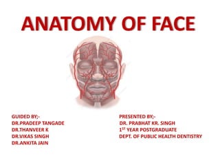
1. PRESENTED BY;-
DR. PRABHAT KR. SINGH
1ST YEAR POSTGRADUATE
DEPT. OF PUBLIC HEALTH DENTISTRY
ANATOMY OF FACE
GUIDED BY;-
DR.PRADEEP TANGADE
DR.THANVEER K
DR.VIKAS SINGH
DR.ANKITA JAIN
2. CONTENTS
• Introduction
• Boundaries of Face
• Superficial fascia
• Bones of the Face
• Muscles of the Face
• Nerve Supply of the Face
• Applied Anatomy
3. • Branches of Facial Artery
• Venous Drainage of Face
• Tributaries of Facial Vein
• Dangerous Area of Face
• Lymph Drainage of Face
• Conclusion
• References
4. INTRODUCTION
• Very Vascular
• Due to rich vascularity face blush and blanch.
• Facial skin is rich in sebaceous gland and
sweat gland.
• Wounds of face bleed profusely but heal
rapidly.
• No deep fascia is present in the face.
5. FACE (Countenance)
Boundaries
• Extends superiorly to
the hair line, inferiorly
to the chin and base of
mandible, and on each
side to auricle
• Forehead is common to
both scalp and face.
6. SUPERFACIAL FASCIA
• Muscle of facial
expression lie in
superficial fascia.
• Embryologically muscles
develops from
Mesoderm of 2nd
Branchial arch,
therefore supplied by
Facial nerve.
• Deep fascia is absent in
face.
7. Bones of the Face
• The facial skeleton
consists of 14 stationary
bones with mandible.
• These 14 bones form the
basic shape of the face,
and are responsible for
providing attachments for
muscles that moves the
jaw and control facial
expressions.
8.
9. Muscles of the Face
(Muscles of Facial Expression)
• The muscles of the face develop from the 2nd
pharyngeal arch and are innervated by branches
of the facial nerve [VII].
• They are in the superficial fascia,& takes origin
from either bone or fascia, and insert into the
skin.
• These muscles control expressions of the face.
• They act as sphincters and dilators of the orifices
of the face (i.e. orbits, nose, and mouth).
10. Muscles of the Face
• Orbital group
• Nasal group
• Oral group
• Other muscle
12. Orbital group
• Two muscles are
–Corrugator
supercilii-
Origin – Superciliary arch
–Orbicularis oculi
–(3 parts)
13. Orbicularis oculi
• 3 parts-
• Orbital part(outer)
–Originate from Medial part of
medial palpebral ligament and
form concentric rings, return to
point of origin
Action –closes the lids tightly
14. • Palpebral part(Inner)
– Originate from Lateral part of medial palpebral
ligament
– Insert into lateral palpebral raphe
Action-closes the lids gently
• Lacrimal part(Small)
– Originate from Lacrimal fascia& Lacrimal bone
– Insert into upper &lower tarsi
Action-dilate lacrimal sac
15. Nasal group
• Three muscles are
associated with the
nasal group:
– Naris (compressor &
dilator)
– Origin- margins of Nose
– Procerus
– Origin –Root of nose
– Depressor septi
Origin-Incisive fossa.
16. Oral group
• The muscles in the oral group
move the lips and cheek:
–Orbicularis oris(extrinsic &
intrinsic)
–Buccinator(upper, middle
and lower fibers)
17. Orbicularis oris (extrinsic& intrinsic)
• Origin: from maxilla
above incisor teeth
• Insertion: into
mucous membrane
of lip.
• Action: closes the
mouth
18. Orbicularis Oris consist of :
• Extrinsic part
• Intrinsic part
The Extrinsic part is arranged in 3 strata –
Deepest stratum
Intermediate stratum
Superficial stratum
The Intrinsic part consist of Oblique fibres extends
from skin to mucous membrane of lips.
20. • Middle fibers
– Origin –from
pterygomandibular
raphe
– Insertion-decussate
before passing to lips
• Action- it aids in mastication
by prevent accumulation of
food in vestibule of mouth.
• Blowing of air from mouth
21. PLATSYMA (Muscle of Neck)
• Origin– upper part of
Pectoral and Deltoid
fascia
• Insertion– base of
mandible, skin of lower
face and lip
• Action– Releases pressure
of skin on the subjacent
veins, depress mandible,
pulls angle of mouth
downwards.
23. Sensory Nerves of the Face
• The skin of the face is supplied
by the trigeminal nerve (V),
except for the small area over
the angle of the mandible and
the parotid gland which is
supplied by the Ant.Div. of
Great auricular nerve (C2&3).
• The trigeminal nerve (V)
divides into three major
divisions-
• Ophthalmic (V1)
• Maxillary (V2) and
Mandibular (V3) nerves
26. Motor Nerves of the Face
• Motor supply:
– Facial nerve
• Facial nerve divides into five
terminal branches in
substance of Parotid gland
for muscles of facial
expression:
– Temporal
– Zygomatic
– Buccal
– Marginal mandibular
– Cervical
27.
28. Applied Anatomy
• Trigeminal Neuralgia
– Maxillary and mandibular nerve are involved
– Excruciating pain in the region of distribution of these nerve
• In infranuclear lesions of facial nerve
• (at stylomastoid foramen) (Eg, Bell’s palsy)-
• whole face of the affected side(ipsilateral) is paralysed
– c/f
• Affected side is motionless
• Loss of wrinkles
• Eye cannot be closed
• In smiling the mouth is drawn to normal side
• During mastication food accumulates in vestibule of mouth
29.
30.
31. 31
Branches of Facial artery(Anaesthetists artery)
1- Submental artery :
arises at the lower border
of the body of mandible
to supply skin of chin +
lower lip.
2- Inferior labial artery :
arises near angle of
mouth to run medially in
the lower lip and
anastomoses with its
fellow of opposite side.
32. • 3- Superior labial artery : runs
medially in the upper lip and gives
branches to the septum + ala of
nose.
• 4- Lateral nasal artery : supplies skin
on the side & dorsum of nose.
33. 33
Venous Drainage of Face
Facial vein :
-is formed at the medial angle of eye by
union of supraorbital & supratrochlear
veins.
-it is connected to cavernous sinus
through superior ophthalmic vein. This
connection is of great clinical importance
because it provides a pathway for spread
of infection from face to cavernous sinus.
–It is joined by anterior division of
retromandibular vein to form common
facial vein to end into the internal jugular
vein.
34.
35. 35
Tributaries of Facial vein
It recevies tributaries that
correspond to the branches of
facial artery.
It is joined to pterygoid
venous plexus ( a venous
network lying around pterygoid
muscles) by deep facial vein
and to the cavernous sinus by
superior ophthalmic vein.
37. Why is it called the
Dangerous Area of Face ?
• The presence of loose areolar tissue
containing the emissary veins allows
the spread of infection through the
emissary veins into the cavernous
sinus which further leads to cavernous
sinus thrombosis.
38. 38
Lymph Drainage of the Face
Lymph from forehead + anterior
part of face drains into
Submandibular Lymph Nodes,
Buccal lymph nodes .
Lateral part of face + lateral
parts of eyelids drain into Parotid
Lymph Node.
Lower lip + chin are drained into
Submental Lymph Node.
39. CONCLUSION
• The human face is a fascinating study of
physiology and psychology. Face is the mirror of
one’s personality. It is our most useful and most
underestimated tool for communication.
• Face is the most beautiful and attractive part of
the body which is most likely to develop
malformations. So, the knowledge of normal
anatomy of face will aid in understanding the
potential reasons for preventing or treating of
anomalies.
40. REFERENCES
• BD Chaurasia’s Human Anatomy (Vol.2,8th
Edition)
• Textbook of Anatomy- Vishram Singh
(Vol.2,2nd Edition)
• Burket’s Oral Medicine (12 Edition)
• Internet (Google,pintrest)