Polymer-Supported N-Tritylaziridinyl(diphenyl)methanol as an Effective Cataly...
Potent HDAC inhibitors identified from substituted 2-piperazinyl-5-pyrimidylhydroxamic acids
1. Identification of a series of substituted 2-piperazinyl-5-pyrimidylhydroxamic
acids as potent histone deacetylase inhibitors
Patrick Angibaud a,*, Kristof Van Emelen b
, Laurence Decrane a
, Sven van Brandt b
, Peter ten Holte b
,
Isabelle Pilatte a
, Bruno Roux a
, Virginie Poncelet a
, David Speybrouck a
, Laurence Queguiner a
,
Sandrine Gaurrand a
, Ann Mariën c
, Wim Floren c
, Lut Janssen c
, Marc Verdonck b
, Jacky van Dun c
,
Jacky van Gompel d
, Ron Gilissen d
, Claire Mackie d
, Marc Du Jardin d
, Jozef Peeters d
, Marc Noppe d
,
Luc Van Hijfte a
, Eddy Freyne c
, Martin Page c
, Michel Janicot c
, Janine Arts c
a
Ortho-Biotech Oncology Research & Development, Department of Medicinal Chemistry, campus de Maigremont BP615, 27106, Val de Reuil, France
b
Johnson & Johnson Pharmaceutical Research & Development (J&JPRD), Turnhoutseweg, 30, 2340 Beerse, Belgium
c
Ortho-Biotech Oncology R&D, Turnhoutseweg, 30, 2340 Beerse, Belgium
d
J&JPRD, ADME-Tox department, Turnhoutseweg, 30, 2340 Beerse, Belgium
a r t i c l e i n f o
Article history:
Received 17 July 2009
Revised 25 October 2009
Accepted 27 October 2009
Available online 30 October 2009
Keywords:
HDAC inhibitors
Histone deacetylase
Anti-cancer agents
Petasis
Pyrimidylhydroxamic acid
a b s t r a c t
Pursuing our efforts in designing 5-pyrimidylhydroxamic acid anti-cancer agents, we have identified a
new series of potent histone deacetylase (HDAC) inhibitors. These compounds exhibit enzymatic HDAC
inhibiting properties with IC50 values in the nanomolar range and inhibit tumor cell proliferation at sim-
ilar levels. Good solubility, moderate bioavailability, and promising in vivo activity in xenograft model
made this series of compounds interesting starting points to design new potent HDAC inhibitors.
Ó 2009 Elsevier Ltd. All rights reserved.
Two enzyme families play a crucial role in controlling the acet-
ylation status of histones, thereby altering chromatin structure and
impacting transcription processes: histones acetyl transferases
(HATs) which catalyze the transfer of an acetyl group to lysine res-
idues of chromatin and histone deacetylases (HDACs) which re-
move an acetyl group from those residues, thus leading to a
changed chromatin structure impacting transcriptional activity.1,2
Inhibiting the HDAC enzymes results in increasing the acetylation
status of chromatin which has been linked to cell cycle arrest and
apoptosis as well as down regulation of genes involved in tumor
progression, invasion, and angiogenesis.3–6
In the past decade,
HDAC inhibition has emerged as an attractive target for the devel-
opment of new anti-cancer agents,7
resulting in the approval of the
HDAC inhibitor (HDACi) vorinostat for Cutaneous T-Cell Lym-
phoma (CTCL),8,9
and in the development of several other hydroxa-
mic acids such as, for example, belinostat10
or panobinostat.11
In recent years, a number of additional nonhistone HDAC sub-
strates have also been identified adding growing evidence that
HDACi exert their pleiotropic effect not only through epigenetic
control.12
Recent data have also demonstrated that HDACi could
target novel therapeutic applications such as neurodegenerative
diseases and inflammation.13–15
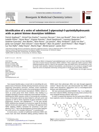
We have previously identified R306465 (Fig. 1) as a highly po-
tent class I selective HDACi, showing oral anti-tumor activity in hu-
man xenograft-bearing athymic nude mice.16
In order to expand and optimize this first generation HDACi, we
have explored a series of analogues on the basis that replacing the
0960-894X/$ - see front matter Ó 2009 Elsevier Ltd. All rights reserved.
doi:10.1016/j.bmcl.2009.10.118
* Corresponding author.
E-mail address: pangibau@prdfr.jnj.com (P. Angibaud).
N
N
O
N
H
HO
N
N
2
N
N
O
N
H
HO
N
N R1
R
3
N
N
O
N
H
HO
N
N
S
1
O
O
R306465
Figure 1. Johnson & Johnson first generation of HDACi: R306465 1, alkyl analogue
2, and targeted compounds 3.
Bioorganic & Medicinal Chemistry Letters 20 (2010) 294–298
Contents lists available at ScienceDirect
Bioorganic & Medicinal Chemistry Letters
journal homepage: www.elsevier.com/locate/bmcl
2. sulfonamide moiety would remove one key element hampering
solubility. We first postulated that substituting the SO2 moiety
by a carbon atom would restore the basicity of the nitrogen atom
and re-introduce the possibility of making salts, thus impacting
compound solubility. Indeed replacement of the sulfonyl moiety
by a CH2 in 2 provided an HDACi of similar in vitro potency (see
Table 1) and improved solubility (0.3 mg/mL at pH 4)17
as com-
pared to R306465 (<0.1 mg/mL at pH 4.8).
In addition, the piperazine secondary amine gave us the oppor-
tunity to use the Petasis multicomponent reaction18
as an option to
further introduce chemical diversity on the cap-part of this pyrim-
idyl-based hydroxamic acid series. First attempts were conducted
using 2-phenyl-ethenylboronic acid 6a in order to test the chemis-
try route as 6a is known for its good reactivity in the Petasis reac-
tion. Furthermore we postulate that the resulting double bond in 3
(Fig. 1) would mimic the first phenyl group of the naphtyl moiety
in 2. Finally, it enabled us to introduce a hydroxyl group to allow
for further chemical modifications.
Reacting the 2-(piperazin-1-yl)pyrimidylethyl ester 4,19
with
the glycolaldehyde dimer 5 and various boronic acids 6a–f in eth-
anol at room temperature provided us with esters 7a–f. Introduc-
tion of the hydroxamic acid moiety was then realized in three steps
using O-(tetrahydro-2H-pyran-2yl)-hydroxylamine as hydroxyl-
amine source. The first step encompasses the cleavage of the ester
into acids or carboxylates, then a classical coupling reaction en-
abled us to introduce the protected hydroxyl amine. This allowed
purification of the amides 8a–f by chromatography on silica gel.
This synthetic scheme was preferred from direct reaction of the
hydroxylamine with the ester as reported previously for other
HDACi,20
as, in our hands, hydroxamic acids where difficult to sep-
arate from the corresponding acids in case of incomplete reaction.
Tetrahydropyranyl (THP) protection was smoothly removed in
acidic media using trifluoroacetic acid which allowed easy isola-
tion, upon crystallization, of compounds 3a–f as trifluoroacetates.
Enantiomeric separation of 3a by liquid chromatography on chi-
ral phase proved to be difficult. We therefore chose to separate the
two enantiomers of the corresponding esters 7a1, 7a2 and to intro-
duce the hydroxamic acid moiety exactly as described above.
Alternatively, THP-protected hydroxamate 11 (Scheme 2) was
prepared as a key intermediate to rapidly test piperazine substitu-
tion. In one pot, the 2-(piperazin-1-yl)pyrimidylethyl ester 4 was
first saponified into its acid which was reacted with 9 to provide
the Fmoc-protected piperazine acid 10 with a moderate yield of
40%. THP-hydroxylamine was then introduced as in Scheme 1
and the Fmoc protection removed by reaction with piperazine in
CH2Cl2 at room temperature to provide 11. Petasis multicompo-
nent reaction could then be performed on 11, however with low
yields, and removal of the THP by trifluoroacetic acid then gave
compounds 3g–i.
All compounds were tested for their ability to inhibit histone
deacetylase activity of a mixture of HDAC enzyme obtained from
a nuclear HeLa cell extract and to inhibit ovarian carcinoma
(A2780) cell proliferation.21,22
Table 1
Enzymatic and antiproliferative potency of compounds 3a–i
Compounds R HDAC Cell proliferation A2780
IC50, nMa
± SD IC50, nMa
± SD
1 NA 6 ± 2 39 ± 17
2 NA 1.5 ± 0.2 66 ± 8
3a H 2.0 ± 0.1 25 ± 8
3b –4–Ph 0.9 ± 0.1 29b
3c –4–OCH3 0.95 ± 0.1 18 ± 1
3d –4–Cl 1.2 ± 0.2 17 ± 6
3e –3–F 1.2 ± 0.3 36 ± 14
3f –2–F 2b
<30b
3g –4–CH3 1.0 ± 0.1 18 ± 2
3h –4–CF3 2.1 ± 0.3 65 ± 18
3i –4–F 1.2 ± 0.3 32 ± 12
3a1 Hc
1.0 ± 0.1 40 ± 5
3a2 Hd
1.8 ± 0.3 30 ± 7
3i1 –4–Fc
1.05 ± 0.1 27 ± 13
3i2 –4–Fd
1.2 ± 0.1 31 ± 12
NA = not applicable.
a
Mean results from experiments performed in triplicate.
b
Single experiment.
c
(+) enantiomer.
d
(À) enantiomer.
a,b
4
EtO
O
N
N
N
NH
+
HO
O
N
N
N
N
9
Fmoc
N
H
O
N
N
N
N
H
O
c,d
N
H
O
N
N
N
N
OH
R
HO
10
11
3g: R=-4-CH3
3h: R=-4-CF3
3i: R=-4-F
O
O
O
N
O
O
e,f
3
O
Scheme 2. Reagents and conditions: (a) (i) NaOH 1 N, THF, rt, 16 h then (ii), Na2CO3,
H2O, rt, 5 h, crystallized in CH3CN, 40%; (c) H2N–O–THP, EDC, HOBT, NEt3, THF,
CH2Cl2, rt, 68%; (d) Piperidine, CH2Cl2, rt, 59%. (e) EtOH, rt, 15–29%; (f) CF3CO2H,
MeOH, rt, 64–81%.
a
4
EtO
O
N
N
N
NH
+
O
O OH
HO
+ (HO)2B
R
EtO
O
N
N
N
N
5 6a-f
OH
R
b, c
N
H
O
N
N
N
N
OH
R
OO
d
N
H
O
N
N
N
N
OH
R
HO
7a-f
8a-f
3a-f
3a: R=H
3b: R=-4-Ph
3c: R=-4-OCH3
3d: R=-4-Cl
3e: R=-3-F
3f: R=-2-F
Scheme 1. Reagents and conditions: (a) EtOH, rt, 42–64%; (b) LiOH or NaOH, THF,
H2O, rt, 89–96%; (c) H2N–O–THP, 1-ethyl-3-(30
-dimethylaminopropyl)carbodiimide
(EDC), 1-hydroxy-1,2,3-benzo-triazole (HOBT), NEt3, THF, CH2Cl2, rt, 36–90%; (d)
CF3CO2H, MeOH, rt, 72–84%.
P. Angibaud et al. / Bioorg. Med. Chem. Lett. 20 (2010) 294–298 295
3. Results from Table 1 above clearly show that replacement of the
naphtalene-2-sulfonyl moiety by phenylbutenol group provided
compounds of similar in vitro potency. Substitution of the phenyl
ring does not seem to have significant impact on enzymatic or
anti-proliferative potency despite introduction of electron-with-
drawing or electron-donating substituent. Since hydroxamic acids
are very efficient Zn2+
chelators, this preliminary results may indi-
cates that additional interactions generated by the phenyl ring,
probably orientated towards the solvent region of the tube-shaped
catalytic site described for a bacterial histone deacetylase by Finnin
et al.,23
are not key drivers of potency. It is also interesting to note
that stereochemistry of the carbon linked to the piperazinyl moiety
has virtually no influence on in vitro potency. Thermodynamic sol-
ubility of compound 3a (>10 mg/mL at pH 4.6 and 1.1 mg/mL at pH
7.4) was greatly increased as compared to R306465 (<0.1 mg/mL at
pH 4.8 and 7.7), thus indicating achievement of our first goal. This
property was also confirmed with compounds 3i1, 3i2, and 21, all
showing a solubility >5 mg/mL at pH 4. As introduction of the hy-
droxyl group was not disturbing interaction with the catalytic site
we were interested in further investigation structure–activity rela-
tionships (SAR) at that position and especially in testing introduc-
tion of solubility enhancing substituents. For the sake of
comparison we replaced the hydroxyl by an hydrogen (compound
3j) using reductive amination of 4 with commercially available
butanone 12, followed by introduction of the hydroxamic acid
moiety in three steps as described before (Scheme 3). Compound
3j showed a similar potency as 3a (Table 2) confirming that the hy-
droxyl moiety is not making key interactions with the catalytic
site.
Alkylation of ester 7a with methyliodide provided methoxy
intermediate 13 and further methoxy analogue 3k showing a
similar potency as 3a (Scheme 4). Bulkier aminogroups were intro-
duced (compounds 3l and 3m) by substitution of the methanesulf-
onic acid ester obtained by reaction of methanesulfonylchloride
onto 7a, followed by introduction of the hydroxamic acid moiety
on the resulting esters 14 and 15 (Scheme 4). Compounds 3l and
3m showed $10-fold drop in antiproliferative IC50 as compared
with 3a (Table 2).
Petasis multicomponent reaction is also a powerful tool to pre-
pare alpha-amino acids using glyoxalic acid and boronic acids. We
could therefore rapidly access amides 3n and 3o using the pathway
depicted in Scheme 5. However for these two compounds we also
noticed a $5-fold drop in antiproliferative activity. For compounds
3l–o, enzymatic potency in inhibiting a mixture of HDAC enzyme,
obtained from a nuclear HeLa cell extract, stayed comparable to 3a
with the exception of 3l being slightly weaker (Table 2) but still in
the nanomolar range.
As cyclic amines such as 3l and 3m or amides were not suitable,
we thought to replace the hydroxyl group of 3a by a small, proton-
able amino moiety. We therefore prepared the phtalimide precur-
sor 17 by Mitsunobu reaction on ester 7a (Scheme 6). However
reaction of 17 with hydrazine in the classical conditions did not
give us the awaited 4-phenyl-2-piperazinyl-but-3-enylamine 18
but rather the 3-phenyl-1-(piperazinylmethyl)-allylamine deriva-
tive 19. Formation of 19 might be the result of the rearrangement
of a transient aziridinium similarly as already proposed on piperi-
dines bearing beta-amino alcohol chain by Frizzle et al.24
We took
a,b,c,d
EtO
O
N
N
N
NH
+
Ph
O
N
N
N
N Ph
O
HOHN
4 12 3j
Scheme 3. Reagents and conditions: (a) 4-phenylbut-3-ene-2-one, Ti(OEt)4, 1,2-
dichloroethane, rt, 8%; (b) NaOH, EtOH, H2O, 80 °C; (c) H2N–O–THP, EDC, HOBT,
NEt3, THF, CH2Cl2, rt, 70%; (d) CF3CO2H, MeOH, rt, 44%.
Table 2
Evaluation of the importance of the hydroxyl moiety (R = H)
N
NO
NHHO
N N
R1
Ph
Compounds R1
HDAC Cell proliferation
A2780
IC50,
nMa
± SD
IC50, nMa
3j –CH3 1.7 ± 0.2 20b
3a –CH2OH 2 ± 0.1 25 ± 8
3k –CH2OCH3 1.5 ± 0.2 45 ± 11
3l –CH2–1-morpholino 11 ± 2 315 ± 154
3m –CH2–4-methylpiperazin–1yl 6.9 ± 0.4 392 ± 189
3n –C(O)-morpholine 7 ± 1 124 ± 26
3o –C(O)NHMe 3.3 ± 0.1 159 ± 11
21 NA 3.8 ± 0.7 281 ± 137
NA = not applicable.
a
See Table 1.
b
Single experiment.
b,c or
b,d
EtO2C
N
N
N
N
OH
Ph
EtO2C
N
N
N
N
OMe
Ph
a
EtO2C
N
N
N
N
N
Ph
X
14: X= O
15: X= N-CH3
N
N
N
N
Y
Ph
3k:Y=OCH3
3l:Y=
3m:Y=
O
HOHN
N O
N NCH3
e,f,g
e,f,g
137a
Scheme 4. Reagents and conditions: (a) NaH, CH3I, THF, 0 °C rt, 34%; (b) MsCl, NEt3,
CH3CN, 5 °C rt; (c) morpholine, K2CO3, CH3CN, 80 °C, 84%; (d) N-methylpiperazine,
K2CO3, CH3CN, 80 °C, 33%; (e) LiOH, THF, H2O, rt, 83%-quantitative; (f) H2N–O–THP,
EDC, HOBT, NEt3, THF, CH2Cl2, rt, 35–89%; (g) CF3CO2H, MeOH, rt, 44–57%.
aEtO
O
N
N
N
NH
+
H
O
OH
+ (HO)2B
Ph
O
EtO2C
N
N
N
N
OH
Ph
O
b or c
N
N
N
N
NR1
R2
Ph
O
N O NH
O
HOHN
3n: 3o:
4
16
d,e,f
N
R2
R1
Scheme 5. Reagents and conditions: (a) glyoxalic acid, 2-phenyl-ethenylboronic
acid, EtOH, 60 °C, 87%; (b) morpholine, EDC, HOBT, NEt3, THF, CH2Cl2, rt, 38%; (c)
CH3NH2ÁHCl, EDC, HOBT, NEt3, THF, CH2Cl2, rt, 57%; (d) LiOH or NaOH, THF, H2O, rt,
71–73%; (f) H2N–O–THP, EDC, HOBT, NEt3, THF, CH2Cl2, rt, 71–74%; (g) CF3CO2H,
MeOH, rt, 75–87% .
296 P. Angibaud et al. / Bioorg. Med. Chem. Lett. 20 (2010) 294–298
4. this opportunity to prepare hydroxamic acid 21, first protecting the
amino moiety by Fmoc and then introducing THP-protected
hydroxylamine after saponification of the ester function. Com-
pound 21 displayed similar HDAC inhibiting potency on enzyme
as 3a showing that variation of linker size could be tolerated. This
observation prompted us to examine the influence of longer dia-
mine linkers (Scheme 7 and Table 3).
Esters 25, 26, and 27 were, respectively, prepared by condensa-
tion of boc-protected 4-aminopiperidine, 4-(aminomethyl)piperi-
dine, and boc-protected-2-amino-methylmorpholine onto 2-meth-
anesulfonylpyrimidine-5-carboxylic acid ethyl ester 22.19
Then
Petasis reaction was performed onto 25–27 and hydroxamic acid
functionality was introduced as described in Scheme 1 (Scheme 7).
All ligands were found to be equipotent with regard to enzy-
matic activity whereas Linker B (4-aminopiperidine) and linker D
(2-methylamino-morpholine) were slightly less potent in the cel-
lular assay.
As demonstrated by the results from Tables 1–3, a wide range of
Petasis substitution on the 2-piperazinyl-5-pyrimidyl-hydroxamic
acid moiety gave birth to a novel series of potent in vitro HDAC
inhibitors. Compounds 3a and 3i2 showed an acceptable pharma-
cokinetic profile (see Table 4) with a bioavailability in rat of
19–22%, similar to the bioavailability of 1 (R306465).
Compound 3a, 3i1, and 3i2 were evaluated in an animal model
allowing noninvasive real-time evaluation of the response to HDAC
inhibitors using whole-body imaging.25
Human A2780 ovarian
carcinoma cells were engineered to express ZsGreen fluorescent
protein under control of the p21waf1,cip1
promoter, sub-cutaneously
grafted to athymic nude mice, and induction of fluorescence
in vivo was measured after 3 days of treatment. Results are reported
as increased fluorescence intensity as compared to R306465 tested
in same conditions and normalized to 1 (Fig. 2). This allowed rapid
comparison of compounds 3a, 3i1, and 3i2 with R306465 as regards
to their anti-tumor potency. Preliminary results indicate that the
three compounds may possess an in vivo efficacy in that model supe-
rior to R306465. Compounds 3i1 and 3i2 showed also similar in-
crease in fluorescence intensity (3–4-fold compared to R305465)
confirming that their stereochemistry had no influence. 21 was cho-
sen as a representative of compounds possessing decreased antipro-
liferative activity as compared to R306465 and showed reduced
fluorescence than R306465.
Compound 3i2, has been tested towards a broad panel of re-
combinant HDAC enzymes in vitro and compared to compound 1
(R306465) (see Table 5).
With the exception of HDAC4, 10 and 11 where 3i2 showed a
10–50-fold drop in inhibiting potency compared to R306465, the
overall profile of both compounds are very similar, even if 3i2
seems slightly less potent (1–3-fold). Compound 3i2 showed
activity towards all HDAC enzyme tested with some specificity
for class I inhibition, a class of HDACs where activity is key for
uncontrolled proliferation of cancer cells.26
Lowest in vitro potency
was observed towards HDAC4, 6, and 7.
aEtO
O
N
N
N
N
OH
Ph
+ NH
O
O
EtO
O
N
N
N
N Ph
N
O
O
N
N
NH2
Ph
but N
N
NH2
Ph
HO
O
N
N
N
N
Ph
HN
Fmoc
c,d e,f,g
N
H
O
N
N
N
N
Ph
NH2
HO
7a 17
18
19
19
20
21
b
Scheme 6. Reagents and conditions: (a) 1H-isoindole-1,3(2H)-dione, P(nBu)3, DIAD, CH2Cl2, rt, 52%; (b) NH2NH2ÁHBr, EtOH, rt, 98%; (c) LiOH, THF, H2O, rt; (d) 1[[(9H-fluoren-
9-ylmethoxy)carbonyl]-oxy]-2,5-pyrrolidinedione, THF, rt, 82%; (e) H2N–O–THP, EDC, HOBT, NEt3, THF, CH2Cl2, rt, 76%; (f) piperidine, CH2Cl2, rt, 56%; (g) CF3CO2H, MeOH, rt,
87%.
Table 3
Evaluation of several linker moieties (R = H)
Compounds Linker HDAC Cell proliferation A2780
IC50, nMa
IC50, nMa
3a A 6 ± 2 25 ± 8
28 B 7 ± 5 219b
29 C 1.5 ± 0.2 47b
30 D 8b
295b
a
See Table 1.
b
Single experiment.
N
N
EtO2C
SO2Me
HN
H
N
Boc
HN
NH2
HN
O
H
N
Boc
N
N
EtO2C
N
NH2
N
N
EtO2C
N N
N
EtO2C
N
O
NH2
NH2
a, b a, b
c
d,e,f,g
N
NO
OH HN
Linker
OH
Ph
N N N NH N
HN
N O
NH
A B C D
22
25 26 27
3a 28 29 30
23 24
Scheme 7. Reagents and conditions: (a) 23 or 24, K2CO3, CH3CN, rt, 53–75%; (b)
CF3CO2H, CH2Cl2, rt, 35–96%; (c) 4-aminomethyl-piperidine, K2CO3, CH3CN, rt, 33%;
(d) 5 + 6a, EtOH, rt, 47–73%; (e) NaOH or LiOH, THF, H2O, rt; (f) H2N–O–THP, EDC,
HOBT, NEt3, THF, CH2Cl2, rt, 4–54%; (g) CF3CO2H, MeOH, rt, 72–85%.
P. Angibaud et al. / Bioorg. Med. Chem. Lett. 20 (2010) 294–298 297
5. In conclusion, we have identified a series of novel 2-piperazi-
nyl-5-pyrimidyl-hydroxamic acids HDACi that are characterized
by high potency on HDAC HeLa cellular extract and cellular anti-
proliferation assay and have demonstrated improved solubility as
compared to our starting molecule 1. First in vivo evaluation of
leads 3a and 3i2 indicates superior potential as compared to
R306465. These results warrants more in depth evaluation of this
series.
References and notes
1. Johnstone, R. W. Nat. Rev. Drug Disc. 2002, 1, 287.
2. Grant, S.; Easley, C.; Kirkpatrick, P. Nat. Rev. Drug Disc. 2007, 6, 21.
3. Ten Holte, P.; van Emelen, K.; Janicot, M.; Jong, P. C.; de Bono, J. S.; Arts, J.. In
Top. Med. Chem; Bradbury, R., Ed.; Springer: Berlin, Heidelberg, 2007; Vol. 1, pp
293–331.
4. Bolden, J. E.; Peart, M. J.; Johnstone, R. W. Nat. Rev. Drug Disc. 2006, 5, 769.
5. Monneret, C. Anticancer Drugs 2007, 18, 363.
6. Paris, M.; Porcelloni, M.; Binashi, M.; Fattori, D. J. Med. Chem. 2008, 51, 1505.
7. Prince, H. M.; Bishton, M. J.; Harrison, S. J. Clin. Cancer Res. 2009, 15, 3958.
8. Marks, P. A. Oncogene 2007, 26, 1351.
9. Marks, P. A.; Breslow, R. Nat. Biotechnol. 2007, 25, 84.
10. Gimsing, P. Expert Opin. Investig. Drugs 2009, 18, 501.
11. Atadja, P. Cancer Lett. 2009, 280, 233.
12. Budillon, A.; Di Genarro, E.; Bruzzese, F.; Rocco, M.; Manzo, G.; Caraglio, M. Rec.
Patents Anticancer Drug Disc. 2007, 2, 119.
13. Kazantsev, A. G.; Thompson, L. M. Nat. Rev. Drug Disc. 2008, 7, 854.
14. Sun, Y.; Reddy, P. Curr. Enz. Inh. 2007, 3, 207.
15. Glauben, R.; Sonnenberg, E.; Zeitz, M.; Siegmund, B. Cancer Lett. 2008, 280,
154.
16. Arts, J.; Angibaud, P.; Mariën, A.; Floren, W.; Janssens, B.; King, P.; van Dun, J.;
Janssen, L.; Geerts, T.; Tuman, R. W.; Johnson, D. L.; Andries, L.; Jung, M.;
Janicot, M.; Van Emelen, K. Br. J. Cancer 2007, 97, 1344.
17. Thermodynamic solubility was determined at different pHs by accurately
weighting around 2.5 mg of compound, attempting to dissolve it in
phosphate-citrate buffers and filtering (if necessary) over a filter disc. An
aliquot of the filtrate was diluted with an appropriate solvent (0.1 N HCl/
acetonitrile, 1/1) and submitted for concentration measurement using a
generic UPLC method.
18. Petasis, N. A.; Goodman, A.; Zavialov, I. A. Tetrahedron 1997, 53, 16463.
19. Angibaud, P.; Arts, J.; Van Emelen, K.; Poncelet, V.; Pilatte, I.; Roux, B.; Van
Brandt, S.; Verdonck, M.; DeWinter, H.; Ten Holte, P.; Marien, A.; Floren, W.;
Janssens, B.; Van Dun, J.; Aerts, A.; Van Gompel, J.; Gaurrand, S.; Queguiner, L.;
Argoullon, J. M.; Van Hijfte, L.; Freyne, E.; Janicot, M. Eur. J. Med. Chem. 2005, 40,
597.
20. Mai, A.; Esposito, M.; Sbardella, G.; Massa, S. Org. Prep. Proced. Int. 2001, 33,
391.
21. Histone Deacetylase assays on a pool of HDAC enzymes obtained from a
nuclear HeLa cell extract or on HDAC isoforms were performed at Reaction
Biology Corporation (www.reactionbiology.com, Malvern PA) using the
‘Spectrum’ assay platform.
22. Van Brandt, S.F.A.; Van Emelen, K.; Angibaud, P.R.; Marconnet-Decrane, L.F.B.
WO2006/010749, 2006. Enzymatic test conditions reported page 49 and
cellular antiproliferative test conditions described pages 49–50 were slightly
modified to adapt to 384 well plates.
23. Finnin, M. S.; Donigian, J. R.; Cohen, A.; Richon, V. M.; Rifkind, R. A.; Marks, P.
A.; Breslow, R.; Pavletich, N. P. Nature 1999, 401, 188.
24. Frizzle, M. J.; Caille, S.; Marshall, T. L.; McRae, K.; Nadeau, K.; Guo, G.; Wu, S.;
Martinelli, M. J.; Moniz, G. A. Org. Process. Res. Dev. 2007, 11, 215.
25. Belien, A.; De Schepper, S.; Floren, W.; Janssens, B.; Marien, A.; King, P.; Van
Dun, J. Mol. Cancer Ther. 2006, 5, 2317.
26. Glaser, K. B.; Li, J.; Staver, M. J.; Wei, R. Q.; Albert, D. H.; Davidsen, . K. Biochem.
Biophys. Res. Commun. 2003, 310, 529.
Table 4
Rat DMPK properties of compounds 3a and 3i2
Compounds AUC0-inf
a
(ng h/mL) (IV) CLa
(L/h/kg) (IV) Vdss
a
(L/kg) (IV) T1/2 (h) 2 h to 8 ha
(IV) AUC0-inf
b
(ng h/mL) (po) F (%)
1 2654 0.8 0.6 1.5 3381 20
3a 413 5.9 5.8 2.6 316 19
3i2 521 4.6 1.6 0.8c
466 22
a
Study in male Sprague-Dawley rats dosed at 2.5 mg/kg in water at pH 4.4.
b
Study in male Sprague-Dawley rats dosed at 10 mg/kg in water at pH 4.5.
c
Half life determined from 1 h till 4 h post dose.
Figure 2. Folds increase of fluorescence for compounds 3a, 3i1 and 3i2 as compared
to R306465 (normalized at 1) following 3 days dosing po at 40 mg/kg in athymic
nude mice bearing s.c. A2780-p21waf1,cip1
ZsGreen ovarian xenograft.
Table 5
Comparison of activity towards all HDAC enzymes21
Class HDAC R306465 3i2
IC50, nMa
± SD IC50, nMa
± SD
I 1 2.5 ± 0.4 7.1 ± 05
I 2 9 ± 3 17.9 ± 2.5
I 3 9 ± 3 30 ± 3
I 8 27 ± 13 30 ± 2
IIa 4 4.6 ± 0.35 207 ± 23
IIa 5 7.1 ± 2.3 21.8 ± 2.6
IIa 7 46 ± 8 102 ± 11.5
IIa 9 28 ± 1 40.7 ± 3.2
IIb 6 66 ± 4 224 ± 33
IIb 10 4.3 ± 1.1 42.6 ± 1.5
IV 11 2.1 ± 0.6 20.7 ± 1.4
a
Mean data from experiments performed in triplicate.
298 P. Angibaud et al. / Bioorg. Med. Chem. Lett. 20 (2010) 294–298