1. Optimization of Substrate Mediated Gene Delivery from Columnar Nanostructures by Tuning
Polyethylenimine/DNA Nanoparticle Formulation and Column Spacing.
Patrick T. Mulcahy1, Albert Nguyen2, Angela K. Pannier2
1 Graduate Studies University of Nebraska-Lincoln Lincoln, Lincoln, NE 68588
2 Department of Biological Systems Engineering, University of Nebraska-Lincoln Lincoln, Lincoln, NE 68588
Methods
• Spacing of slanted column thin films (SCTFs) was
controlled by adjusting diameter of diblock
copolymer-Poly(styrene-b-2-vinyl pyridine)-micelles
coated onto silicon and glass cell culture surfaces as
monolayer films. Micelles were loaded with gold salt
that deposited after removal of polymer by oxygen
plasma. These gold deposits served as nucleation
points for titanium column nanostructures formed by
GLAD (30, 80, and 150nm spacing)
• Tune N/P ratio (5, 10, 15, 20), DNA amount (6 and
12 µg/mL), and formulation medium (Opti-mem vs.
ddH20) to achieve DNA/PEI nanoparticle diameter
less than 100nm, determined by zeta-sizer (Nano-
ZS90, Malvern).
• Nanoparticle formulations were tested by bolus
delivery to NIH/3T3 fibroblast cells cultured on
polystyrene in DMEM (supplemented with 10% fetal
calf serum (FCS) and 1% penicillin/streptomycin) to
determine optimum formulation conditions for
transfection.
• Nanoparticle formulations were also tested for
adsorption (Hoechst assay) to cell culture surfaces
(polystyrene, glass, and titanium SCTFs on silicon of
various spacing)
Results (cont.)Introduction
Acknowledgements&References
This work was supported by:
The Pannier Lab
National Science Foundation
Center for Nanohybrid Functional Materials, UNL
1)Kasputis, Tadas. "Use of Precisely Sculptured Thin Film
(STF) Substrates with Generalized Ellipsometry to
Determine Spatial Distribution of Adsorbed Fibronectin to
Nanostructured Columnar Topographies and Effect on Cell
Adhesion." Acta Biomateriala, May 2015. Web. 01 Aug.
2016.
Objectives
Methods (cont.) Results (cont.)
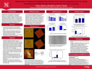
Figure 1) AFM of gold particles after micelles were
removed from the surfaces. The average distance from
center to center was 105nm (top row) and 86nm (middle
row). Micelles did not form well on glass (bottom left)
Copolymers form micelles and are loaded with gold salt
(bottom right).
Figure 2) Complex sizing at different parameters
Figure 6) Transfection efficiency of complexes
made in Optimem on SCTFs with different column spacing
• Micelle method can be used control spacing of
SCTFs
• Increased transfection efficiency was measured
in SMD to SCTFs of wider spacing. This
increase does not appear to be due to
increased loading, but rather due to the wider
spaced SCTF nanostructures themselves.
• The specific mechanisms by which increased
SCTF spacing enhances SMD transfection
efficiency requires further investigation to
improve SMD optimization and understanding
cell-substrate interactions with nanotopography.
Conclusion
Gene delivery is the transfer of exogenous nucleic
acids into cells, with applications in biomedical
research, such as gene therapy. More efficient
methods of nonviral gene delivery are sought because
of transfection inefficiency relative to viral transduction.
Surface mediated delivery (SMD) is more efficient than
bolus delivery of nonviral gene carriers. Nanostructured
surfaces have been found to influence cell behavior(1),
and perhaps can be designed to enhance non viral
gene delivery to clinically relevant efficiencies.
1. Use micelle monolayer film to control spacing of
slanted column nanostructures fabricated by
glancing angle deposition (GLAD), at the CNFM at
UNL.
2. Devise DNA/PEI nanoparticle formulation that
results in transfection competent complexes with
particle diameter less than 100nm, that adsorb
efficiently to cell culture surfaces
3. Assess SMD transfection efficiency of nanoparticle
formulations on nanocolumnar surfaces.
5
2
0
0
5 0
1 0 0
1 5 0
S izin g o f D N A p a rtic le s a t
v a rio u s N /P ra tio a t 1 2 u g /m L
fo rm e d in w a te r
N /P ra tio
Complexsize(nm)
5
1
0
1
5
2
0
0
2 0
4 0
6 0
8 0
1 0 0
S iz in g o f D N A p a rtic le s a t
v a rio u s N /P ra tio a t 6 u g /m L
fo rm e d in w a te r
N /P
Complexsize(nm)
5
1
0
1
5
2
0
0
5 0 0
1 0 0 0
1 5 0 0
S iz in g o f D N A p a rtic le s a t
v a rio u s N /P ra tio a t 6 u g /m L
fo rm e d in O p ti-m e m
N /P
Complexsize(nm)
• Cell culture surfaces sterilized under UV light (1hr)
and rinsed with phosphate buffered saline (PBS)
prior to adsorption of DNA/PEI nanoparticles
(adsorption of an initial fibronectin layer was tested
to determine effect on subsequent nanoparticle
loading). For SMD, NIH/3T3 cells were seeded at
8000 cells/well and cultured at 37o C at 5% CO2.
pEGFPLuc plasmid was used for transfection for
fluorescent and luminescent reporter proteins. At
48hrs after seeding, cells were assessed for
transfection efficiency by LUC/BCA assay.
Results (cont.)
O
p
tim
e
m
W
a
te
r
0
2 0 0
4 0 0
6 0 0
8 0 0
S iz e o f D N A c o m p le x e s a t
1 2 u g /m L D N A ,N /P 2 0 ,
1 m g /m L P E I s to c k s o lu tio n
Complexsize(nm)
1
0
5
n
m
8
5
n
m
3
0
n
m
P
S
c
o
n
tro
l
0
5 0 0 0 0
1 0 0 0 0 0
1 5 0 0 0 0
2 0 0 0 0 0
T ra n s fe c tio n o f D N A c o m p le x e s m a d e in w a te rRLU/mg-cm
2
1
0
5
8
5
3
0
0
2 0
4 0
6 0
8 0
A d s o rp tio n o f D N A c o m p le x m a d e in
O p ti-m e m w ith v a rie d s u rfa c e s
%DNAadsorbedtosurface
Figure 5) DNA adsorption to SCTFs
with varying column spacing
Figure 4) Transfection efficiency of complexes
made in water on SCTFs with different column spacing
1
0
5
n
m
8
5
n
m
3
0
n
m
c
o
n
tro
l
0
5 0 0 0 0
1 0 0 0 0 0
1 5 0 0 0 0
T ra n s fe c tio n o f D N A c o m p le x e s m a d e in O p ti-m e m
RLU/mg-cm
2
*
Figure 3) Adhesion of cells to glass surface A)
without fibronectin. B) with fibronectin
BA