

Empfohlen
Weitere ähnliche Inhalte
Was ist angesagt?
Was ist angesagt? (20)
Andere mochten auch
Andere mochten auch (20)
Ähnlich wie Ehrlichia
Ähnlich wie Ehrlichia (20)
Mehr von Osama Zahid
Mehr von Osama Zahid (20)
Kürzlich hochgeladen
Kürzlich hochgeladen (20)
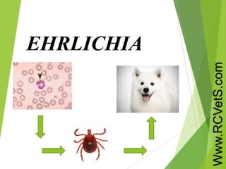
Ehrlichia
- 2. Introduction Small gram negative, obligate, intracellular parasites These are tiny organisms measuring 0.2-2.4micromtrs. Which have affinity towards WBC particularly mononucslear phagocytes
- 3. Clusters of Ehrlichia multiply in host cell vacuoles to form large mulbery shaped aggregates called MORULAE Ehrlichia inclusions like morulae are visible in cytoplasm of infected cell after 5-7 days
- 4. Ehrlichia Species Ehrlichia sennetsu Ehrlichia caffeensis Ehrlichia phagocytophila Ehrlichia cannus
- 5. EHRLICHIA SENNETSU Endemic in JAPAN and SOUTH EAST ASIA It causes GLANDULAR FEVER It shows lymphoid hyperplasia and atypical lymphocytosis No arthropod vector identified Human infection is suspected to be caused by ingestion of fish carrying infected flukes
- 6. EHRLICHIA PHAGOCYTOPHILA Causes human GRANULOCYTIC EHRLICHIOSIS Transmitted by IXODES ticks Deer, cattle and sheep are suspecte reservoirs Leucopenia and thrombocytopenia observed in patients
- 7. EHRLICHIA CAFFEENSIS Cause human MONOCYTIC EHRLICHIOSIS Transmitted by Amblyomma ticks Deers and rodents reservoirs Leucopenia and thrombocytopenia increased liver enzymes Most dangerous can cause multisystem failure and fatality
- 8. EHRLICHIOSIS Ehrlichiosis is infection of WBC that is characterised by mulbery shaped aggregates called morulae in infected cells These morulae are visiible after 5-7days of infection
- 9. Pathophysiology It is not completely known Like RICKETTSIA sps EHRLICHIA gain access to blood via bite from infected tick
- 10. AMBLYOMMA AMERICANAM(lone star tick) E.chaffeensis IXODES PERSUKATUS DERMACENTOR VARIABILIS (dog tick wood tick)
- 11. The major antigen determinants are surface membrane protien These are complexes consisting of : 1)thermolabile 2)thermostable Key protien bands associated are: E.phagocytophia - 27,29,44 KD bands E.caffeensis - 40,44,65 KD bands
- 12. LIFE CYCLE
- 13. Mortality and morbidity Great majority of EHRLICHIOSIS are asymptomatic Most cases present as mild to moderate acute febrile illness In immunocompromised persons ehrliosis may be severe manifesting as ROCKY MOUNTAIN SPOTTED FEVER may be fatal
- 14. Clinical features Rash and pedal edema
- 15. Patients with Ehrlichiosis usually present with head ache, myalgia, fever, shaking chills. Nausea and vomiting are common Abdominal pain is uncommon and is typically mild Skin rash due to ehrlichiosis is rare. When present as macculopapular rash rather than peticheal
- 16. Cont… Some patients develop heptomegaly Lymphadenopathy is observed in <25% Splenomegaly is uncommon Patients with severe ehrlichiosis develop thrombocytopenia and disseminated intravascular coaggulation(DIC) which can result in hemorrhage into skin
- 17. Distribution Ehrlichiosis occurs worldwide and frequensy parallels distribution of appropriate tick vector for transmission of ehrlichia and mammalian host In USA it occurs in states of CALIFORNIA, TEXAS and SOUTH EAST NORTHERN REGIONS OF CAENTRY World wide it occurs in JAPAN, SOUTH EAST ASIA
- 18. Lab diagnosis Diagnosis rests on 1)single elevated IgG IFA antibody titre 2)demonstration of incr. in acute and convalescent IFA ehrlichia titre Difficult to culture Detection with PCR
- 19. Blood smear for cytoplasmic inclusions CBP for thrombocytopenia and neutropenia Atypical lymphocytes in blood Serum transaminases are mild high DIC may be diagnosed with cutaneous bleeding Lumbar puncture to rule out meningitis
- 21. Prevention