
Empfohlen
Weitere ähnliche Inhalte
Was ist angesagt?
Was ist angesagt? (20)
Ähnlich wie protozoa.pptx
Ähnlich wie protozoa.pptx (20)
Kürzlich hochgeladen
Kürzlich hochgeladen (20)
protozoa.pptx
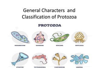
- 1. General Characters and Classification of Protozoa
- 3. General Characters: Phylum Protozoa are microscopic, eukaryotic, unicellular, solitary or colonial, commonly motile and phagocytic heterotrophic animalcules without tissues and organs. They have one or more nuclei. Almost about 50,000 species are known till date. Anton Van Leeuwenhoek (1676) was the first to observe protozoa (Vorticella convellaria) and called them animalcules. Gold fuss coined the term Protozoa (1818).
- 4. They may be free living (Amoeba) or symbiotic (Parasitic, mutualistic or commensalistic) Body symmetry is symmetrical (Actinopodeans) or radial (sessile forms) or bilateral (Giardia) or absent (Amoeba). Locomotion is brought about by pseudopodia or flagella or cilia or myonemes.
- 5. Nutrition is holozoic or holophytic or osmotrophic (Saprophytic or parasitic). Digestion is intracellular. Some forms like Euglena are mixotrophic (perform more than one type of nutrition) Excretion occurs by diffusion across general body surface or by contractile vacuoles. Contractile vacuoles serve mainly for Osmoregulation and are common in freshwater forms. Excretion
- 6. Exchange of respiratory gases takes place by diffusion through the general body surface. Respiration is aerobic or anaerobic in some parasitic forms. Asexual reproduction takes place by binary fission or multiple fission or plasmotomy or budding.
- 7. Sexual reproduction takes place by conjugation or by fusion of gametes (Syngamy). Many forms undergo encyctment to tide over unfavorable conditions Somotoplasm and germplasm are not differentiated. Hence they are immortal (exempt from natural death). Syngamy Sexual Reproduction Sexual Reproduction Encyctment
- 8. Classification of Phylum Protozoa Classification given by BM Honigberg and the society of Protozoologists divides this phyla into 4 subphyla.
- 10. Superclass 1: Mastigophora (Gr. Mastix=whip; phoros=bearing) The body of animals belonging to this super class is covered by pellicle. The locomotory organelles are flagella. Asexual reproduction occurs by longitudinal binary fission. This super class includes 2 classes: Class 1: Phytomastigophora (Gr. Phyton=plant; Mastix=whip; phoros=bearing) They have chromatophores with chlorophyll. The nutrition in these organisms is mainly holophytic which takes place by phototrophy. These are free living organisms. The reserve food in is starch. These organisms may have 1 or 2 flagella. Ex: Euglena, Ceratium, Noctiluca Ceratium Euglena Noctiluca
- 11. Class 2: Zoomastigophora (Gr. Zoon=animal; Mastix=whip; phoros=bearing) These organisms do not have chlorophyll bearing chromatophores. These are mostly parasitic. The nutrition in these organisms is holozoic or saprozoic. The reserved food is glycogen. They may have one to many flagella. Ex: Leishmania, Trypanosoma, Trichomonas, Trichonympha Trichonympha
- 12. Superclass 2: Opalinata The organisms belonging to this super class live as commensals or parasites in the gut of anurans (Frog). Their body is covered by oblique rows of cilia-like flagella. These organisms may have 2 or many nuclei. They undergo asexual reproduction by binary fission or by syngamy. Sexual reproduction takes place by anisogamy. Ex: Opalina, Zelleriella Zelleriella
- 13. Superclass 3: Sarcodina (Gr. Sarcode=fleshy) The locomotion is brought about by pseudopodia. Their body is amoeboid without definite pellicle. The nutrition is holozoic or saprozoic. This super class is further divided into 3 classes: Class 1: Rhizopodea (Gr. Rhizo= root; poda – foot). The pseudopodia of the animals in this class are in the form of lobopodia, filopodia or reticulopodia without axial filaments. This class includes amoebas, foraminiferans and mycetozoans. These animals are mostly free living and a few are also parasitic. In foraminiferans the body is covered by porous calcareous shell. Ex: Amoeba, Entamoeba, Elphidium
- 14. Class 2: Piroplasmea (pear shape) The animals belonging to this class are parasitic. Locomotory structures and Spores are absent. These are the small parasites in the red blood cells of vertebrates. Ex: Babesia Class 3: Actinopodea (Gr. Actis=ray; podos=foot) The pseudopodia of the animals belonging to this class are in the form of axopodia with axial filaments, radiating from the spherical body. These are planktonic. This class includes Heliozoans, Radiolarians and acanthareans. Radiolarians and acanthareans are marine forms whereas heliozoans are both marine and fresh water forms. Skeletons of radiolarians have siliceous shells. The shells of dead radiolarians accumulate on the ocean floor to form radiolarian ooze. Ex: Collozoum, Actinophrys, Acanthometra
- 15. radiolarian ooze under microscope at X250 magnification.
- 16. SUBPHYLUM II: SPOROZOA The animals are exclusively endoparasites. Special locomotory organelles are absent in these animals. Sometimes pseudopodia are present which are useful only for ingestion of food. Merozoites bear anterior apical complex that helps penetrate host cells. This subphylum includes 3 classes: Class 1: Telosporea The Sporozoites are long in these animals. Reproduction is both asexual and sexual. They are blood and gut parasites of vertebrates. Sexual reproduction is by isogamy or anisogamy. Ex: Monocyctis, Eimera, Plasmodium Eimera
- 17. Class 2: Toxoplasmea In this class reproduction is only asexual type which takes place by internal budding where two daughter cells are produced within the mother cell and the mother cell is finally destroyed in the process of reproduction. Spores are absent. Ex: Toxoplasma Class 3: Haplosporea The spores in this class are amoeboid. Also reproduction is only asexual type taking place through multiple fissions. Ex: Haplosporidium, Ichthyosporidium
- 18. SUBPHYLUM III: CNIDOSPORA (Gr. Cnide=needle; spora=seed) The animals are parasitic. Spores are present with one or more polar filaments. Polar filaments are special and unique features of these animals. When these spores infect a host, the polar filament is discharged and it gets attached to the host tissue. This subphylum includes 2 classes: Class 1: Myxosporidea The spores of the animals of this class are large and develop from several nuclei. These are generally extracellular parasites. The spores of this class have two polar filaments and have two to three valves. Ex: Myxobolus polar filaments
- 19. Class 2: Microsporidea The spores of the animals of this class are small and are developed from only one nucleus. These spores have single valve. These are generally intracellular parasites. Many of the animals of this class have a single polar filament. Ex: Nosema bombycis polar filament
- 20. SUBPHYLUM IV: CILIOPHORA (La. Cilium=eye lid with lashes; phoros=bearing) Ciliophorans are complex of all the protozoans. The locomotory organelle of all the animals of this subphylum is cilia. These cilia also help in feeding at some stage of the life cycle of the animals. The nuclei of these organisms are dimorphic. Macronucleus is vegetative and polyploid. Micronucleus is reproductive and diploid. Asexual reproduction takes place by binary fission. Sexual reproduction takes place by conjugation. Only one class is included in this subphylum: Class 1: Ciliatea
- 21. Class 1: Ciliatea The locomotory organelles of these animals are numerous hair-like cilia. One or more contractile vacuoles are present in these forms. The nucleus is dimorphic including both macro nucleus and micronucleus. Ex: Paramoecium, Vorticella, Balatidium Paramoecium Balatidium
- 22. TYPE STUDY ELPHIDIUM or POLYSTOMELLA Phylum : Protozoa Class : Rhizopoda Order :Foraminifera.
- 23. Elphidium is a marine form and found creeping on Sea weeds to a depth of 1800 feet. Structure : Elphidium is also called 'Polystomella is a 'dimorphic rhizopod'. It is a unicellular microscopic protozoan, and" 1 mm in diameter. It is pale yellow in colour. The body is covered by a biconvex shell. The first formed chamber is proloculum. The shell contains spirally arranged V shaped chambers. Hence it is called "polythalamus or multilocular. They overlap one another.
- 24. These chambers show perforation. The cytoplasm of the whole body of the animal is not divided into compartments but it is continuous. The cytoplasm extends through the pores of the shell to form a layer over the shell. The cytoplasm is produced into a number reticulopodia. which will form a network. From hinder end of each chamber cytoplasmic processes will develop. They are directed backwards. They are called retral processes'. 1. The cytoplasm contains one or many nuclei. 2. Cytoplasm contains food vacuoles which takes up the process of digestion.
- 25. Dimorphism: Polystomella exhibits dimorphism. The individual occurs in two distinct forms. 1. Megalosperic form. 2. Microspheric form. 1. Megalospheric form: Its proloculum is big in size. A single large nucleus is present in one of the chambers. It takes up sexual reproduction. 2. Microspheric form: Its proloculum is small in size. Many nuclei are present in the cytoplasm. This form reproduces by asexual reproduction. proloculum Megalospheric Microspheric
- 27. Locomotion and Nutrition: Polystomella show slow creeping movements with the help of reticulopodia. It is a holozoic feeder. These reticulopodia will capture the prey. When it comes in contact with the prey, it kills the prey by secreting toxic substance. The prey is digested in the food vacuole, the digested food is absorbed by the cytoplasm.
- 28. Reproduction and Life Cycle of Elphidium: Elphidium exhibits an alternation of generation in its life cycle. The megalospheric forms alternate with the microspheric forms. The microspheric forms always develop by the conjugation or syngamy and megalospheric forms develop without conjugation or syngamy. The microspheric form reproduces asexually by fission to produce a number of amoebulae. The inner cytoplasmic mass containing several nuclei creeps out of the shell and remains as a lump around it. A small amount of cytoplasm collects around each nucleus.
- 29. As a result, a large number of amoeboid cells (amoebulae) are formed. Each amoebula secretes the proloculum, forms rhizopodia, then it grows and forms other chambers of the shell to become a megalospheric form.
- 30. The megalospheric form reproduces sexually by syngamy or conjugation. During sexual reproduction in megalospheric forms, nucleus first breaks up into many small nuclei and the cytoplasm collects around each of these nuclei. The nuclei divide twice giving rise to a large number of tiny cells. The cells develop flagella and come out of the shell. The biflagellate cells are haploid and known as isogametes. The isogametes of two different individuals fuse (conjugate) in pairs to form zygotes. (The fusion of similar size gametes is known as isogamy). The zygotes, thus, formed develop into microspheric forms.
- 33. Name of the Disease Causal organism Vector Pathogenesis Disease symptoms Malaria Plasmodium falciparum, P. vivax, P. malariae, P. ovale Female Anopheles mosquito The parasite attacks the liver and RBCs. It multiplies within liver cells, enters the bloodstream and ruptures RBCs. It releases a toxic substance called ‘hemozoin’, which causes fever. The sporozoite is the infectious stage Fever, headache, vomiting, abdominal pain and it may lead to fatal conditions if not treated like organ failure and convulsions Amoebiasis or Amoebic dysentery Entamoeba histolytica None. It gets transmitted by contaminated food or water Invades intestinal mucosa and spreads to other parts like liver. Causes dysentery and liver abscesses. The infected stage is trophozoites Abdominal pain, loose bowel movement, bloody stool, loss of appetite, nausea, fever African Sleeping sickness or Trypanosomia sis Trypanosoma brucei gambiense, T. brucei rhodesiense Tsetse fly B-lymphocyte proliferation leading to tissue damage High fever, muscle and joint pain, irritability, swollen lymph nodes, skin rashes. If left untreated, neurological problems develop, which become fatal
- 34. Trichomoniasis Trichom oniasis vaginalis Sexually transmitted disease (STD) Destroys epithelial cells and cytotoxic substances are released. Vaginal pH increases and the number of leukocytes also increases in response to the toxic substance released by the pathogen Itching and burning in genital organs and discharge. Mostly asymptomatic in males, but in females it may lead to many complications such as complication during pregnancy and after birth. Toxoplasmosis Toxoplas ma gondii Transmission by contaminate d water and soil or get attached to fur of animals Sporozoites penetrate the intestinal cells and multiply in the intestine. It invades the lymphatic system and blood and damages the tissue leading to necrosis Redness of eye, blurred vision, flu-like symptoms Balantidiasis Balantidi um coli Pigs Excystation occurs in the small intestine. Sporozoites migrate to the colon Ulcer due to lesion in the colon, colitis, blood and mucus in the stool,
- 35. Giardiasis Giardia lamblia or duodenalis None. It gets transmitted by contaminated food or water Mucosal damage is related to the mucosal inflammation and release of lectin or proteinases. Malabsorption may also be due to inhibition of pancreatic enzymes and depletion of bile concentration The parasite is present in the duodenum. Watery or foul- smelling diarrhoea, nausea, flatulence, weight loss Leishmaniasis or Kala-azar Leishmania donovani Female Sandflies (of the genus Phlebotomus) The flagellated promastigotes of the parasite bind to macrophages present in the skin. There is marked suppression of cell- mediated immunity Enlarged liver and spleen, fever, skin turns dark
- 36. Leishmania donovani Classification: Kingdom: Protista Phylum: Protozoa Subphylum: Sarcomastigophora Super class: Mastigophora Class: Zoomastigophora Genus: Leishmania Species: donovani
- 37. Leishmaniasis is a vectorborne disease that is transmitted by sand flies and caused by obligate intracellular protozoa of the genus Leishmania. Leishmania is a digenetic parasite which requires two hosts to complete its life cycle. These two parasites are named as primary and secondary hosts. The primary host is the principal host which is a vertebrate or man. In the primary host the parasite feeds and multiplies itself asexually. On the other hand the secondary host is the intermediate host or vector which is usually an invertebrate or a blood sucking insect. In the case of Leishmania it is sand-fly (Tse-tse fly). This sand fly belongs to the genus Phlebotomus.
- 38. Life Cycle of Leishmania 1. People are infected when they are bitten by an infected female sand fly. Sand flies inject a form of the protozoa (called promastigotes) that can cause infection. 2. Promastigotes are ingested by certain immune cells called macrophages. 3. In these cells, promastigotes develop into another form (called amastigotes). 4. Amastigotes enter the cells of reticulo-endothelial system (consists of the organs like spleen, bone marrow, liver and lymph nodes) and undergo slow multiplication by binary fission and because of this the respective organs become significantly enlarged. When the number of parasites reaches 50 to 200 or more the macrophages get ruptured to liberate the parasites. 5–6. When a sand fly bites an infected person or animal, it becomes infected by ingesting blood containing macrophages with amastigotes inside.
- 39. 7. In the middle part of the fly's gut (midgut), amastigotes develop into promastigotes. 8. In the fly's midgut, promastigotes multiply, develop, and migrate to the fly's mouth parts. They are injected when the fly bites another person, completing the cycle.
- 40. DUMDUM FEVER or Kala-azar. Occurrence: Dumdum fever is the serious oriental disease of man caused by Leishmania donovani . It is also known as black fever or Kala-azar. The word kala-azar is derived from two Indian words Kala meaning black and azar meaning sickness. This fever is prevalent in India, China, Mediterranean countries and some parts of Arica and South America. The very early symptoms of dumdum fever include swelling, high fever, enlargement of organs or reticulo-endothelial system like spleen and liver. These symptoms are followed by general weakness, anemia due to reduction in the number of blood cells and darkening of the skin. In the advanced stages the skin of the infected person becomes completely dry, rough and dark with lot of pigmentation, hair becomes brittle and fall out. If the patient does not receive proper treatment it can lead to his death in 2 years.
- 42. Classification: Kingdom: Protista Phylum: Protozoa Subphylum: Sarcomastigophora Super class: Sarcodina Class- Rhizopoda Genus: Entamoeba Species: histolytica Entamoeba histolytica
- 43. The life cycle of E. histolytica (histo–lytic = tissue destroying) begins and ends inside one single host, i.e., an individual human being. Infection is also called as Amoebiasis or Amoebic dysentery. Life Cycle of Entamoeba histolytica: 1) Cyst – Stage 1 – Infection occurs by ingestion of mature cysts through fecally contaminated water or food. Due to protection from walls, cysts survive several days and sometimes weeks. They are responsible for transmission. 2) Excystation – Stage 2 – Excystation is the process by which cysts transform into trophozoites. When the cysts enter the ileum of the small intestine of the host, the process of excystation begins. Trophozoites are released in the small intestine and from here they migrate to the large intestine.
- 44. 3) Trophozoite – Stage 3 – Trophozoites are unicellular parasites that measure from 14 to 18 mm in diameter. They multiply in the small intestine by binary fission to produce cysts that exit via human stool. Several trophozoites remain inside the lumen of the small intestine. The rest attach themselves to the intestinal mucosa, enter the bloodstream and further grow in the extraintestinal regions of the host like lungs, liver, brain. E. histolytica, is pathogenic; infection can be asymptomatic, or it can lead to amoebic dysentery or amoebic liver abscess. Symptoms can include fulminating dysentery, bloody diarrhea, weight loss, fatigue, abdominal pain, and amoeboma. The amoeba can 'bore' into the intestinal wall, causing lesions and intestinal symptoms, and it may reach the blood stream. A common outcome of this invasion of tissues is a liver abscess, which can be fatal if untreated.
- 45. Life-cycle of Entamoeba histolytica
- 46. Classification: Kingdom: Protista Phylum: Protozoa Subphylum: Sprozoa Class- Teleospora Genus: Plasmodium Species: falciparum Plasmodium falciparum
- 48. Life cycle of Plasmodium: Plasmodium parasites have a complex life cycle that includes three stages namely Gametocytes, Sporozoites and Merozoites. 1) Gametocytes – Stage 1 – The male gametocytes called microgametocytes and female gametocytes called macrogametocytes are transmitted through an anopheles mosquito during a blood meal. The gametocytes within the mosquito fuse to form a sporozoite. 2) Sporozoites – Stage 2 – When the infected mosquito feeds on humans, the sporozoites are transmitted through the saliva into the bloodstream. After that, the sporozoites enter the liver cells and here they mature into schizonts. Later, these rupture and release merozoites.
- 49. 3) Merozoites – Stage 3 – The merozoites enters RBCs, here they grow to form a ring shaped trophozoites and multiply while destroying all blood cells in that process. When the red blood cells are destroyed by the merozoites, it releases a toxin that causes bone-shaking chills and fever. Extreme cold chills and fever are classic symptoms of malaria in human beings. Dizziness, muscle pain, abdominal pain, nausea, vomiting, mild diarrhea, and dry cough are also generally associated. High heartrate, jaundice, enlarged liver, and enlarged spleen are also diagnosed. Certain merozoites develop into gametocytes, which is later ingested into the bloodstream by a mosquito and the whole cycle starts again. Some other species of plasmodium known to spread Malaria are P. vivax, P. ovale, P. malariae, and P. knowlesi.
- 50. Trypanosomiasis, human African (sleeping sickness) Classification: Kingdom: Protista Phylum: Protozoa Subphylum: Sarcomastigophora Super class: Mastigophora Class: Zoomastigophora Genus: Trypanosoma brucei Species: gambiense
- 51. Human African trypanosomiasis, also known as sleeping sickness, is a vector-borne parasitic disease. It is caused by infection with protozoan parasites belonging to the genus Trypanosoma. They are transmitted to humans by tsetse fly bites which have acquired their infection from human beings or from animals harbouring human pathogenic parasites. Two subspecies that are morphologically indistinguishable cause distinct disease patterns in humans: T. b. gambiense, causing chronic African trypanosomiasis (“West African sleeping sickness”) and T. b. rhodesiense, causing acute African trypanosomiasis (“East African sleeping sickness”). The third subspecies T. b. brucei is a parasite primarily of cattle and occasionally other animals, and under normal conditions does not infect humans.
- 53. Life Cycle of Trypanosoma brucei gambiense 1. When an infected tsetse fly bites a person (or animal), it injects a form of the protozoa that can cause infection (called trypomastigotes) into the skin. The protozoa move to the lymphatic system and bloodstream. 2. Inside the person, they change forms and travel to organs and tissues throughout the body, including lymph and spinal fluid. 3. The protozoa multiply in the bloodstream and other body fluids. They circulate in the bloodstream. 5. A fly ingests the protozoa when it bites an infected person. 6. Inside the fly, the protozoa change forms and multiply. 7–8. The protozoa travel to the fly's salivary glands, multiply, and change into trypomastigotes—the form that is injected when the fly bites a person.
- 54. African trypanosomiasis symptoms occur in two stages: the hemolymphatic stage and the neurological stage. Hemolymphatic phase Incubation period is 1–3 weeks for T. b. rhodesiense, and longer in T. b. gambiense infection. The first/initial stage, known as the hemolymphatic phase, is characterized by non-specific, generalised symptoms like: fever, headaches , joint pains, itching, weakness, fatigue, weight loss, lymphadenopathy, and hepatosplenomegaly. Invasion of the circulatory and lymphatic systems by the parasite is associated with severe swelling of lymph nodes, often to tremendous sizes.
- 55. Neurological phase The second phase of the disease, the neurological phase (also called the meningoencephalic stage, begins when the parasite invades the central nervous system by passing through the blood–brain barrier. Progression to the neurological phase occurs after an estimated 21– 60 days in case of T. b. rhodesiense infection, and 300–500 days in case of T. b. gambiense infection. Sleep-wake disturbances are a leading feature of neurological stage and gave the disease its common name African sleeping sickness. Infected individuals experience a disorganized and fragmented sleep-wake cycle. Neurological symptoms include: tremor, general muscle weakness, hemiparesis, paralysis of a limb, abnormal muscle tone, gait disturbance, ataxia, speech disturbances, paraesthesia, hyperaesthesia, anaesthesia, visual disturbance, abnormal reflexes, seizures, and coma. Parkinson-like movements might arise due to non-specific movement disorders and speech disorders.