
Empfohlen
Weitere ähnliche Inhalte
Ähnlich wie III B.Sc.Clinical Laboratory Technology PRACTICAL.pptx
Ähnlich wie III B.Sc.Clinical Laboratory Technology PRACTICAL.pptx (20)
Kürzlich hochgeladen
Kürzlich hochgeladen (20)
III B.Sc.Clinical Laboratory Technology PRACTICAL.pptx
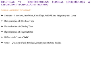
- 1. PRACTICAL: VI – BIOTECHNOLOGY, CLINICAL MICROBIOLOGY & LABORATORY TECHNOLOGY (17BZMP506) CLINICAL LABORATORYTECHNOLOGY Spotters – Autoclave, Incubator, Centrifuge, WIDAL and Pregnancy test (kits) Determination of Bleeding Time Determination of Clotting Time Determination of Haemoglobin Differential Count of WBC Urine – Qualitative tests for sugar, albumin and ketone bodies.
- 2. autoclave
- 3. PRINCIPLE: The autoclave works under the principle of moist heat sterilization where steam under pressure is used to sterilize the material present inside the chamber. The high pressure increases the boiling point of water and thus helps achieve a higher temperature for sterilization. To be effective, the autoclave must reach and maintain a temperature of 121° C for at least 30 minutes by using saturated steam under at least 15 psi of pressure. APPLICATIONS: They are used to decontaminate specific biological waste and sterilize media, instruments, and labware. Regulated medical waste that might contain bacteria, viruses, and other biological materials are recommended to be inactivated by autoclaving before disposal. In medical labs, autoclaves are used to sterilize medical equipment, glassware, surgical equipment, and medical wastes. Autoclaves are used for the sterilization of culture media, autoclavable containers, plastic tubes, and pipette tips.
- 4. INCUBATOR
- 6. PRINCIPLE: An incubator is based on the principle that microorganisms require a particular set of parameters for their growth and development and also dry heat sterilization where heat waves are used to sterilize the material present inside the chamber. All incubators are based on the concept that when organisms are provided with the optimal condition of temperature, humidity, oxygen, and carbon dioxide levels, they grow and divide to form more organisms. In an incubator, the thermostat maintains a constant temperature that can be read from the outside via the thermometer. The temperature is maintained by utilizing the heating and no-heating cycles. During the heating cycle, the thermostat heats the incubator, and during the no-heating period, the heating is stopped, and the incubator is cooled by radiating heat to the surrounding. Insulation from the outside creates an isolated condition inside the cabinet, which allows the microbes to grow effectively. APPLICATIONS: Incubators are used to grow microbial culture or cell cultures. Incubators also provide a controlled condition for sample storage before they can be processed in the laboratories. In Clinical laboratories, incubator is used to sterilize the laboratory instruments such as glasswares, culture plates, etc.
- 7. CENTRIFUGE
- 8. PRINCIPLE: The centrifuge works using the sedimentation principle, where the centrifugal force causes denser substances and particles to move outward in the radial direction. In a solution, particles whose density is higher than that of the solvent sink (sediment), and particles that are lighter than it floats to the top. APPLICATIONS: To separate two miscible substances Fractionation of subcellular organelles (including membranes/membrane fractions) In clinical laboratories , centrifuge is used to separate the urine components like crystals, parasites , etc to detect the kidney disorders. It is used to separate the blood components such as plasma and formed elements and plasma is used to perform biochemical tests like detection of blood glucose, urea, protein, cholesterol, etc.
- 10. PRINCIPLE: It works under the principle of antigen-antibody reaction. Individual infected with Salmonella Typhi or S. paratyphi produce antibodies against either somatic (O) antigens and/or flagellar antigens (H). These produced antibodies in serum, if exposed to bacterial suspension carrying homologous antigens, result in agglutination. The agglutination reaction can be seen as visible clumping. In the test, the patient’s serum is mixed with killed bacterial suspension of Salmonella carrying specific O, H, AH and BH antigens and observed for agglutination reaction. If the patient’s serum contains specific antibodies against the antigens, clumping is evident which indicates positive test. Absence of agglutination indicates a negative result. APPLICATIONS: It is a rapid test for screening enteric fever in endemic areas. When culture facilities are not available, Widal test is handy. It can detect infection caused by both Salmonella Typhi and Salmonella Paratyphi.
- 12. PRINCIPLE: The hCG Card Pregnancy Test is a rapid chromatographic immunoassay for the qualitative detection of human chorionic gonadotropin in urine to aid in the early detection of pregnancy. The test utilizes a combination of antibodies including a monoclonal hCG antibody to selectively detect elevated levels of hCG. The assay is conducted by adding a urine specimen to the specimen well of the test device and observing the formation of colored lines. The specimen migrates via capillary action along the membrane to react with the colored conjugate. Positive specimens react with the specific antibody-hCG-colored conjugate to form a colored line at the test line region of the membrane. Absence of this colored line suggests a negative result. To serve as a procedural control, a colored line will always appear in the control line region indicating that proper volume of specimen has been added and membrane wicking has occurred. APPLICATIONS: It enables the user to find out whether a women is pregnant or not within the confines of the home. Women who are trying to get pregnant need not rush to the doctor everytime they miss a period. The test enables women to seek antenatal care as soon as possible. Detection of hCG in the urine sample of male indicates testicular cancer
- 13. Sterilize the tip of a finger with surgical spirit Make a deep prick on the finger using a sterile lancet. Start the stop-watch immediately. Start gently touching the pricked finger with filter paper in a circular fashion Stop the watch when no more blood spot comes on the filter paper Note the time taken for the bleeding to stop. DETERMINATION OF BLEEDING TIME – DUKE’S METHOD
- 14. DETERMINATION OF BLEEDING TIME – DUKE’S METHOD PRINCIPLE: It is a standard test performed by puncturing the skin with a lancet and measuring the time it takes for the bleeding to stop. MATERIALS REQUIRED: Sterile lancet, Spirit, Cotton, Filter paper, Stop watch. PROCEDURE: •Sterilize the tip of a finger with surgical spirit. •Make a deep prick on the finger using a sterile lancet. •Start the stop-watch immediately. •Start gently touching the pricked finger with filter paper till blood spots continue to be made on the filter paper. •Stop the watch when no more blood spot comes on the filter paper and note the time taken for the bleeding to stop. NORMAL VALUE: upto 5 mins. RESULT: The bleeding time was found to be _______________.
- 16. DISCUSSION: Bleeding time is used to screen for bleeding disorders and quantify platelet function in patients taking aspirin or non-steroidal anti-inflammatory medications. This test is widely used not just for evaluation of platelet function but also to assess the effects of medications and medical devices such as cardiopulmonary bypass or dialysis machines on homeostasis status. The bleeding time is prolonged in the following conditions: •Thrombocytopaenia: Decreased platelet count impairs primary hemostasis. •Von Willebrand Disease: Von Willebrand disease is an inherited deficiency in the quantity or function of the platelet aggregation protein Von Willebrand factor. •Disseminated Intravascular Coagulation: DIC is a symptom of other severe disease processes like sepsis, burns, trauma, pregnancy, and malignancy. •Bernard-Soulier Syndrome: It is a rare autosomal recessive genetic defect in glycoprotein Ib causing giant platelets with perceived thrombocytopenia. •Chronic liver disease •Abnormality in the walls of the blood vessels
- 18. DETERMINATION OF CLOTTING TIME – SABRAZE’s CAPILLARY TUBE METHOD PRINCIPLE: It is a standard test performed by puncturing the skin with a lancet and measuring the time it takes for the bleeding to form a clot. MATERIALS REQUIRED: Sterile lancet, Spirit, Cotton, Capillary tube, Stop watch. PROCEDURE: •Sterilize the tip of a finger with surgical spirit. •Make a deep prick on the finger using a sterile lancet. •Start the stop-watch immediately. •The blood coming out of the puncture is sucked into a capillary glass tube. Then the tube is kept undisturbed horizontally for about 30 seconds. •A small bit of the capillary tube is broken off every 30 seconds until a fine thread of clotted blood appears while the capillary tube is broken. •When the fibrin thread appears between the broken ends of the capillary tube, stop the watch and the time is noted. NORMAL VALUE: upto 6 mins. RESULT: The clotting time was found to be _______________.
- 20. DISCUSSION: Clotting time depends on the availability of coagulation factors. In coagulation disorders like haemophilia, clotting time is prolonged but bleeding time remains normal. Clotting time is higher in females as compared to males. This is because of increased estrogen in females which prolongs Clotting time and decreases plasma fibrinogen level. Clotting time is also prolonged in the following conditions: •Vitamin K deficiency •Haemophilia A •Haemophilia B •Liver diseases Overdosage of anticoagulants like heparin and warfarin You tube video link for clotting time demonstration: https://youtu.be/71GNTwUuaNI
- 21. DETERMINATION OF HAEMOGLOBIN – ACID HAEMATIN METHOD PRINCIPLE: N/10 HCl converts haemoglobin into soluble unstable acid haematin. The colour intensity of the acid haematin after dilution is compared with standard brown glass in the comparator APPARATUS REQUIRED: Sahli’s haemoglobinometer It contains haemoglobin/Sahli’s tube, Colour comparator, haemoglobin/Sahli’s pipette and stirrer Haemoglobin/Sahli’s pipette – pipette has one mark indicating 20cu.mm (no bulb)
- 22. Haemoglobin/Sahli’s tube – it is graduated on both sides. One side is graduated in gram percentage from 2 – 22 (g%). Other side is graduated as percentage from 20 – 140 (%)
- 23. Colour comparator – central slot accommodates hemoglobin tube and on either side non-fading brown tinted glass pieces for colour matching are present Stirrer – thin glass rod for stirring the solution
- 24. N/10 HCl- To make 500 ml of N/10 HCl mix Concentrated HCl – 4.5ml Distilled water – 500ml Distilled water
- 25. Procedure Add N/10 HCl to the hemoglobinometer tube up to its lowest mark i.e 2g% (If N/10 HCl is taken above the mark the color of undiluted solution is lighter than standard and if N/10 HCl is taken less then all the hemoglobin is not converted). Sterilize the index finger using surgical spirit. Make a deep prick on the tip of the finger using a sterile lancet. Take blood up to 20 cu. mm mark on the pipette and transfer it to the haemoglobinometer tube containing N/10 HCl. Leave the solution for 10 minutes (for the conversion of hemoglobin to acid haematin). After 10 minutes add distilled water drop by drop and mix it by stirrer until the colour matches with the colour of comparator. While matching the colour, glass rod should be removed from the solution. The lower meniscus of the solution should be taken as the result which expresses hemoglobin content as g%.
- 26. Normal value: Adult Male -13 to 18 g% Adult Female -12 to 16 g% Children -12 to14 g% Infants -16 to 22 g% Result: The haemoglobin present in the blood sample was found to be _______ g% Clinical Significance Conditions where Hb is decreased Anaemia Auto immune diseases Blood loss (bleeding, internal hemorrhage) Parasitic infection Lead poisoning Dietary deficiency (iron, copper, vitamins) Mal-absorption of nutrients Chronic disease
- 27. Conditions where Hb is increased High altitude Strenous physical exercise Severe vomiting or diarrhoea Smoking Polycythaemia vera Obstructive lung disease Congestive heart disease Tumours which cause increase in haemoglobin such as Renal cell carcinoma, Liver tumors (Hepato cellular carcinoma), Cerebellar hemangioblastoma Sickle cell disease and thalassemia are genetic disorders caused by errors in the genes for hemoglobin, a substance composed of a protein and iron molecule that is responsible for carrying oxygen within the red blood cell. These disorders can cause fatigue, jaundice, and episodes of pain ranging from mild to very severe. Youtube video link: https://youtu.be/qMTJ05l855Q
- 28. DIFFERENTIAL COUNT OF WBC AIM: The aim of leukocyte differential test is to measure the percentage of each type of white blood cell (WBC) present in the blood sample. It also reveals if there are any abnormal or immature cells. MATERIALS REQUIRED: Grease free glass slide lancet surgical spirit cotton Leishman’s stain distilled water microscope
- 29. PROCEDURE: •Sterilize the tip of a finger with surgical spirit. •Make a deep prick on the finger using a sterile lancet. •The first drop of the blood should be wiped off. •The second drop of the blood was placed on the one corner of grease free glass slide, holding another slide at 45◦ angle the smear was made. •The smear should be uniform thickness so that the WBCs are clearly visible. •Add few drops of stain over the smear and leave it for 4-5 mins. •Excess stain was removed by showing the one corner of the slide under running tap water. •100 leucocytes were counted by moving the slides in zig-zag direction and the results were tabulated in the tabular column. NORMAL RANGE: Neutrophil – 60 – 70% Eosinophil – 2-4% Basophil - 0-2% Lymphocyte – 20-30% Monocyte – 1-2%
- 30. PRINCIPLE OF LEISHMAN STAINING Leishman Stain is a neutral stain for blood smears which was devised by the British surgeon W. B. Leishman (1865–1926). It consists of a mixture of eosin (an acidic stain), and Methylene blue (a basic stain) in Methyl alcohol and is usually diluted and buffered during the staining procedure. It stains the different components of blood in a range of shades between red and blue. It is based on a methanolic mixture of “polychromed” Methylene blue and eosin. The methanolic stock solution is stable and also serves the purpose of directly fixing the smear eliminating a prefixing step. COMPOSITION OF LEISHMAN STAIN 1. Eosin powder 2. Methylene blue 3. Methanol
- 31. 1 2 3 4 6 5
- 35. RESULT: The Differential count of WBCs Neutrophil- Eosinophil- Basophil- Lymphocyte- Monocyte- TABULAR COLUMN:
- 36. DISCUSSION: White blood corpuscle, a cellular component of the blood that lacks hemoglobin, has a nucleus, is capable of motility and defends the body against infections and diseases by ingesting foreign materials and cellular debris by destroying infectious agents and cancer cells. They are basically classified into two types based on the presence and absence of cytoplasm as Granulocytes and Agranulocytes. The Granulocytes includes Neutrophil, Eosinophil and Basophil, whereas Agranulocytes includes Lymphocyte and Monocyte. Granulocytes: •Neutrophil: It has multilobed nucleus (3-5 lobes) hence it is also known as PolyMorpho Nuclear Leucocyte (PMNL). It phagocyte the infectious microbes and also destroying them with the help of enzymes. A high neutrophil level can indicate an acute bacterial infection, inflammation, tissue death such as after a heart attack, stress on the body or chronic leukemia. A neutrophil count may be low after an adverse drug reaction or chemotherapy treatments. Illnesses such as myelodysplastic syndrome, autoimmune disorders, bone marrow cancers and aplastic anemia.
- 37. •Eosinophil: It has bilobed/horse-shoe shaped nucleus. It is active during parasite infections and allergic reactions, stops substances or other foreign materials from harming the body. They release chemical compounds such as histamine, serotonin to destroy the infectious microbes. A high eosinophil count tends to result from an allergic reactions such as asthma, eczema or a reaction to a medication, Inflammatory disorders such as celiac disease or inflammatory bowel disease (IBD). Low levels of eosinophil do not tend to show any issues but stress or steroid use may show eosinophil count to be low. •Basophil: It has bilobed/inverted ‘S’ shaped nucleus. They release chemical compounds such as histamine, serotonin, prostaglandins etc., to destroy the infectious microbes. An increase in basophil count can point to certain types of leukemia, including chronic myeloid leukemia, severe allergic reactions, inflammatory disorders such as rheumatoid arthritis or ulcerative colitis A low basophil count does not typically suggest a medical condition. However, stress, allergic reactions, steroid use and hyperthyroidism can cause a basophil count to be low.
- 38. Agranulocytes: •Lymphocyte: It has spherical nucleus. Active leucocyte classified into two types based on the site of maturation as B-lymphocyte and T-lymphocyte. B-lymphocytes uses antibodies to stop bacteria or viruses from entering the body. Hence, it is involved in antibody-mediated/humoral immunity. Whereas, T-lymphocytes kills off the body cells if they have been compromised by a virus or cancer cells. Hence it is involved in cell- mediated immunity. A high lymphocyte level can indicate an acute viral infection such as chicken pox, herpes or hepatitis, bacterial infection such as tuberculosis or pertussis or a condition such as lymphocytic leukemia or lymphoma. A low lymphocyte level can point to an autoimmune disorder such as lupus or rheumatoid arthritis. HIV, tuberculosis, hepatitis or the flu. •Monocyte: It has a large bean shaped nucleus. Diapedesis is a condition commonly seen in monocyte, it becomes a macrophage in the body’s tissues by phagocyte the infectious microbes and getting rid of dead cells while increasing immune system strength. A high monocyte count can result from a chronic infections such as tuberculosis or a fungal infection, IBD, monocytic leukemia, rheumatoid arthritis. Low count of monocyte indicates hairy cell leukemia or bone marrow damage. Youtube video: https://youtu.be/ovgiLLkp9QY
- 39. QUALITATIVE TEST FOR URINE SUGAR – BENEDICT’s TEST AIM: To detect the presence of glucose in the urine using Benedict’s reagent. PRINCIPLE: When Benedict’s solution and simple carbohydrates are heated, the solution changes to orange red/ brick red. This reaction is caused by the reducing property of simple carbohydrates. The copper (II) ions in the Benedict’s solution are reduced to Copper (I) ions, which causes the colour change. The red copper(I) oxide formed is insoluble in water and is precipitated out of solution. This accounts for the precipitate formed. As the concentration of reducing sugar increases, the nearer the final colour is to brick-red and the greater the precipitate formed. Sometimes a brick red solid, copper oxide, precipitates out of the solution and collects at the bottom of the test tube. MATERIALS REQUIRED: Test tubes and test tube holder Boiling water bath Urine sample and Benedict’s reagent PROCEDURE: Take 5 ml of Benedict’s reagent (CuSO4) in a clean glass test tube. Add 1 ml urine sample and mix it well. The solution is then heated in a boiling water bath for 3-5 minutes. Observe for colour change in the solution of test tubes.
- 40. RESULT INTERPRETATION OF BENEDICT’S TEST: If the colour upon boiling is changed into green, then there would be 0.1 to 0.5 percent sugar in solution (+) If it changes to yellow, then 0.5 to 1 percent sugar is present (++) If it changes to orange, then it means that 1 to 1.5 percent sugar is present (+++) If colour changes to red, then 1.5 to 2.0 percent sugar is present (++++) If colour changes to brick red, it means that more than 2 percent sugar is present in solution.
- 41. CLINICAL SIGNIFICANCE: In a healthy individual, almost all of the glucose filtered by the renal glomerulus is reabsorbed in the proximal convoluted tubule. The amount of glucose reabsorbed by the proximal tubule is determined by the body's need to maintain a sufficient level of glucose in the blood. If the concentration of blood glucose becomes too high (160-180 mg/dL), the tubules no longer reabsorb glucose, allowing it to pass through into the urine. Conditions in which glucose levels in the urine are above 100 mg/dL and detectable include: 1. Diabetes mellitus and other endocrine disorders 2. impaired tubular reabsorption due to advanced kidney disease 3. pregnancy - glycosuria developing in the 3rd trimester may be due to latent diabetes mellitus 4. central nervous system damage 5. pancreatic disease 6. disturbances of metabolism such as, burns, infection or fractures
- 42. QUALITATIVE TEST FOR URINE ALBUMIN – HEAT & ACETIC ACID TEST AIM: To detect the presence of albumin in the urine by Heat and Acetic acid test. PRINCIPLE: Proteins are denatured and coagulated on heating to give white colour cloudy precipitate. MATERIALS REQUIRED: Test tubes and test tube holder Spirit lamp Urine sample and Acetic acid PROCEDURE: Take a clean glass test tube and fill 2/3rd of the test tube with urine sample. Heat only the upper part of the test tube over the flame, keeping lower part as control. Presence of phosphate, carbonates, proteins gives white colour cloudy precipitation. Add 3 drops of acetic acid and if the cloudy precipitate persists it indicates the presence of protein in the urine sample (because acetic acid dissolves the phosphates and carbonates.
- 43. RESULT: Cloudiness appears on the upper portion of urine sample – Presence of Albuminuria Absence of cloudiness - Absence of Albuminuria
- 44. CLINICAL SIGNIFICANCE: The presence of an increased amount of protein in a urine specimen is often the first indicator of renal disease. Proteinuria may signal severe kidney damage, be a warning of impending kidney involvement, or be transient and unrelated to the renal system. Further quantitative testing of urine for protein may be needed to determine the significance of the proteinuria. Proteinuria related to kidney impairment may be due to glomerular membrane damage caused by toxic agents, immune complexes found in lupus erythematosus, or streptococcal glomerulonephritis. The amount of protein present in urine samples from patients with glomerular damage usually ranges from 10-40 mg/dL. If the urinary protein is due to a disorder that affects tubular reabsorption, the urine protein quantities will be much greater. In patients with multiple myeloma, proteinuria is due to the excretion of the Bence Jones protein. This low molecular weight protein produced by a malignant clone of plasma cells circulates in the blood and is filtered in the kidneys in quantities exceeding the tubular capacity. This excess protein is excreted in the urine.
- 45. QUALITATIVE TEST FOR URINE KETONE BODIES – ROTHERA’s TEST AIM: To detect the presence of ketone bodies in the urine by Rothera’s test. PRINCIPLE: Acetoacetic acid and acetone react with alkaline solution of sodium nitroprusside to form a purple colored complex. This method can detect above 1-5 mg/dl of acetoacetic acid and 10-20 mg/dl of acetone. MATERIALS REQUIRED: Test tubes and test tube holder Urine sample Rothera’s powder: Sodium nitroprusside – 0.75gm and Ammonium sulphate – 20gm (mix and pulverize) Liquor Ammonia (Ammonium hydroxide) PROCEDURE: Transfer about 5 ml of urine sample to a clean glass test tube. Add 1 gm of Rothera’s powder mixture and mix well. Layer over the urine, transfer 1-2 ml of concentrated ammonium hydroxide using a dropper. Observe the pink-purple ring at the interface.
- 46. RESULT: Immediate formation of purple permanganate coloured ring at the interface: Ketone bodies present (Positive) No formation of purple permanganate coloured ring at the interface: Ketone bodies absent (Negative)
- 47. CLINICAL SIGNIFICANCE: Ketone bodies are usually absent in urine. The presence of ketones in the urine probably indicates that the body is using fats rather than carbohydrates for energy. High levels of ketones may be present in the urine of individuals with uncontrolled diabetes because the body's ability to metabolize carbohydrates is defective. Detecting the presence of ketones in the urine is a valuable aid to managing and monitoring individuals with diabetes mellitus. Ketonuria is an indication that the insulin dose needs to be increased. Electrolyte imbalance and dehydration occur when ketones accumulate in the blood. If these conditions are not corrected by adjusting the dose of insulin, the patient may develop ketoacidosis and ultimately diabetic coma. Low levels of ketones may be detected during conditions of physiological stress such as fasting, rapid weight loss, frequent strenuous exercise or prolonged vomiting. The presence of ketones in these situations is due to either inadequate intake of carbohydrates or increased loss of carbohydrates.