SlideShare ist ein Scribd-Unternehmen logo
1 von 8
Downloaden Sie, um offline zu lesen
Description of the movements associated with muscular activity in the rhopalial complex in Carybdea
marsupialis (juvenile)
Introduction
Cubomedusae are well-known for their ability to adapt their swimming behavior in response to photic or
visual stimuli (Satterlie, 2002). Carybdea marsupialis, along with several other species of its class, uses
the two lens eyes, the two slit eyes, and the two pit eyes found in the rhopalia to sense its surrounding
environment. In the adult form, the rhopalia of Carybdea marsupialis are located in four heart-shaped
rhopalial cavities – the rhopalial niches – found at the center of each side of the box-shaped bell.
However, in the juvenile form, the rhopalia are not yet encased in niches and the bell is not fully
developed, thus easing the observation of the rhopalial complex. The rhopalia are attached to the bell
by rhopalial stalks that have the ability to swing to virtually any direction, thus eventually allowing the
jellyfish to cover a wider field of view than that with fixed rhopalia. It has been previously suspected that
slight movements of the rhopalial complex were eventually associated with the water currents
generated by the contraction of the bell. In addition, it was thought that the weight of the statolith
located underneath the lower lens eye was weighting down the rhopalium and amplifying the bouncing
movements of the rhopalium, thus acting like the weight of a pendulum. However, it has been
suggested that the bell contractions in a close relative of Carybdea marsupialis, Tripedalia cystophora,
induced a voluntary angular shift of the rhopalium to counteract the putative image fading in the upper
lens eye (Bielecki et al. 2013). Such results may indicate a clear control of the different structures
present in the rhopalial complex through the use of muscle structures.
Here, the movements of the rhopalia and the deformation of the stalk is examined in some detail to
describe the different movements of these structures that are associated with voluntary muscular
contractions. Evidences of such movements, whether induced by external stimuli or not, are presented
here as preliminary research for further investigations of the muscle structures and the nervous
system(s) controlling both the rhopalial stalk, and the rhopalium by itself.
Methods
• Direct observation
Each specimen had been isolated in individual small containers with a minimum amount of water in
order to minimize slight water currents generated by either the tools used to generate the physical
stimuli or by the contractions of the bell of the observed individuals. Due to the shape of the juvenile
jellyfish, and because they exhibited a highly developed protective behavior, known as crumpling
(Satterlie et al. 2005), all the specimens were observed laterally, or from their oral side (top of the bell
down).
Two types of stimuli were tested on individual rhopalium: 1) physical stimuli produced by physical
contact with the surface of the rhopalium and 2) visual stimuli generated through two different
methods. A shiny pair of thin tweezers and/or a micro improvised optic fiber flashlight with different
filters were waved in front of the biggest (and most reachable) lower lens eye at variable distances from
the rhopalium. However, it has to be noted that the background lighting was provided by a halogen
lamp emitting a broad spectrum of wavelength, thus eventually masking or blending the light produced
by the micro optic fiber flashlight. Green and purple-blue filters were used to match the sensitivity
spectrum of Carybdea marsupialis that are well-known to exhibit different behaviors under these
lighting conditions.
Balance and inclination were also tested to investigate an eventual intentional reaction to a position
change. However, the difficulty of the manipulation and the crumbling response of the individuals were
not suitable for a correct and accurate observation of the reaction of the specimens.
• Measurements
Several variables were investigated to understand the mechanics of the rhopalial movements. It
appeared that the rhopalial stalk could bend and swing the rhopalia in any direction. Both, the inside-
out and lateral angular shift have been measured digitally, image per image using Adobe Photoshop CS6.
The slight bouncing movements of the rhopalium itself were also established using Adobe Photoshop
CS6 to measure the angular shift between the position at rest and the final position after the bouncing
movement. The position at rest of the rhopalia in all cases were visually estimated based on the average
straight shape of the rhopalial stalk in the three individuals investigated. The total rotation of the
rhopalia at the rhopalium-stalk junction has also been digitally measured with Adobe Photoshop CS6.
Results
• Bending of the stalk
Bending of the stalk was the initial easily observable feature among the variety of movements produced
in the rhopalial complex. Direct observations of the bouncing movement of the rhopalia on the stalks
indicted that the latter had the ability to bend towards the center of the bell, and laterally (parallel to
the edges of the box-shaped bell). At the resting position, the rhopalia point downward (lower-lens eye
down) and slightly away from the bell with an angle of 8-10° relative to the aboral-oral axis (n=3) (Figure
1). Several stages in the inside-out bending process have been observed (figure 2): 1) the side of the
stalk facing inward shortens, thus initiating the bending process; 2) the outer side of the region of the
stalk near the rhopalium-stalk junction bends inward; 3) the stalk has finally bent along its entire length
at the final stage of the bending process. Measurements of the total angular shift on three different
juvenile individuals suggest that the stalk can bend inward with an angle of about 52° relative to the
initial (at rest) axis of symmetry of the stalk – originating from the base of the stalk and extending to the
rhopalium-stalk junction (figures 3 and 4). Additional independent bouncing of the rhopalia at the
junction – discussed later – allows the rhopalial complex to bend inward at angles greater than 70°. In
certain cases, the bending process was clearly associated with a response to external stimuli (either by
touching the rhopalium or waving the shiny tweezers in front of the lower-lens eye). However a limited
amount of replicates were successfully recorded, therefore, the conclusions are yet to be clearly
established.
Figure 1 Rhopalium and rhopalial stalk at
resting position. The rhopalium hangs down
(lower-lens eye (lle) facing downward), away
from the center of the bell with an angle of 8-
10° relative to the aboral-oral axis.
Figure 2 Representation of the inward bending process of the rhopalial stalk. In the resting position, the rhopalium hangs
down and the rhopalial stalk remains straight (a). Three main stages can be observed throughout the bending process: The
side of the stalk facing inward shortens (arrowhead), thus initiating the bending process (b); the outer side of the region of
the stalk near the rhopalium-stalk junction is also bending inward (c); the stalk is finally bending along its entire length
(arrowhead) at the final stage of the bending process(d). The rhopalium has moved to about 50° from the resting position.
The rhopalium possesses two pit eyes (pe), two slit eyes (se), one upper lens eye (ule), one lower lens eye facing down in the
resting position (lle), and a statolith (st).
Figure 3 Example of a rhopalium of a juvenile medusae being moved by the rhopalial stalk bending inward. Note that at the
resting position, the rhopalium points downward with an angle of about 8-10° relative to the aboral-oral axis. The total
angular shift of the stalk is about 52° (n=3) (b) relative to the initial (at rest) axis of symmetry of the stalk (a). The solid white
line in figure 3a) represents the axis of symmetry of the stalk originating from its base and extending to the rhopalium-stalk
junction. The solid white line in figure 3b) depicts the final angular shift produced through the bending of the stalk. The
white dashed lines emphasize the outline of the rhopalial stalk for better visualization of the shape of the stalk.
Figure 4 Illustration of the inside-out bending process of the rhopalial stalk without additional bouncing of the rhopalium at
the rhopalium-stalk junction. At the resting position, the rhopalia point downward (lower lens-eye down) and slightly
toward the outside with an angle of about 8-10° relative to the aboral-oral axis. The stalk can bend inward with an angle of
about 50° relative to the initial (at rest) axis of symmetry of the stalk originating from its base to the rhopalium-stalk
junction. The rhopalium has moved to about 50° from the resting position. The rhopalium possesses two pit eyes (pe), two
slit eyes (se), one upper lens eye (ule), one lower lens eye facing down in the resting position (lle), and a statolith (st).
Lateral bending of the stalk has also been observed and no significant differences in the bending process
have been identified compared to the inside-out bending process. However, unlike for the inside-out
bending where the stalk can only bend toward the center of the bell, the stalk can move on either side
of the axis formed by the relaxed rhopalial stalk. Lateral movements of the rhopalial complex observed
in three juveniles indicated that the angular shift of the stalk from the resting position to either side was
about 60° without any difference between the two sides. The total swing span of the stalk has been
calculated to be 120° (figures 5 and 6). Additional bouncing of the rhopalia at the rhopalium-stalk
junction allows the rhopalial complex to bend on either side at angles greater than 86°.
Figure 5 Examples of the rhopalium of a juvenile medusae being moved by the rhopalial stalk bending from right to left. At
the resting position, the rhopalial stalk is straight (b). The same four bending stages observed in the inside-out bending apply
in the lateral bending process (a and c)). The shortening of one side of the stalk and the relaxation of the opposite side of this
stalk allow the rhopalium to swing from either side of the initial (at rest) axis of symmetry of the stalk originating from its
base to the rhopalium-stalk junction. The stalk can bend on both sides with an angle of about 60° relative to its axis of
symmetry, thus allowing a total angle of freedom of about 120°. The black dashed lines emphasize the outline of the
rhopalial stalk.
Figure 6 Illustration of the lateral bending process of the rhopalial stalk without additional bouncing of the rhopalium at the
rhopalium-stalk junction. The shortening of one side of the stalk (arrowhead) and the relaxation of the opposite side of this
stalk allow the rhopalium to swing from either side of the initial (at rest) axis of symmetry of the stalk originating from its
base and extending to the rhopalium-stalk junction. The stalk can bend on both sides with an angle of about 60° relative to
its axis of symmetry, thus allowing a total angle of freedom of about 120°.
These two movements suggest that the rhopalia can swing in any direction, allowing the rhopalium to
eventually achieve a complete rotation around the axis of symmetry formed by the rhopalial stalk in the
relaxed position (video).
• Independent movements of the rhopalium itself
Further tedious observations of the rhopalial complex indicated that the rhopalium had the ability to
move independently from the stalk. Indeed, two types of movements associated with muscular activity
were identified: 1) the rotation of the rhopalium at the center of rotation formed by the rhopalium-stalk
junction and 2) a slight independent bouncing of the rhopalium at the same junction.
In the resting position, the rhopalium is aligned with the stalk – upper-lens eye, lower-lens eye, and
statolith being aligned with the stalk. The rhopalium has been observed to accomplish a maximum
rotation of about 134° in total (figure 7). No evidences of a particular shortening or stiffening of the stalk
as been identified, but more accurate observations need to be performed to establish the accuracy of
these results.
Figure 7 Example of the rotation process achieved by the rhopalium. The maximum observed rotation was equal to 134°.
Note that image b) depicts the position of the rhopalium at rest. The white dashed arrows indicate the orientation of the
rhopalium to emphasize the rotation (a, b, and c). Here, the rhopalium is viewed from behind, so only the statolith (st) and
the rhopalium-stalk junction can be seen.
The rhopalium also seemed to have the ability to bounce on the rhopalium-stalk junction independently
from the stalk. Even though high definition recording would probably provide more accurate results
regarding the independence of the rhopalium from the stalk, the preliminary results indicated that the
rhopalium could bend on either side of the axis of symmetry formed by the stalk with an angle of 16°.
The total angle shift from one side to the other was calculated to be about 32°. Such movement occur
in any direction around the rhopalium-stalk junction (figure 8).
Figure 8 Illustration of the independent bouncing of the rhopalia at the extremity of the rhopalial stalk. The rhopalia can
bounce to about 16° degrees on either side relative to the initial position at rest without significant bending of the stalk.
Figure 1a) represents the inside-out bouncing viewed from a side of the rhopalial complex and figure 1b) the lateral bouncing
viewed from bellow. Note that the center of the bouncing movement is located at the rhopalium-stalk junction (black
arrowhead).
Discussion
The immobilization of the individuals and the avoidance of undesirable slight water currents clearly
helped to establish that rhopalial movements were not artifacts of random surrounding features, but
were instead achieved through the use of muscle structures in the rhopalial stalk. Several different types
of movements were examined in both the rhopalial stalk and the rhopalia. The most noticeable
movements are performed by the rhopalial stalk. The ability of this structure to bend from the outside
of the bell toward the inside, and to bend laterally along the edges of the box-shaped bell allows the
rhopalia to cover a wider field of view. This is especially true when the rhopalia achieve complete
rotations around the axis of symmetry formed by the stalk at rest. It has been observed that the bending
process starts from the region of the stalk near the rhopalium-stalk junction. The bending process is
initiated by the shortening of the side of the stalk facing the direction towards with the stalk bends.
What has not been clearly determined is whether or not the opposite side of the stalk lengthens to
ease the bending processes or if it bends through a passive relaxation of muscle structures. I addition,
the causes of the return to the initial resting position of stalk, has yet to be determined. Muscle activity
may be involved in that process but the natural rigidity of the stalk may eventually contribute to the
return to the initial position of the rhopalial system – similarly to the mesoglea allowing the bell to relax.
Such movements have been observed multiple times as responses to external stimuli such as the waving
of the shiny tweezers in front of the rhopalia, but touching these structures has not proven to be
efficient in observing a voluntary contraction of the stalk. The results obtained with the micro fiber-optic
flashlight are not conclusive but further investigation may eventually provide more accurate results.
Closer examination of the four rhopalia of the three juvenile individuals suggested that each rhopalium
not only had the capacity to move independently from the three other rhopalia, but also that there is a
certain degree of freedom between the rhopalium itself and the stalk to which it is attached. Indeed,
this independent movement of the rhopalium may add sixteen additional degrees to the total angular
ship exhibited by the rhopalial stalk. This eventually increase the field of view of each rhopalium without
further applying significant stress on the rhopalial stalk. The rhopalium also have the ability to rotate at
the center of rotation formed by the rhopalium-stalk junction. Such rotation mostly allows the re-
orientation of the upper-lens eye toward either an object or a source of light. However, more
information is needed regarding this mechanism. Indeed, the possible implication of mechanisms
present in the stalk that would influence the rotation process still remains unclear due to a lack of both
direct visual information and physiological knowledge about the rhopalial system.
Little is known about the mechanics of rhopalial movements, but deeper analysis of the rhopalial
structures may provide significant clues about the importance for juvenile Carybdea marsupialis to
develop such rhopalial mobility. Since external stimuli delivered throughout the experiments
successfully triggered a response, such mobility might be associated with a protective response of
juvenile medusae against potential threats – to the same order as the crumbling response.
References
Bielecki, J., J.T. Høeg and A. Garm. 2013. Fixational Eye Movements in the Earliest Stage of Metazoan
Evolution. PLoS ONE 8: e66442.
Satterlie, R.A. 2002. Neuronal control of swimming in jellyfish: a comparative story. Can. J. Zoology 80:
1654-1669.
Other readings scavenged on the web
Garm, A. et al. 2006. Rhopalia are integrated parts of the central nervous system in box jellyfish. Cell.
Tissue. Res.325: 333-343.

Weitere ähnliche Inhalte

Was ist angesagt?

Articulators in dentistry
Articulators in dentistryArticulators in dentistry
Articulators in dentistrySonali Harjani
 
Orientation relation with facebow and hinge axis and abvance in facebow
Orientation relation with facebow and hinge axis and abvance in facebowOrientation relation with facebow and hinge axis and abvance in facebow
Orientation relation with facebow and hinge axis and abvance in facebowPratik Hodar
 
Facebow george/ oral surgery courses  
Facebow george/ oral surgery courses  Facebow george/ oral surgery courses  
Facebow george/ oral surgery courses  Indian dental academy
 
Orientation jaw relation
Orientation jaw relationOrientation jaw relation
Orientation jaw relationRavi banavathu
 
Orientation jaw relations & face bow
Orientation jaw relations & face bowOrientation jaw relations & face bow
Orientation jaw relations & face bowRohan Bhoil
 
Orientation /orthodontic courses by Indian dental academy 
Orientation /orthodontic courses by Indian dental academy Orientation /orthodontic courses by Indian dental academy 
Orientation /orthodontic courses by Indian dental academy Indian dental academy
 
Orientation jaw relation/ dentistry jobs
Orientation jaw relation/ dentistry jobsOrientation jaw relation/ dentistry jobs
Orientation jaw relation/ dentistry jobsIndian dental academy
 
Orientation jaw relation/cosmetic dentistry courses
Orientation jaw relation/cosmetic dentistry coursesOrientation jaw relation/cosmetic dentistry courses
Orientation jaw relation/cosmetic dentistry coursesIndian dental academy
 
Occlusion n articulatirs/prosthodontic courses
Occlusion n articulatirs/prosthodontic coursesOcclusion n articulatirs/prosthodontic courses
Occlusion n articulatirs/prosthodontic coursesIndian dental academy
 
Orientation jaw relation 4 / dental implant courses by Indian dental academy 
Orientation  jaw  relation 4 / dental implant courses by Indian dental academy Orientation  jaw  relation 4 / dental implant courses by Indian dental academy 
Orientation jaw relation 4 / dental implant courses by Indian dental academy Indian dental academy
 
Face bow / dental lab technology courses
Face bow / dental lab technology coursesFace bow / dental lab technology courses
Face bow / dental lab technology coursesIndian dental academy
 
Occlusion n articulatirs/ orthodontic continuing education
Occlusion n articulatirs/ orthodontic continuing educationOcclusion n articulatirs/ orthodontic continuing education
Occlusion n articulatirs/ orthodontic continuing educationIndian dental academy
 
Orientation jaw relation/ dentistry jobs
Orientation jaw relation/ dentistry jobsOrientation jaw relation/ dentistry jobs
Orientation jaw relation/ dentistry jobsIndian dental academy
 

Was ist angesagt? (20)

Orientation jaw relation
Orientation jaw relationOrientation jaw relation
Orientation jaw relation
 
Face bow/endodontic courses
Face bow/endodontic coursesFace bow/endodontic courses
Face bow/endodontic courses
 
Articulators in dentistry
Articulators in dentistryArticulators in dentistry
Articulators in dentistry
 
Orientation relation with facebow and hinge axis and abvance in facebow
Orientation relation with facebow and hinge axis and abvance in facebowOrientation relation with facebow and hinge axis and abvance in facebow
Orientation relation with facebow and hinge axis and abvance in facebow
 
Facebow george/ oral surgery courses  
Facebow george/ oral surgery courses  Facebow george/ oral surgery courses  
Facebow george/ oral surgery courses  
 
Orientation jaw relation
Orientation jaw relationOrientation jaw relation
Orientation jaw relation
 
Tmj instrumentation
Tmj instrumentation Tmj instrumentation
Tmj instrumentation
 
Facebow
FacebowFacebow
Facebow
 
Orientation jaw relations & face bow
Orientation jaw relations & face bowOrientation jaw relations & face bow
Orientation jaw relations & face bow
 
Orientation /orthodontic courses by Indian dental academy 
Orientation /orthodontic courses by Indian dental academy Orientation /orthodontic courses by Indian dental academy 
Orientation /orthodontic courses by Indian dental academy 
 
Orientation jaw relation/ dentistry jobs
Orientation jaw relation/ dentistry jobsOrientation jaw relation/ dentistry jobs
Orientation jaw relation/ dentistry jobs
 
Face bow
Face bowFace bow
Face bow
 
Orientation jaw relation/cosmetic dentistry courses
Orientation jaw relation/cosmetic dentistry coursesOrientation jaw relation/cosmetic dentistry courses
Orientation jaw relation/cosmetic dentistry courses
 
Occlusion n articulatirs/prosthodontic courses
Occlusion n articulatirs/prosthodontic coursesOcclusion n articulatirs/prosthodontic courses
Occlusion n articulatirs/prosthodontic courses
 
Facebow / dental courses
Facebow / dental coursesFacebow / dental courses
Facebow / dental courses
 
ARTICULATORS
ARTICULATORSARTICULATORS
ARTICULATORS
 
Orientation jaw relation 4 / dental implant courses by Indian dental academy 
Orientation  jaw  relation 4 / dental implant courses by Indian dental academy Orientation  jaw  relation 4 / dental implant courses by Indian dental academy 
Orientation jaw relation 4 / dental implant courses by Indian dental academy 
 
Face bow / dental lab technology courses
Face bow / dental lab technology coursesFace bow / dental lab technology courses
Face bow / dental lab technology courses
 
Occlusion n articulatirs/ orthodontic continuing education
Occlusion n articulatirs/ orthodontic continuing educationOcclusion n articulatirs/ orthodontic continuing education
Occlusion n articulatirs/ orthodontic continuing education
 
Orientation jaw relation/ dentistry jobs
Orientation jaw relation/ dentistry jobsOrientation jaw relation/ dentistry jobs
Orientation jaw relation/ dentistry jobs
 

Andere mochten auch

Activitat aurasma
Activitat aurasmaActivitat aurasma
Activitat aurasmaasansa83
 
Mahoney Monique Cadbury Coco
Mahoney Monique Cadbury CocoMahoney Monique Cadbury Coco
Mahoney Monique Cadbury CocoMonique Mahoney
 
Darkness and light
Darkness and lightDarkness and light
Darkness and lightkrowland
 
bow chicka wow HOW: Moving from WHAT to HOW
bow chicka wow HOW: Moving from WHAT to HOWbow chicka wow HOW: Moving from WHAT to HOW
bow chicka wow HOW: Moving from WHAT to HOWAaron Maurer
 
The Future of Software is Open Source
The Future of Software is Open SourceThe Future of Software is Open Source
The Future of Software is Open SourceAngelo Corsaro
 
Yeot Spring Lab #3 Solder 101
Yeot Spring Lab #3  Solder 101Yeot Spring Lab #3  Solder 101
Yeot Spring Lab #3 Solder 101Aaron Maurer
 
The Open Splice.Org Community
The Open Splice.Org CommunityThe Open Splice.Org Community
The Open Splice.Org CommunityAngelo Corsaro
 
Spring 2015 yeot lab 1
Spring 2015 yeot lab 1Spring 2015 yeot lab 1
Spring 2015 yeot lab 1Aaron Maurer
 
Crowdsourcing for Educators
Crowdsourcing for EducatorsCrowdsourcing for Educators
Crowdsourcing for EducatorsAaron Maurer
 
Participatory research report
Participatory research reportParticipatory research report
Participatory research reportRey Tagum
 
Lauren and alana language arts project
Lauren and alana language arts projectLauren and alana language arts project
Lauren and alana language arts projectAaron Maurer
 

Andere mochten auch (20)

Eskom job descrption
Eskom job descrptionEskom job descrption
Eskom job descrption
 
Activitat aurasma
Activitat aurasmaActivitat aurasma
Activitat aurasma
 
Unit 13 Pecha Kucha
Unit 13 Pecha KuchaUnit 13 Pecha Kucha
Unit 13 Pecha Kucha
 
Mahoney Monique Cadbury Coco
Mahoney Monique Cadbury CocoMahoney Monique Cadbury Coco
Mahoney Monique Cadbury Coco
 
Darkness and light
Darkness and lightDarkness and light
Darkness and light
 
Day in our lives
Day in our livesDay in our lives
Day in our lives
 
bow chicka wow HOW: Moving from WHAT to HOW
bow chicka wow HOW: Moving from WHAT to HOWbow chicka wow HOW: Moving from WHAT to HOW
bow chicka wow HOW: Moving from WHAT to HOW
 
Day In Our Lives
Day In Our LivesDay In Our Lives
Day In Our Lives
 
St Charles 0106
St Charles 0106St Charles 0106
St Charles 0106
 
The Future of Software is Open Source
The Future of Software is Open SourceThe Future of Software is Open Source
The Future of Software is Open Source
 
Wear It! Across The Country (Updated Sept. 2009)
Wear It! Across The Country (Updated Sept. 2009)Wear It! Across The Country (Updated Sept. 2009)
Wear It! Across The Country (Updated Sept. 2009)
 
Yeot Spring Lab #3 Solder 101
Yeot Spring Lab #3  Solder 101Yeot Spring Lab #3  Solder 101
Yeot Spring Lab #3 Solder 101
 
Inflatable Life Jacket Education Presentation
Inflatable Life Jacket Education PresentationInflatable Life Jacket Education Presentation
Inflatable Life Jacket Education Presentation
 
The Open Splice.Org Community
The Open Splice.Org CommunityThe Open Splice.Org Community
The Open Splice.Org Community
 
Spring 2015 yeot lab 1
Spring 2015 yeot lab 1Spring 2015 yeot lab 1
Spring 2015 yeot lab 1
 
Eagles
EaglesEagles
Eagles
 
Crowdsourcing for Educators
Crowdsourcing for EducatorsCrowdsourcing for Educators
Crowdsourcing for Educators
 
Participatory research report
Participatory research reportParticipatory research report
Participatory research report
 
Lauren and alana language arts project
Lauren and alana language arts projectLauren and alana language arts project
Lauren and alana language arts project
 
Stepping into Scala
Stepping into ScalaStepping into Scala
Stepping into Scala
 

Ähnlich wie Rhopalial movements in Carybdea marsupialis

Presentation1.pptx, radiological anatomy of the shoulder joint.
Presentation1.pptx, radiological anatomy of the shoulder joint.Presentation1.pptx, radiological anatomy of the shoulder joint.
Presentation1.pptx, radiological anatomy of the shoulder joint.Abdellah Nazeer
 
Radiological anatomy of the Head and Neck
Radiological anatomy of the Head and NeckRadiological anatomy of the Head and Neck
Radiological anatomy of the Head and NeckRAVINDRANATH GANDRAKOTA
 
Solid-state plastic deformation in the dynamic interior of a differentiated a...
Solid-state plastic deformation in the dynamic interior of a differentiated a...Solid-state plastic deformation in the dynamic interior of a differentiated a...
Solid-state plastic deformation in the dynamic interior of a differentiated a...Carlos Bella
 
growthrotation-160510082813.pdf
growthrotation-160510082813.pdfgrowthrotation-160510082813.pdf
growthrotation-160510082813.pdfNonSub1
 
squint-150215112527-conversion-gate01.pptx
squint-150215112527-conversion-gate01.pptxsquint-150215112527-conversion-gate01.pptx
squint-150215112527-conversion-gate01.pptxpreethiraja9
 
Reflection And Refraction Of Light
Reflection And Refraction Of LightReflection And Refraction Of Light
Reflection And Refraction Of LightBhavesh Singh
 
4-fetal spine Dr Ahmed Esawy
4-fetal spine Dr Ahmed Esawy4-fetal spine Dr Ahmed Esawy
4-fetal spine Dr Ahmed EsawyAHMED ESAWY
 
interpretations_of_radiographs2022.ppt
interpretations_of_radiographs2022.pptinterpretations_of_radiographs2022.ppt
interpretations_of_radiographs2022.pptRekhaPathak7
 
Shoulder labral tears MRI
Shoulder labral tears MRIShoulder labral tears MRI
Shoulder labral tears MRIDr. Mohit Goel
 
Slit lamp biomicroscopy
Slit lamp biomicroscopySlit lamp biomicroscopy
Slit lamp biomicroscopySSSIHMS-PG
 
Skull anatomy and positioning
Skull anatomy and positioningSkull anatomy and positioning
Skull anatomy and positioningAmit Rauniyar
 
Applications of conic and vectors
Applications of conic and vectorsApplications of conic and vectors
Applications of conic and vectorsNauman Shafqat
 
Rainbows, billiards and chaos
Rainbows, billiards and chaosRainbows, billiards and chaos
Rainbows, billiards and chaosAlberto Tufaile
 
Conic sections in daily life
Conic sections in daily lifeConic sections in daily life
Conic sections in daily lifeAditya Garg
 

Ähnlich wie Rhopalial movements in Carybdea marsupialis (20)

Presentation1.pptx, radiological anatomy of the shoulder joint.
Presentation1.pptx, radiological anatomy of the shoulder joint.Presentation1.pptx, radiological anatomy of the shoulder joint.
Presentation1.pptx, radiological anatomy of the shoulder joint.
 
Radiological anatomy of the Head and Neck
Radiological anatomy of the Head and NeckRadiological anatomy of the Head and Neck
Radiological anatomy of the Head and Neck
 
Sella Turcica (RAD32)
Sella Turcica (RAD32)Sella Turcica (RAD32)
Sella Turcica (RAD32)
 
Solid-state plastic deformation in the dynamic interior of a differentiated a...
Solid-state plastic deformation in the dynamic interior of a differentiated a...Solid-state plastic deformation in the dynamic interior of a differentiated a...
Solid-state plastic deformation in the dynamic interior of a differentiated a...
 
growthrotation-160510082813.pdf
growthrotation-160510082813.pdfgrowthrotation-160510082813.pdf
growthrotation-160510082813.pdf
 
Growth rotation
Growth  rotationGrowth  rotation
Growth rotation
 
squint-150215112527-conversion-gate01.pptx
squint-150215112527-conversion-gate01.pptxsquint-150215112527-conversion-gate01.pptx
squint-150215112527-conversion-gate01.pptx
 
Squint
SquintSquint
Squint
 
Reflection And Refraction Of Light
Reflection And Refraction Of LightReflection And Refraction Of Light
Reflection And Refraction Of Light
 
4-fetal spine Dr Ahmed Esawy
4-fetal spine Dr Ahmed Esawy4-fetal spine Dr Ahmed Esawy
4-fetal spine Dr Ahmed Esawy
 
interpretations_of_radiographs2022.ppt
interpretations_of_radiographs2022.pptinterpretations_of_radiographs2022.ppt
interpretations_of_radiographs2022.ppt
 
polarimetry
polarimetrypolarimetry
polarimetry
 
Shoulder labral tears MRI
Shoulder labral tears MRIShoulder labral tears MRI
Shoulder labral tears MRI
 
Slit lamp biomicroscopy
Slit lamp biomicroscopySlit lamp biomicroscopy
Slit lamp biomicroscopy
 
Skull anatomy and positioning
Skull anatomy and positioningSkull anatomy and positioning
Skull anatomy and positioning
 
Applications of conic and vectors
Applications of conic and vectorsApplications of conic and vectors
Applications of conic and vectors
 
Rainbows, billiards and chaos
Rainbows, billiards and chaosRainbows, billiards and chaos
Rainbows, billiards and chaos
 
Conic sections in daily life
Conic sections in daily lifeConic sections in daily life
Conic sections in daily life
 
Seismic Waves .pptx
Seismic Waves .pptxSeismic Waves .pptx
Seismic Waves .pptx
 
Reflection
ReflectionReflection
Reflection
 

Rhopalial movements in Carybdea marsupialis

  • 1. Description of the movements associated with muscular activity in the rhopalial complex in Carybdea marsupialis (juvenile) Introduction Cubomedusae are well-known for their ability to adapt their swimming behavior in response to photic or visual stimuli (Satterlie, 2002). Carybdea marsupialis, along with several other species of its class, uses the two lens eyes, the two slit eyes, and the two pit eyes found in the rhopalia to sense its surrounding environment. In the adult form, the rhopalia of Carybdea marsupialis are located in four heart-shaped rhopalial cavities – the rhopalial niches – found at the center of each side of the box-shaped bell. However, in the juvenile form, the rhopalia are not yet encased in niches and the bell is not fully developed, thus easing the observation of the rhopalial complex. The rhopalia are attached to the bell by rhopalial stalks that have the ability to swing to virtually any direction, thus eventually allowing the jellyfish to cover a wider field of view than that with fixed rhopalia. It has been previously suspected that slight movements of the rhopalial complex were eventually associated with the water currents generated by the contraction of the bell. In addition, it was thought that the weight of the statolith located underneath the lower lens eye was weighting down the rhopalium and amplifying the bouncing movements of the rhopalium, thus acting like the weight of a pendulum. However, it has been suggested that the bell contractions in a close relative of Carybdea marsupialis, Tripedalia cystophora, induced a voluntary angular shift of the rhopalium to counteract the putative image fading in the upper lens eye (Bielecki et al. 2013). Such results may indicate a clear control of the different structures present in the rhopalial complex through the use of muscle structures. Here, the movements of the rhopalia and the deformation of the stalk is examined in some detail to describe the different movements of these structures that are associated with voluntary muscular contractions. Evidences of such movements, whether induced by external stimuli or not, are presented here as preliminary research for further investigations of the muscle structures and the nervous system(s) controlling both the rhopalial stalk, and the rhopalium by itself. Methods • Direct observation Each specimen had been isolated in individual small containers with a minimum amount of water in order to minimize slight water currents generated by either the tools used to generate the physical stimuli or by the contractions of the bell of the observed individuals. Due to the shape of the juvenile jellyfish, and because they exhibited a highly developed protective behavior, known as crumpling (Satterlie et al. 2005), all the specimens were observed laterally, or from their oral side (top of the bell down). Two types of stimuli were tested on individual rhopalium: 1) physical stimuli produced by physical contact with the surface of the rhopalium and 2) visual stimuli generated through two different methods. A shiny pair of thin tweezers and/or a micro improvised optic fiber flashlight with different
  • 2. filters were waved in front of the biggest (and most reachable) lower lens eye at variable distances from the rhopalium. However, it has to be noted that the background lighting was provided by a halogen lamp emitting a broad spectrum of wavelength, thus eventually masking or blending the light produced by the micro optic fiber flashlight. Green and purple-blue filters were used to match the sensitivity spectrum of Carybdea marsupialis that are well-known to exhibit different behaviors under these lighting conditions. Balance and inclination were also tested to investigate an eventual intentional reaction to a position change. However, the difficulty of the manipulation and the crumbling response of the individuals were not suitable for a correct and accurate observation of the reaction of the specimens. • Measurements Several variables were investigated to understand the mechanics of the rhopalial movements. It appeared that the rhopalial stalk could bend and swing the rhopalia in any direction. Both, the inside- out and lateral angular shift have been measured digitally, image per image using Adobe Photoshop CS6. The slight bouncing movements of the rhopalium itself were also established using Adobe Photoshop CS6 to measure the angular shift between the position at rest and the final position after the bouncing movement. The position at rest of the rhopalia in all cases were visually estimated based on the average straight shape of the rhopalial stalk in the three individuals investigated. The total rotation of the rhopalia at the rhopalium-stalk junction has also been digitally measured with Adobe Photoshop CS6. Results • Bending of the stalk Bending of the stalk was the initial easily observable feature among the variety of movements produced in the rhopalial complex. Direct observations of the bouncing movement of the rhopalia on the stalks indicted that the latter had the ability to bend towards the center of the bell, and laterally (parallel to the edges of the box-shaped bell). At the resting position, the rhopalia point downward (lower-lens eye down) and slightly away from the bell with an angle of 8-10° relative to the aboral-oral axis (n=3) (Figure 1). Several stages in the inside-out bending process have been observed (figure 2): 1) the side of the stalk facing inward shortens, thus initiating the bending process; 2) the outer side of the region of the stalk near the rhopalium-stalk junction bends inward; 3) the stalk has finally bent along its entire length at the final stage of the bending process. Measurements of the total angular shift on three different juvenile individuals suggest that the stalk can bend inward with an angle of about 52° relative to the initial (at rest) axis of symmetry of the stalk – originating from the base of the stalk and extending to the rhopalium-stalk junction (figures 3 and 4). Additional independent bouncing of the rhopalia at the junction – discussed later – allows the rhopalial complex to bend inward at angles greater than 70°. In certain cases, the bending process was clearly associated with a response to external stimuli (either by touching the rhopalium or waving the shiny tweezers in front of the lower-lens eye). However a limited amount of replicates were successfully recorded, therefore, the conclusions are yet to be clearly established.
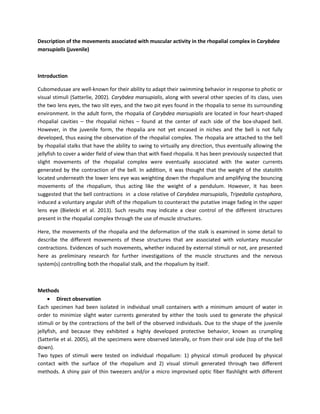
  • 3. Figure 1 Rhopalium and rhopalial stalk at resting position. The rhopalium hangs down (lower-lens eye (lle) facing downward), away from the center of the bell with an angle of 8- 10° relative to the aboral-oral axis. Figure 2 Representation of the inward bending process of the rhopalial stalk. In the resting position, the rhopalium hangs down and the rhopalial stalk remains straight (a). Three main stages can be observed throughout the bending process: The side of the stalk facing inward shortens (arrowhead), thus initiating the bending process (b); the outer side of the region of the stalk near the rhopalium-stalk junction is also bending inward (c); the stalk is finally bending along its entire length (arrowhead) at the final stage of the bending process(d). The rhopalium has moved to about 50° from the resting position. The rhopalium possesses two pit eyes (pe), two slit eyes (se), one upper lens eye (ule), one lower lens eye facing down in the resting position (lle), and a statolith (st).
  • 4. Figure 3 Example of a rhopalium of a juvenile medusae being moved by the rhopalial stalk bending inward. Note that at the resting position, the rhopalium points downward with an angle of about 8-10° relative to the aboral-oral axis. The total angular shift of the stalk is about 52° (n=3) (b) relative to the initial (at rest) axis of symmetry of the stalk (a). The solid white line in figure 3a) represents the axis of symmetry of the stalk originating from its base and extending to the rhopalium-stalk junction. The solid white line in figure 3b) depicts the final angular shift produced through the bending of the stalk. The white dashed lines emphasize the outline of the rhopalial stalk for better visualization of the shape of the stalk. Figure 4 Illustration of the inside-out bending process of the rhopalial stalk without additional bouncing of the rhopalium at the rhopalium-stalk junction. At the resting position, the rhopalia point downward (lower lens-eye down) and slightly toward the outside with an angle of about 8-10° relative to the aboral-oral axis. The stalk can bend inward with an angle of about 50° relative to the initial (at rest) axis of symmetry of the stalk originating from its base to the rhopalium-stalk junction. The rhopalium has moved to about 50° from the resting position. The rhopalium possesses two pit eyes (pe), two slit eyes (se), one upper lens eye (ule), one lower lens eye facing down in the resting position (lle), and a statolith (st). Lateral bending of the stalk has also been observed and no significant differences in the bending process have been identified compared to the inside-out bending process. However, unlike for the inside-out bending where the stalk can only bend toward the center of the bell, the stalk can move on either side of the axis formed by the relaxed rhopalial stalk. Lateral movements of the rhopalial complex observed in three juveniles indicated that the angular shift of the stalk from the resting position to either side was about 60° without any difference between the two sides. The total swing span of the stalk has been
  • 5. calculated to be 120° (figures 5 and 6). Additional bouncing of the rhopalia at the rhopalium-stalk junction allows the rhopalial complex to bend on either side at angles greater than 86°. Figure 5 Examples of the rhopalium of a juvenile medusae being moved by the rhopalial stalk bending from right to left. At the resting position, the rhopalial stalk is straight (b). The same four bending stages observed in the inside-out bending apply in the lateral bending process (a and c)). The shortening of one side of the stalk and the relaxation of the opposite side of this stalk allow the rhopalium to swing from either side of the initial (at rest) axis of symmetry of the stalk originating from its base to the rhopalium-stalk junction. The stalk can bend on both sides with an angle of about 60° relative to its axis of symmetry, thus allowing a total angle of freedom of about 120°. The black dashed lines emphasize the outline of the rhopalial stalk. Figure 6 Illustration of the lateral bending process of the rhopalial stalk without additional bouncing of the rhopalium at the rhopalium-stalk junction. The shortening of one side of the stalk (arrowhead) and the relaxation of the opposite side of this stalk allow the rhopalium to swing from either side of the initial (at rest) axis of symmetry of the stalk originating from its base and extending to the rhopalium-stalk junction. The stalk can bend on both sides with an angle of about 60° relative to its axis of symmetry, thus allowing a total angle of freedom of about 120°. These two movements suggest that the rhopalia can swing in any direction, allowing the rhopalium to eventually achieve a complete rotation around the axis of symmetry formed by the rhopalial stalk in the relaxed position (video).
  • 6. • Independent movements of the rhopalium itself Further tedious observations of the rhopalial complex indicated that the rhopalium had the ability to move independently from the stalk. Indeed, two types of movements associated with muscular activity were identified: 1) the rotation of the rhopalium at the center of rotation formed by the rhopalium-stalk junction and 2) a slight independent bouncing of the rhopalium at the same junction. In the resting position, the rhopalium is aligned with the stalk – upper-lens eye, lower-lens eye, and statolith being aligned with the stalk. The rhopalium has been observed to accomplish a maximum rotation of about 134° in total (figure 7). No evidences of a particular shortening or stiffening of the stalk as been identified, but more accurate observations need to be performed to establish the accuracy of these results. Figure 7 Example of the rotation process achieved by the rhopalium. The maximum observed rotation was equal to 134°. Note that image b) depicts the position of the rhopalium at rest. The white dashed arrows indicate the orientation of the rhopalium to emphasize the rotation (a, b, and c). Here, the rhopalium is viewed from behind, so only the statolith (st) and the rhopalium-stalk junction can be seen. The rhopalium also seemed to have the ability to bounce on the rhopalium-stalk junction independently from the stalk. Even though high definition recording would probably provide more accurate results regarding the independence of the rhopalium from the stalk, the preliminary results indicated that the rhopalium could bend on either side of the axis of symmetry formed by the stalk with an angle of 16°. The total angle shift from one side to the other was calculated to be about 32°. Such movement occur in any direction around the rhopalium-stalk junction (figure 8).
  • 7. Figure 8 Illustration of the independent bouncing of the rhopalia at the extremity of the rhopalial stalk. The rhopalia can bounce to about 16° degrees on either side relative to the initial position at rest without significant bending of the stalk. Figure 1a) represents the inside-out bouncing viewed from a side of the rhopalial complex and figure 1b) the lateral bouncing viewed from bellow. Note that the center of the bouncing movement is located at the rhopalium-stalk junction (black arrowhead). Discussion The immobilization of the individuals and the avoidance of undesirable slight water currents clearly helped to establish that rhopalial movements were not artifacts of random surrounding features, but were instead achieved through the use of muscle structures in the rhopalial stalk. Several different types of movements were examined in both the rhopalial stalk and the rhopalia. The most noticeable movements are performed by the rhopalial stalk. The ability of this structure to bend from the outside of the bell toward the inside, and to bend laterally along the edges of the box-shaped bell allows the rhopalia to cover a wider field of view. This is especially true when the rhopalia achieve complete rotations around the axis of symmetry formed by the stalk at rest. It has been observed that the bending process starts from the region of the stalk near the rhopalium-stalk junction. The bending process is initiated by the shortening of the side of the stalk facing the direction towards with the stalk bends. What has not been clearly determined is whether or not the opposite side of the stalk lengthens to ease the bending processes or if it bends through a passive relaxation of muscle structures. I addition, the causes of the return to the initial resting position of stalk, has yet to be determined. Muscle activity may be involved in that process but the natural rigidity of the stalk may eventually contribute to the return to the initial position of the rhopalial system – similarly to the mesoglea allowing the bell to relax. Such movements have been observed multiple times as responses to external stimuli such as the waving of the shiny tweezers in front of the rhopalia, but touching these structures has not proven to be efficient in observing a voluntary contraction of the stalk. The results obtained with the micro fiber-optic flashlight are not conclusive but further investigation may eventually provide more accurate results. Closer examination of the four rhopalia of the three juvenile individuals suggested that each rhopalium not only had the capacity to move independently from the three other rhopalia, but also that there is a certain degree of freedom between the rhopalium itself and the stalk to which it is attached. Indeed,
  • 8. this independent movement of the rhopalium may add sixteen additional degrees to the total angular ship exhibited by the rhopalial stalk. This eventually increase the field of view of each rhopalium without further applying significant stress on the rhopalial stalk. The rhopalium also have the ability to rotate at the center of rotation formed by the rhopalium-stalk junction. Such rotation mostly allows the re- orientation of the upper-lens eye toward either an object or a source of light. However, more information is needed regarding this mechanism. Indeed, the possible implication of mechanisms present in the stalk that would influence the rotation process still remains unclear due to a lack of both direct visual information and physiological knowledge about the rhopalial system. Little is known about the mechanics of rhopalial movements, but deeper analysis of the rhopalial structures may provide significant clues about the importance for juvenile Carybdea marsupialis to develop such rhopalial mobility. Since external stimuli delivered throughout the experiments successfully triggered a response, such mobility might be associated with a protective response of juvenile medusae against potential threats – to the same order as the crumbling response. References Bielecki, J., J.T. Høeg and A. Garm. 2013. Fixational Eye Movements in the Earliest Stage of Metazoan Evolution. PLoS ONE 8: e66442. Satterlie, R.A. 2002. Neuronal control of swimming in jellyfish: a comparative story. Can. J. Zoology 80: 1654-1669. Other readings scavenged on the web Garm, A. et al. 2006. Rhopalia are integrated parts of the central nervous system in box jellyfish. Cell. Tissue. Res.325: 333-343.