
Empfohlen
Weitere ähnliche Inhalte
Was ist angesagt?
Was ist angesagt? (20)
Ähnlich wie pressure ulcer.pptx
Ähnlich wie pressure ulcer.pptx (20)
Mehr von Nilofar Loladiya
Kürzlich hochgeladen
Kürzlich hochgeladen (20)
pressure ulcer.pptx
- 2. An ulcer is a discontinuity or break in a bodily membrane that impedes normal function of the affected organ. According to Robbins's pathology, "ulcer is the breach of the continuity of skin, epithelium or mucous membrane caused by sloughing out of inflamed necrotic tissue." WHAT IS AN ULCER? PRESSURE ULCER: Mrs Nilofar 2
- 3. WHAT IS A PRESSURE ULCER?
- 4. PRESSURE ULCER: Mrs Nilofar 4 WHY PRESSURE ULCER DEVELOPS?
- 5. PRESSURE ULCER: Mrs Nilofar 5 HOW PRESSURE ULCER DEVELOPS? Risk factors act on the soft tissues overlying the bony prominences This pressure exceeds normal capillary pressure Reduced tissue perfusion Ischemic Necrosis PRESSURE SORE Occlusion and tearing of small blood vessels
- 6. • A perpendicular load of force exerted on a unit of area (this could be a patients body weight bearing down on a hip or sacrum). • It causes local capillary occlusion (reduction in blood supply) and compresses the structures between the skin surface and bone. Pressure PRESSURE ULCER: Mrs Nilofar 6
- 7. • Pressure exerted by bony prominences on the body that stop capillary flow to the tissues. • Deprives tissues of oxygen and nutrients causing cell death. • The damage can often be caused under the skin, but not become obvious until the skin above it has broken down. PRESSURE ULCER: Mrs Nilofar 7
- 8. • This is where pushing or pulling the skin means more than one layer of skin slides against each other and • this can cause damage to these layers or they may become detached from each other all together. Shearing PRESSURE ULCER: Mrs Nilofar 8
- 9. Pressure and shear PRESSURE ULCER: Mrs Nilofar 9
- 10. This is where two surfaces rub together • Skin and bed sheets, or a chair cushion • poorly fitting clothing • manual handling aids. Hot, moist skin is likely to experience even more damage from friction than more healthy skin. Friction PRESSURE ULCER: Mrs Nilofar 10
- 11. PRESSURE ULCER: Mrs Nilofar 11 Pressure, shear and Friction
- 12. • A pressure ulcer is an ulcer related to some form of pressure and should not be confused with ulcers relating to disease (like cancer), vascular flow (venous or arterial) or neuropathy (like in persons with diabetes) • You should be able to see a “cause and effect” relating to pressure with the ulcer. – Redness or discoloration over a bony area related to sitting or lying – Redness or discoloration on the skin related to pressure from a device or a wheelchair pedal PRESSURE ULCER: Mrs Nilofar 12
- 13. How pressure injuries are diagnosed ? 13 Tools include skin visualization techniques and risk assessment tools The Braden Scale for Predicting Pressure Ulcer Risk, is a tool that was developed in 1987 by Barbara Braden and Nancy Bergstrom. The purpose of the scale is to assess a patient's risk of developing a pressure ulcer. The Norton risk-assessment scale which was published in 1962. The scale is used in the evaluation of pressure injury risk based on factors such as mobility or physical condition. PRESSURE ULCER: Mrs Nilofar
- 14. PRESSURE ULCER: Mrs Nilofar 14
- 15. PRESSURE ULCER: Mrs Nilofar 15
- 16. PRESSURE ULCER: Mrs Nilofar 16 Most Common Sites SUPINE POSITION
- 17. PRESSURE ULCER: Mrs Nilofar 17 Most Common Sites PRONE POSITION
- 18. PRESSURE ULCER: Mrs Nilofar 18 Most Common Sites LATERAL POSITION
- 19. PRESSURE ULCER: Mrs Nilofar 19 Most Common Sites
- 20. • Sacrum (tail bone)- 1st -Semi-fowlers’ position -Slouching in bed or chair -higher risk in tube fed or incontinent Most Common Sites PRESSURE ULCER: Mrs Nilofar 20 • Heels- 2nd -Immobile or numb legs -Leg traction -Higher risk with PVD & DM neuropathy • Trochanter (hip bone) -Side lying • Ischium (sitting erect bone) -highest risk paraplegics
- 21. Medical devices and equipment PRESSURE ULCER: Mrs Nilofar 21
- 22. STAGE 1: Non-blanching. • Grade 1: • Non-blanchable erythema of intact skin. • Discolouration of the skin, warmth, oedema, induration or hardness. PRESSURE ULCER: Mrs Nilofar 22
- 23. • Grey/purple hue to skin • Induration present • May be mushy or boggy instead • Often cannot visualize damage until top layers of skin sloughed PRESSURE ULCER: Mrs Nilofar 23 STAGE 1: Non-blanching.
- 24. • Grade 2: • Partial thickness skin loss • involving epidermis, dermis, or both. The ulcer is superficial and presents as an abrasion or blister. STAGE 2: Broken skin PRESSURE ULCER: Mrs Nilofar 24
- 25. PRESSURE ULCER: Mrs Nilofar 25 STAGE 2: Broken skin
- 26. • Grade 3: • Full thickness skin loss • involving damage to or necrosis of subcutaneous tissue that may extend down to, but not through, underlying fascia. STAGE 3: Sub-cutaneous involvement PRESSURE ULCER: Mrs Nilofar 26
- 27. PRESSURE ULCER: Mrs Nilofar 27 STAGE 3: Sub-cutaneous involvement
- 28. • Grade 4: • Extensive destruction, tissue necrosis or damage to muscle, bone of supporting structures, with or without full thickness skin loss. STAGE 4: Deep tissue involvement PRESSURE ULCER: Mrs Nilofar 28
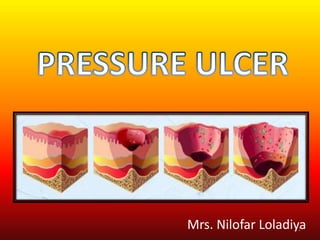
- 29. Pressure Ulcer Stages 29 Skin is intact but red. Skin is broken but there is no depth to the wound. Skin is broken but there is obvious depth to the wound, fat tissue may be noted. Skin is broken, muscle or bone may be visible. Severe tissue loss is noted, wound may appear as empty hole. PRESSURE ULCER: Mrs Nilofar Unstageable Stage 1 Stage 4 Stage 3 Stage 2
- 30. • Unusual changes in skin color or texture • Swelling • Pus-like draining • An area of skin that feels cooler or warmer to the touch than other areas • Tender areas 30 Signs and Symptoms of a pressure injury ANY Changes in skin appearance should always be reported. PRESSURE ULCER: Mrs Nilofar
- 31. PRESSURE ULCER: Mrs Nilofar 31
- 32. PRESSURE ULCER: Mrs Nilofar 32
- 33. PRESSURE ULCER: Mrs Nilofar 33 Age, nutritional and Hydration status, including weight, weight loss, and serum albumin levels, if indicated. history of pre-existing chronic diseases (e.g., diabetes mellitus, acquired immune deficiency syndrome, guillain-barré syndrome, peripheral and/or cardiovascular disease)., Cancer (history of radiation therapy) Client’s awareness of the sensation of pressure. Assess for fecal and urinary incontinence. Assess client’s ability to move (shift weight while sitting, turn over in bed, move from the bed to a chair). Assess for environmental moisture (excessive perspiration, high humidity, wound drainage). Post-OP status
- 34. PRESSURE ULCER: Mrs Nilofar 34 Assess the amount of shear (pressure exerted laterally) and friction (rubbing) on the client’s skin. Assess the surface that the clients spend a majority of time on (mattress for bedridden clients, cushion for clients in wheelchairs). skin over bony prominences (sacrum, trochanters, scapulae, elbows, heels, inner and outer malleolus, inner and outer knees, back of head). tool for pressure ulcer risk assessment: Braden/Norton scale. Assess the client’s level of pain, Assess and stage the pressure ulcers: Measure the size of the ulcer, and note the presence of undermining. Assess the condition of wound edges and surrounding tissue.
- 35. PRESSURE ULCER: Mrs Nilofar 35 Nursing Diagnosis Impaired Skin Integrity Possibly evidenced by Destruction of skin layers. Disruption of skin surfaces. Drainage of pus. Invasion of body structures. Pressure ulcer stages May be related to Chronic disease state. Extreme of ages. Imbalanced nutritional state. Impaired sensation. Immobility. Immunological deficit. Incontinence. Mechanical factors (friction, pressure, shear). Moisture. Poor circulation. Pronounced body prominence. Radiation.
- 36. PRESSURE ULCER: Mrs Nilofar 36 DESIRED OUTCOMES • Client will experience healing of pressure ulcers • Experiences pressure reduction. • Absence of signs of inflammation GOAL SETTING • SHORT TERM: Client will get stage-appropriate wound care • LONG TERM: Control risk factors for prevention of additional ulcers in future
- 37. PRESSURE ULCER: Mrs Nilofar 37 SKIN INSPECTION WOUND CARE PATIENT/FAMILY INVOLVEMENT: SKIN CARE POSITIONING COMFORT DEVICES TREATMENT SPECIAL NEEDS NUTRITION
- 38. • Use the daily check charts to record on a daily basis that every area has been checked: BRADEN/NORTON Scale • If there is a pressure ulcer grade it accordingly. PRESSURE ULCER: Mrs Nilofar 38 SKIN INSPECTION
- 39. • Wound care may be required based on severity • Any patient with a pressure ulcer which is grade 2 or higher should have a wound care plan. • Dates and times should be set for the evaluation of pressure ulcer and wound care plans so that regular updates can take place. PRESSURE ULCER: Mrs Nilofar 39 WOUND CARE
- 40. Encourage patients to maintain their NUTRITION: – Meat, fish, or Protien alternatives. – Fruit and vegetables. (Vit C) – Milk and dairy products. – Plenty of fluids stop the skin becoming dehydrated and can reduce the risk of ulceration. LIFE STYLE CHANGES: SMOKING EXERCISE CHECK SKIN at least once daily, or ask a carer to help. A mirror will help to see hard-to-reach areas. Attend especially to those areas where pressure is heaviest. PATIENT/FAMILY INVOLVEMENT: PRESSURE ULCER: Mrs Nilofar 40
- 41. • Avoid massaging bony parts of the body. This can cause addition damage to skin which may already be delicate. • Bed sheets should have no creases. • Use warm (not too hot) water and mild soap to cleanse. Use a moisturiser to avoid dry skin, and avoid cold or dry air. • Skin should be cleansed as soon as Soiled. Using a soft cloth or sponge should reduce friction. • Control moisture-skin should be kept clean and dry (sweat). PRESSURE ULCER: Mrs Nilofar 41 SKIN CARE
- 42. • A patient who is unable to reposition themselves MUST have a repositioning plan. • Plan on 2 hourly repositioning day and night. Include 30° tilt on bed rest. • Repositioning regimes need to: – Minimise prolonged pressure on bony prominences. – Specify that repositioning takes place regularly – even with pressure-relieving devices in situ. – Establish a means of recording when this repositioning takes place PRESSURE ULCER: Mrs Nilofar 42 POSITIONING
- 43. Do not use ring cushions as these increase rather than reduce pressure. PRESSURE ULCER: Mrs Nilofar 43 COMFORT DEVICES
- 44. PRESSURE ULCER: Mrs Nilofar 44 Pain management-pressure injuries are painful and may require oral medication for pain, especially around treatment times. Barrier ointments should be used after incontinence episodes to protect skin. Oral antibiotics Surgical repair: Tissue Flap, Plastic Surgery Debridement Hyperbaric Oxygen Treatment Topical Human growth factors TREATMENT
- 45. PRESSURE ULCER: Mrs Nilofar 45
- 46. PRESSURE ULCER: Mrs Nilofar 46 Minimize pressure by utilizing edges, frequent turning, and repositioning ever 2 hours. Minimize shear and friction to reduce the damage to tissue. Avoid dragging; causes friction and increases risk for skin damage
- 47. Case Study- Meet John 47 PRESSURE ULCER: Mrs Nilofar • John is diagnosed with Cerebral Palsy, GERD, Diabetes insulin dependent, and chronic constipation. He is verbal, friendly and has a good sense of humor. He really loves going to his day program. • John has slow movements and requires assistance to stand and pivot. If lying in the bed he can roll from side to side using the bed rails.
- 48. 48 • When John becomes upset, he will wiggle out of his wheelchair onto the floor when staff are not looking. On the floor he refuses to let the staff help him back into his wheelchair. • Today, John became very agitated when he could not go to his day program. John wiggled out of his wheelchair onto the floor. Staff tried to get him up, but he would slap at them and try to spit in their face. Finally, the staff gave up and told John to let them know when he was ready to get back in his chair. • John sat in the floor for 3 hours. Finally, he asked for help and staff put him in on the bed. HIs clothes were wet due to incontinence. As staff provided hygiene and put on dry clothes, they noticed that John had two areas of red skin: one on his right cheek and knee. PRESSURE ULCER: Mrs Nilofar
- 49. 49 Incontinent of Urine and/or stool (wears briefs) Requires DME for mobility Spends 4.5hours or more a day in wheelchair Fragile skin Edema related to Congestive Heart Failure, and /or Peripheral Artery Disease. Recent change in weight loss or gain Obesity Anorexia (Prader Willi's) Behavior plan in place addressing putting self in risky situations (Refusing help off floor) Unable to change body position. (Cerebral Palsy, Stroke) PRESSURE ULCER: Mrs Nilofar
- 50. Apply what you've learned Name three interventions that could have prevented John from developing pressure injury: 1.____________________ _______ 2.____________________ _______ 3.____________________ _______ 50 PRESSURE ULCER: Mrs Nilofar
Hinweis der Redaktion
- Pressure injuries are identified through a staging process where the injury is described and classified based on the extent of tissue damage (Edsberg et al, 2016).
- Must palpate Rare photo of damage-usually cannot see the difference in color
- Once identification of a pressure injury has occurred, the next step is commonly called ‘staging’. Staging is when a healthcare professional (physician, nurse, certified wound specialist) examines the skin at and around the site of the injury. The healthcare provider will then determine, based on the criteria listed below, the severity of the injury (National Pressure Injury Advisory Panel, 2019). Matching Activity Handout