1. STRUCTURE AND MEMBRANE LOCATION OF PH-LOW INSERTION PEPTIDE
INTRODUCTION
The pH-low insertion peptide (pHLIP) have shown
themselves as potential biomarkers and drug delivery
scaffolds for tumor cells. Under physiological pH, pHLIPs
are able to bind themselves to a lipid bilayer, and
furthermore, when presented in acidic environments, they
are able to further insert to the interior of a lipid bilayer,
as it is shown in Figure 1. There is a lack of knowledge of
the physical structure of the peptide, thus impeding
further potential research in applications of pHLIP. By
utilizing spectroscopic measurements such as solid-state
nuclear magnetic resonance (ssNMR) spectroscopy and
fluorescence spectroscopy, it is possible to further identify
the structure of the peptide in the surface-binding and
membrane-inserted states.
Nicolas Shu, Michael Chung, Dr. Lan Yao, Dr. Ming An, and Dr. Wei Qiang
Department of Chemistry, Binghamton University, State University of New York. Vestal, New York
METHODS and RESULTS
Solid-state NMR samples were prepared with relative
high peptide-to-lipid molar ratio (i.e. 1:75). To ensure that
the pHLIPs have normal pH-regulated membrane insertion
behavior, we prepared samples with “stepwise addition”
protocols where the peptides were added into liposomes
in portions. Sufficient amount of time was allowed
between each portion to ensure membrane binding. Figure
2 showed that the NMR samples with 1:75 peptide-to-
lipid ratio behaved normally as the samples prepared
with 1:300 ratio.
ACKNOWLEDGEMENTS
This work is supported by the Research Foundation of
SUNY and the Chemistry Department of BU, as well as the
NSF Major Instrumentation Grant. We appreciate the help
from Mr. Matthew Siano.
Figure 1 Illustration of the three states of pHLIPs. (State I) pHLIP in
aqueous buffer. (State II) pHLIP bound to membrane surface. (State III)
pHLIP inserts into the interior of membrane bilayer at pH below a
critical value. It is known that the pHLIP inserts into membrane with its C-
terminus.
Figure 2 Biophysical characterization of ssNMR samples. (A) Circular
dichroism (CD) spectra for NMR samples at pH 7.4 (State II), 5.3 (State
III) and pH 6.4 (intermediate). (B) Traces of Trp fluorescence for
samples at pH 7.9 (red), 7.4 (blue), 6.3 (green), 6.1 (orange) and 5.7
(black). Trp fluorescence shows blue shift upon membrane insertion. (C)
Plots of the peak wavelengths of Trp fluorescence traces vs. pH. The
transition at pH 6.1 shows the critical pH of pHLIP insertion.
Solid-state NMR measurements shows that there is a single
set of helical 13C chemical shifts for residues A10, A13,
L21, L22, L26 and A27 at pH 5.3, which indicated a well-
defined structure for the inserted sate. At pH 7.4, only
weak signal with non-helical conformation was detected for
A10 and A13. At pH 6.4, there seems to be a mixture of
secondary structures, which suggested the co-existence of
multiple populations of pHLIP (Fig. 3). Analysis of
secondary structures showed dominant a-helix state III, non-
helices for state II and mixture of the intermediate pH
value (Fig. 4).
Figure 3 Representative 13C/13C 2D solid-state NMR spectra for (A) pH 5.3, (B) pH 6.4 and (C) pH 7.4.
Multiple Cα/Cβ crosspeaks were shown in panel (B) for the residue A13. For comparison, the position of
crosspeaks from a different pH value were shown as colored crosses.
Figure 4 Plots of
secondary chemical
shits for (A) pH 5.3,
(B) pH 6.4 and (C)
pH 7.4. Typical
helices have positive
Cα and CO and
negative Cβ.
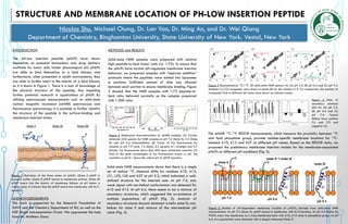
The ssNMR 13C-31P REDOR measurements, which measure the proximity between 13C
and lipid phosphate group, provide residue-specific membrane locations for 13C-
labeled A10, A13 and A27 at different pH values. Based on the REDOR data, we
proposed the preliminary membrane insertion models for the membrane-associated
pHLIPs at different pH conditions (Fig. 5).
Figure 5 Models of pH-dependent membrane location of pHLIPs, derived from solid-state NMR
measurements. At pH 7.4 (State II), pHLIP bound to membrane with its N-terminus. At pH 5.3 (State III),
PHLIPs insert into membrane as a trans-membrane helix with A10, A13 close to phosphate group. At pH
6.4, two populations were detected with a deeply immersed State II’.