SlideShare a Scribd company logo
1 of 11
Download to read offline
180 Nanomed J, Vol. 1, No. 3, Spring 2014
Received: Sep. 19, 2013; Accepted: Nov. 12, 2013
Vol. 1, No. 3, Spring 2014, page 180-190
Received: Apr. 22, 2014; Accepted: Jul. 12, 2014
Vol. 1, No. 5, Autumn 2014, page 298-301
Online ISSN 2322-5904
http://nmj.mums.ac.ir
Original Research
Comparison of MnO2 nanoparticles and microparticles distribution in CNS
and muscle and effect on acute pain threshold in rats
Nahid Nosrati1,2
, Majid Hassanpour-Ezzati1*
, Sayyed Zahra Mousavi2
, Mohammad
safi Rahmanifar1
, Shiva Rezagholiyan2
1
Department of Biology, Basic Sciences School, Shahed University, Tehran, Iran
2
Department of Pharmacology, Faculty of Pharmaceutical Sciences, Islamic Azad University, Tehran, Iran
Abstract
Objective(s): Recently, applications of MnO2 nanoparticles and microparticles in industry,
pharmacology, and medicine have considerably expanded. Mn distribution and clearance
from brain and spinal cord tissue compared with muscle tissue of rats after single
subcutaneous injection of nanoparticles and microparticle of MnO2. Pain sensory threshold of
rat was evaluated as neurologic consequence of the particles on CNS activity of rats.
Materials and Methods: Rats divided to control and two experimental groups. Each
experimental group received a single subcutaneous injection of MnO2 nano- and
microparticles, respectively. Acute pain thresholds of rats were evaluated by tail immersion
method and its weight gain was recorded during these weeks. Samples taken from brain,
spinal cord and muscle tissues of rats, once every 2 week for 8 weeks. The tissue Mn level
was measured by inductively coupled plasma-mass spectrometry method.
Results: Both particles size passed from blood barriers. Unlike brain tissue, manganese
completely cleared from spinal tissue after 8 weeks in both groups. Clearance of Mn from
muscle tissue is not complete in both of the groups. Weight gain of rats in both groups was
slower than control rats. In microparticle group, rats showed progressive analgesia (p<0.05).
In nanoparticle groups, rats showed hyperalgesia for first 4 weeks and analgesia during
remaining weeks.
Conclusion: Change in MnO2 particles size affect on Mn distribution and clearance from
central nervous system. Effect of particles on whole body metabolism varied with its size too.
Finally, comparison of pain response of rats among particle treated groups indicates that
neurobiological mechanism affected by particles is varied with their size during times after
administration.
Keywords: Biodistribution, Clearance, MnO2, Nanoparticles, Pain
*Corresponding author: Majid Hassanpour-Ezzati, Shahed University, Opposite Holy Shrine of Imam
Khomeini, Khalij Fars Expressway, Tehran, Iran.
Tel: +982151212242, Email: hassanpour@shahed.ac.ir
M H E and S Z M equally designed research
Size dependent effect of MnO2 on acute pain threshold in rat
Nanomed J, Vol. 1, No. 3, Spring 2014 181
Introduction
The use of metal oxide nanoparticles is
currently developed in all industries; from
computer to perfume manufacturing (1).
Currently, microparticles and
nanoparticles of manganese dioxide
(MnO2) application in industry, medicine,
and pharmacology is increased and
therefore human exposure with these
particles augmented. The rate of
contamination with MnO2 in environments
is higher than other forms of Mn because
of MnO2 use as a substrate for synthesis of
other Mn-containing compounds (2). Also,
in comparison with other forms of Mn
particles, MnO2 nanoparticles have a
higher oxidation power (3). Steel casting
and dry battery industries are two major
industries in which workers are in contact
with MnO2 nanoparticles. Besides, due to
the use of MnO2 nanoparticles in
composition of methylcyclopentadienyl
manganese tricarbonyl (MMT) as a
gasoline additive, all people that live in big
cities are at risk of its toxic effects (4).
This nanoparticle is also used in MRI
contrast dye (5). Most of the previous
information about MnO2 neurotoxicity is
restricted to its chronic effects (6, 7). The
high oxidative activity of these particles
can cause stimulation of pathological
mechanisms and induce adverse effects on
cell function, like autophagy, induction of
cell apoptosis (8, 9). Indeed, it is reported
that changes in particle size from
micrometer to nanometer can augment the
oxidation power of the particles. This
enhancement of oxidative properties of
particles increased the probability of
undesirable acute effects of nanoparticles
on living organisms. The syndrome
resulting from chronic exposure to
manganese particles in industrial workers
is known as manganism and is similar to
Parkinson’s disease (10). This finding
indicates that dopaminergic neurons are
one of the main targets of MnO2 particles
in CNS. Recent findings show a
relationship between intensity of metal
nanoparticles toxicity with their
concentration and size (11). Therefore, the
information about size-dependent
biodistribution, clearance, and CNS effects
of these compounds has practical
importance for further applications of
these particles (12). Kim and colleagues
reported that the application of a certain
dose of MnO2 nanoparticles in a special
size does not have any toxic effects on
laboratory animals (13). Contrary, others
reported that topical administrations of
MnO2 particles with a diameter less than 2
micrometers to monkeys for one day up to
10 months caused wide distribution of Mn
in the nervous system and appearance of
some neurological symptoms (14). Also, a
linear relation between concentration of
MnO2 nanoparticles and production of
reactive oxygen substances (ROS)
compounds in Hela cells was demonstrated
(15). The oxidative power and easily
passing of nanoparticles through blood-
brain barrier can induce undesirable acute
effects on sensory, motor, and cognitive
aspects of CNS functions (16). It has been
shown that some metal nanoparticles
produced some neural dysfunction even
after single exposure (17). In contrary,
administration of other nanoparticles has
no toxic effects on the function of nervous
system in animals (18). Therefore,
particles on the base of concentration and
size have unique neurotoxic effect and
must be studied separately. ICP-MS
provides a high quality method for
measurement of metal particles
concentration in various tissues; especially
in brain tissues (19). Tail immersion is a
method for assessment of animal pain
threshold. The efficacy of this method is
shown in assessment of pain threshold
changes with respect to dopaminergic
system manipulation (20). In this research,
the Mn level in rat brain, spinal cord, and
muscle tissues were measured once 2
weeks for eight weeks by ICP-MS after
single subcutaneous injections (100 µg/kg)
of MnO2 micro and nanoparticles for
pharmacokinetic comparison of these two
particles. Also, body weight gain of rats
Nosrati N, et al
182 Nanomed J, Vol. 1, No. 3, Spring 2014
during this period was compared between
MnO2 microparticles and nanoparticles
treated groups. Also, pain thresholds of
rats in the two groups were determined by
tail immersion methods during these
weeks for comparison of acute
neurological effects of these particles.
Materials and Methods
Animals
Male albino Wistar rats (Pasteur’s
institute, Tehran, Iran) weighing 150±5 g
were housed in an air-conditioned colony
room on a light/dark cycle (21-23°C and a
humidity of 30-40%) and supplied with
standard diet and tap water ad libitum.
Procedures involving animals and their
care were conducted in conformity with
the NIH guidelines for the care and use of
laboratory animals.
Selecting the dose
The dose of MnO2 nanoparticles and
microparticles selected on the basis of You
and coworkers report (21). Rats were
divided into two groups and treated
subcutaneously with solution containing
nano- and microparticles of MnO2 (100
µg/kg) dissolved in saline, respectively.
Control rats only received normal saline.
MnO2 microparticles (Figure 1) used in
this research were purchased from
MERCK Company, Germany.
Figure 1. A scanning electromicrograph of MnO2
micropaticles.
The MnO2 nanoparticles preparation
MnO2 nanoparticles were prepared via the
hydrothermal procedure proposed by
Zhang et al., with some modification (22).
In practice, 20 ml of KMnO4 (0.2 mM/lit)
were mixed with 16 ml MnO4 (0.125
mM/lit) for 5 minutes. The resulting
mixture was taken directly into a steel
autoclave with Teflon covering and kept
for 16 hours at 160C and then was cooled
at room temperature. The resulting brown
product was collected, washed with
distilled water and ethanol 3 times, and
dried with hot air current 80C for 12
hours. The resulting particles were
scrutinized by an electron microscope and
it was ensured that they were 25 to 85
micrometers in size (Figure 2). Preparing
MnO2 nanoparticles and microparticles
injecting solution.
First, the particles were dissolved in
normal saline solution. The prepared
solutions were sonicated for 30 minutes
before injection.
Figure 2. Scanning electromicrograph of MnO2
nanopaticles.
Tissue Samples Preparation for ICP/MS
measurement
Male Wistar rats (140 ±10 g) were used in
this experiment. Rats were divided into
three groups: control, microparticle, and
nanoparticle groups. The experimental
groups injected subcutaneously with
solution containing MnO2 (100 µg/kg)
microparticles or nanoparticles,
respectively.
Control rats received only normal saline.
Five rats were chose from each group
every two weeks and were deeply
anesthetized with diethyl-ether (Merck).
Size dependent effect of MnO2 on acute pain threshold in rat
Nanomed J, Vol. 1, No. 3, Spring 2014 183
Then, rats scarified and their brains
transformed into ice-cold ACSF solution
(4C). The spinal cords of rats were
pushed from vertebral canal with perfusion
of high-pressure of cold ACSF solution
into the vertebral canal. Muscle tissue
sample was taken from hind paw of rats
(squadriceps femoris).
All samples were frozen with liquid
nitrogen after weighting and kept in a -
20C until ICP-MS measurement.
The tissue Mn was extracted by the
following procedure: 1/2 gram of each
tissue sample was selected and one ml of
HNO3 %63 (Merck, Germany) was added
to it (23). Then, the samples were kept for
24 hours at 4C for complete digestion of
the tissues. The digested samples were
centrifuged at 4500 rpm for 5 minutes. The
upper clear solution was diluted with
extra-pure water and the MnO2 level was
measured by ICP-MS (24).
The ICP-MS measurement of tissue Mn
level
A standard Agilent 7500 ICP-MS, fitted
with a crossflow nebulizer and a quartz
spray chamber was used throughout (25).
Operational parameters are given in Table
1. A solution containing 100 PPM of Mn
(Merck) was used as the standard solution.
Table 1. Operational parameters of ICP/MS for
measurement of Mn level in liver tissue extract.
Parameter Value Units
Argon Gas Flow Rates
Plasma 15.0 L/min
Auxiliary 1.0 L/min
Carrier 1.2 L/min
RF Power 1350 W
Sampling Depth 6.7 mm
Nebulizer Cross-flow
Spray Chamber Double-pass,
quartz
Spray Chamber Temp. 2 °C
Sample Uptake 0.4 ml/min
Tail immersion test
The rat pain threshold was assessed using
tail immersion test. After the adaptation,
rat tail was immersed in warm water
(49°C) and the tail flick response latency
(withdrawal response of tail) was observed
as the end-point response.
Each experiment was repeated 4 times for
each animal with an interval of 2 min and
its average was reported. Cut-off time of
25 sec was considered (26, 27).
Results
Tissue Mn level
The basal level of Mn in brain, spinal cord,
and muscle tissues of control rats is 1
(µg/g tissue wet weight). The maximum
content of Mn in brain tissue of
microparticles group is higher than
nanoparticles group.
In MnO2 microparticles group, Mn level
in the brain tissues reached to its
maximum level 4 weeks after injection
(Figure 3).
Then, the Mn level in this tissue returned
to 77.5% its maximal value after 8 weeks.
In MnO2 nanoparticles group, Mn level in
brain tissue was reached to its maximum
level 2 weeks after injection and its level
reduced to 42% its maximum valve after 8
weeks.Variation of Mn level in spinal cord
tissues during 8 weeks after the injection
of particles is shown in Figure 4.
Mn concentration in spinal cord tissues
following the injection of MnO2
microparticles attained its maximum level
after 4 weeks, but this time for the
nanoparticles receiving group was 2
weeks.
The variation of Mn level in muscle tissues
during 8 weeks after the injection of MnO2
microparticles and nanoparticles is shown
in figure 5. In the MnO2 microparticle
groups, Mn level in muscle tissue increase
very slowly and reach to a plateau after 2th
week.
But in MnO2 nanoparticle group, the tissue
level of Mn increased very fast and
reached to high level in second weeks after
this particle injection.
Nosrati N, et al
184 Nanomed J, Vol. 1, No. 3, Spring 2014
But the tissue Mn level quickly decreased
to a basal level at 4th week and remained
stable till the end of experiment.
Figure 3. Change in Mn level in brain tissues of rat
during 8 weeks after single treatment with MnO2
(100µg/kg) microparticles and nanoparticles.
Groups compared by ANOVA and tukey post-test,
*p<0.05 (n=5).
Figure 4. Comparison of Mn level in spinal cord
tissues of rat (n=5) in MnO2 microparticles or
nanoparticles (100 µg/kg) treated groups during 8
weeks. ANOVA and tukey post-test showed a
significant (p<0.05) difference in Mn tissues level
in between groups.
Weight gain changes
The rate of weight gain in rats in
nanoparticle group in comparison with
control rats significantly (p<0.05) reduced
during 2th to 4th week after injection
(Figure 6).
Weight gain of rats receiving the same
dose of microparticles only at the 4th
weeks was significantly (p<0.05) lower
than control rats (Figure 6).
Tail immersion measurement of acute
pain
The results of tail withdrawal latency of
rats in groups during 8 weeks after
injections are shown in Figure 7.
Figure 5. Change in Mn level in muscle tissues of
rats (n=5) in MnO2 microparticles or nanoparticles
(100µg/kg) groups during 8 weeks after injection.
The ANOVA and Tukey post-test show a
significant difference (p<0.05) between two
groups.
Figure 6. Rat weight gain changes during 8 weeks
after single subcutaneous injections of MnO2
microparticles or nanoparticles (100 µg/kg) in
comparison with control rats (n=5). *control vs.
nanoparticle groups, # control vs. microparticle
groups (p<0.05).
The mean tail withdrawal latency of rats
before the particles injections was 10
seconds.
The cut-off time was chosen 25-second.
The microparticles injection caused a
significant (p<0.05) progressive increase
in tail flick latency of rats during 8 weeks.
This progressive increase in tail filck
latency of rats continued to end of
experiment.
The rats treated with MnO2 nanoparticles
showed a significant (p<0.05) hyperalgesia
in the first four weeks after injection and
analgesia (p<0.05) during the remaining
weeks (Figure 7).
Size dependent effect of MnO2 on acute pain threshold in rat
Nanomed J, Vol. 1, No. 3, Spring 2014 185
Figure 7. Tail flick latency of rats (n=5) following
single subcutaneous injections of MnO2
microparticles or nanoparticles (100 µg/kg) in
comparison with the control group. *Control vs.
experimental groups (p<0.05).
Discussion
Our results showed that basal amount of
Mn in normal rat brain is very low.
Previous research also has shown presence
of very small amount of Mn in brain tissue
of human and other animals (28, 29). The
Mn level in brain tissue of control rats in
this study was proportional with its level
in previous studies (30). Measurement of
Mn level in brain and spinal cord of rats
following administrations of MnO2 micro
and nanoparticles indicate that both types
of particles can pass through blood barrier
and entered to spinal cord. Previous
research has shown Mn can enter and
distributed in brain through olfactory bulb
(31). It is shown that water soluble and
insoluble forms of manganese particles can
easily pass through the BBB (32). In this
study, Mn clearance rate from brain and
spinal cord is not the same for MnO2 nano-
and microparticles receiving groups. The
spinal cord tissues were completely
cleared from Mn after 8 weeks in both
treated groups but brain tissues were not
completely cleared from Mn during this
time. Previously, it was demonstrated that
bioclearance of nanoparticles varies
among different tissues; even in tissues
have not blood barriers (33).
The Mn clearance rate from brain tissues
in microparticle group is faster than
nanoparticles group, but Mn clearance rate
from spinal cord tissues was the same for
nanoparticles and microparticles treated
groups. Sharma and colleagues evaluated
permeability range of blood and spinal
cord barrier in rats after hyperthermia
stimulation with the use of nanoparticles.
Their findings suggest that blood-spinal
cord barrier permeability variation is more
than blood-brain barrier (34).
This report confirms our observation about
difference of nanoparticles clearance rates
from brain and spinal cord. Previously, it
is reported that Mn distributed to all areas
of rat brain after intracerebroventricular
administration of MnO2 nanoparticles
within three weeks and then concentration
of Mn in brain tissue decreased gradually
(35, 36).
The time course of this report is equal to
time that obtained in our study for
reaching of Mn level in brain tissue to
maximum value after MnO2 microparticle
administration. Size-dependent differences
in tissue bioclearance of particles were
shown previously, for example,
comparison of fluorescent polystyrene
bioclearance microparticles (2
micrometers) with its nanoparticles (200
nanometers) after subconjunctival
injections in rats showed that most of
nanoparticles disappeared from the
periocular tissue during 15 days and only
%8 of their initial amount remained in
tissues up to 7 days after the injection, but
micro particles stayed in the injected tissue
for more than two months (37). In
nanoparticle group, the Mn slowly
accumulated in muscle tissues and reached
to plateau at fourth week. In micro
particles group, the concentration of Mn in
muscle tissue show an initial rise in the
second week and then returned to a lower
level and stayed at this level till the end of
study.A similar situation was demonstrated
for clearance of gold nanoparticles from
liver and spleen tissues of rats (38).
Thus, the accumulation and clearance
pattern of Mn in each tissue special pattern
and also changed with particles size. It
should be noted that weight gain changes
was measured as indicator of general
Nosrati N, et al
186 Nanomed J, Vol. 1, No. 3, Spring 2014
health status of animals.
The reduction in weight gain of
microparticle receiving rats continued for
two weeks after the injection, while this
effect in nanoparticle-receiving group was
longer and continued for four weeks after
the injection. Our results show a relation
between tissues level of Mn and weight
changes of rats. Little information was
available about the acute effect of metal
oxide nanoparticles on weight changes of
animals and most of the researchers have
studied the effects of chronic
administration of metal oxide particles on
the weight changes of animals. Besides,
the effect of nanoparticles on weight gain
changes of animals varies from one metal
oxide nanoparticle to another. For
example, intraperitoneal injections of TiO2
nanoparticles for 3-month to rats have not
any significant effects on its weight gain
(39). On the contrary, inhalation of Fe2O3
nanoparticles significantly affects on rat
weight gain (40).
Scientists related these differences in
nanoparticles effect on body weight to
their oxidative power (41). In support of
this statement, it is shown that manganese
oxides can cause impaired mitochondrial
redox activity (18). MnO2 oxidative power
is higher than other form of Mn oxides,
such as Mn3O4 (42).
Therefore, our results indicate MnO2
particles in both size can influence on the
whole body metabolism in rats, moreover
the effect of the nanoparticle is slightly
higher than the microparticles.
Tail flick latency of rats was evaluated in
order to compare effects of the particles on
nervous system activity (43).
Scientists previously are used tail
immersion latency test for evaluation of
analgesia duration after subcutaneously
injected nanoparticles (44).
Tail immersion test considered as a model
of acute thermal pain (45). Acute pain
results from disease, inflammation, or
injury to tissue.
Inflammatory processes contribute to
nerve cell toxicity (46). Our results
indicate that both size of MnO2 particles
influenced on pain thresholds of rats.
The administration of microparticles led to
a gradual, irreversible increase in the pain
thresholds of rats during eight weeks.
However, nanoparticles produced biphasic
response in rat, initial hyperalgesia for four
weeks and late analgesic during last four
weeks. On the other hand it has been
reported that inhalation of MnO2 particles
caused changes in the neurochemistry of
brain dopaminergic system (47) and
severely affect on expression of genes like
MAOA, Park2, and SNCA (48).
Dopamine also plays a clear role in pain
control at the spinal cord level (49, 50) and
dysfunctions in this system in spinal cord
level can produce deficits in pain sensation
(51). Patients with Parkinson's disease
showed a progressive hyperalgesia similar
to rats treated the MnO2 micro particles in
our study (52, 53).
As a conclusion we can say that MnO2
particles effects on acute pain threshold
probably mediated via dopaminergic
systems and progressive nature of their
effect is similar to Parkinson's disease. As
explanation for biphasic effect of MnO2
nanoparticles on rat pain responses and
according to Wang’s finding, a direct
relation exists between the level of Mn
nanoparticles accumulation in central
nervous system and its neurological
symptoms (54).
Tissue Mn concentration increased
considerably after nanoparticle injection
during the first four weeks and then
decreased and remained constant during
the remaining ones.
Thus, it seems that the neural mechanisms
that modulated by MnO2 nanoparticles in
the first four weeks after injection are
different from late-phase mechanisms.
It is shown that manganese ions can
interrupt the transmission of stimulatory
signals in the nervous system through
changes in the activity of calcium channels
at pre-synaptic terminals (26).
Also, neuronal evoked potentials of rats
showed a significant reduction after long-
Size dependent effect of MnO2 on acute pain threshold in rat
Nanomed J, Vol. 1, No. 3, Spring 2014 187
term inhalation of MnO2 nanoparticles
(55).
The reduction in activity of descending
pain modulation tract can induce
hyperalgesia (56).
Thus, it seems that during first four weeks
after injection, nanoparticles activate
central descending mechanisms that
induced hyperalgesia in rats.
Also these mechanisms can mask the
MnO2 nanoparticle analgesic effects in this
phase.
But, at the end of first phase and
termination of these fast effects, the long
term analgesic effect of nanoparticles via
effect on dopaminergic system is
appeared.
In support of our hypothesis that central
dopaminergic system that mediate
analgesic effect of MnO2 nanoparticles, it
is reported that depletion of dopaminergic
neurons in periaqueductal grey areas of rat
brain can induce analgesia (20).
Conclusions
Administrations of microparticles or
nanoparticles of MnO2 caused Mn
accumulation in brain, spinal cord and
muscle tissues of rats after single injection
for long time. Both sizes of MnO2 have
adverse effects on general metabolism of
rats and reduce normal rate of rats weight
gain.
The MnO2 nanoparticle or microparticle
has different effect on pain sensation.
Probably, change in Mn level in CNS with
effect on dopaminergic system mediates its
effects on pain modulating systems of rats.
Acknowledgment
This work was supported by Islamic Azad
University, Tehran, Iran and contribution
of Shahed University of Iran.
We also thanks from Dr Hassani in
ICP/MS lab of Tarbiat Modares University
for his helps.
References
1. Allaker RP, Ren G. Potential impact of
nanotechnology on the control of
infectious diseases. Trans R Soc Trop
Med Hyg. 2008; 102(1): 1-2.
2. Prasad B, Mondal KK. Environmental
impact of manganese due to its leaching
from coal fly ash. J Environ Sci Eng.
2009; 51(1): 27-32.
3. Najafpour MM, Rahimi F, Aro EM, Lee
CH, Allakhverdiev SI. Nano-sized
manganese oxides as biomimetic catalysts
for water oxidation in artificial
photosynthesis: a review. J R Soc
Interface. 2012; 9(75): 2383-2395.
4. Davis CD, Wolf TL, Greger JL. Varying
levels of manganese and iron affect
absorption and gut endogenous losses of
manganese by rats. J Nutr. 1992; 122(6):
1300-1308.
5. Na HB, Lee JH, An K, Park YI, Park M,
Lee IS, et al. Development of a T-1
contrast agent for magnetic resonance
imaging using MnO nanoparticles. Angew
Chem Int Ed Engl. 2007; 46(28): 5397-
5401.
6. Roels H, Meiers G, Delos M, Ortega I,
Lauwerys R, Buchet JP, et al. Lison,
Influence of the route of administration
and the chemical form (MnCl2, MnO2) on
the absorption and cerebral distribution of
manganese in rats. Arch Toxicol. 1997;
71(4): 223-230.
7. Oszlánczi G, Vezér T, Sárközi L, Horváth
E, Kónya Z, Papp A. Functional
neurotoxicity of Mn-containing
nanoparticles in rats. Ecotoxicol Environ
Saf. 2010; 73(8): 2004-2009.
8. Wang SH, Shih YL, Kuo TC, Ko WC,
Shih CM. Cadmium toxicity toward
autophagy through ROS-activatied GSK-
3beta in mesangial cells. Toxicol Sci.
2009; 108(1): 124-131.
9. Nel A, Xia T, Mädler L, Li N. Toxic
potential of materials at the nanolevel.
Science. 2006; 311(5761): 622-627.
10. Bowler RM, Roels HA, Nakagawa S,
Drezgic M, Diamond E, Park R, et al.
Dose-effect relationships between
manganese exposure and neurological,
neuropsychological and pulmonary
function in confined space bridge welders.
Occup Environ Med. 2007; 64(3): 167-
177.
11. Lison D, Lardot C, Huaux F, Zanetti G,
Fubini B. Influence of particle surface
area on the toxicity of insoluble
manganese dioxide dusts. Arch Toxicol.
1997; 71(12): 725-729.
12. Schmidt C, Lautenschlaeger C, Collnot
EM, Schumann M, Bojarski C, Schulzke
JD, et al. Nano- and microscaled particles
for drug targeting to inflamed intestinal
Nosrati N, et al
188 Nanomed J, Vol. 1, No. 3, Spring 2014
mucosa-A first in vivo study in human
patients. J Control Release. 2013; 165(2):
139-145.
13. J Kim JS, Yoon TJ, Yu KN, Kim BG,
Park SJ, Kim HW, et al. Toxicity and
tissue distribution of magnetic
nanoparticles in mice. Toxicol Sci. 2006;
89(1): 338-347.
14. Nishiyama K, Suzuki Y, Fujii N, Yano H,
Ohnishi K, Miyai T. Biochemical changes
and manganese distribution in monkeys
exposed to manganese dioxide dust.
Tokushima J Exp Med. 1977; 24(3-4):
137-145.
15. Li Y, Tian X, Lu Z, Yang C, Yang G,
Zhou X, et al. Mechanism for α-MnO2
nanowire-induced cytotoxicity in Hela
Cells. J Nanosci Nanotechnol. 2010;
10(1): 397-404.
16. Muthu MS, Singh S. Targeted
nanomedicines: effective treatment
modalities for cancer, AIDS and brain
disorders. Nanomedicine (Lond). 2009;
4(1): 105-118.
17. Sarkozy L, Horvath E, Szabo A, Horvath
E, Sapi A, Konya Z, et al. Neurotoxic
effects of metal oxide nanoparticles on the
somatosensory system of rats following
subacute intratracheal application.
CEJOEM. 2008; 14(3): 277-290.
18. Yang Z, Liu ZW, Allaker RP, Reip P,
Oxford J, Ahmad Z, et al. A review of
nanoparticle functionality and toxicity on
the central nervous system. J R Soc
Interface. 2010; 7 Suppl 4: S411-422.
19. Wang J, Chen C, Liu Y, Jiao F, Li W, Lao
F, et al. Potential neurological lesion after
nasal instillation of TiO2 nanoparticles in
the anatase and rutile crystal phases.
Toxicol Lett. 2008; 183(1-3): 72-80.
20. Flores JA, El Banoua F, Galán-Rodríguez
B, Fernandez-Espejo E. Opiate anti-
nociception is attenuated following lesion
of large dopamine neurons of the
periaqueductal grey: critical role for D1
(not D2) dopamine receptors. Pain. 2004;
110(1-2): 205-214.
21. You H, Zhao J, Li L, Yuan J. Effects of
MnO2 Nanoparticles on Liver and Kidney
Cells of Rats. Proceedings of the 5th
International Conference on
Bioinformatics and Biomedical
Engineering (iCBBE) 2011; May 10-12;
Wuhan, China, p. 1-4.
22. Zhang Y, Yang Y, Zhang Y, Zhang T, Ye
M. Heterogeneous oxidation of naproxen
in the presence of α-MnO2 nanostructures
with different morphologies. Appl. Catal.
B. 2012; 127: 182-189.
23. Vanhoe H. A review of the capabilities of
ICP-MS for trace element analysis in body
fluids and tissues. J Trace Elem
Electrolytes Health Dis. 1993; 7(3): 131-
139.
24. Niazi SB, Littlejohn D, Halls DJ. Rapid
partial digestion of biological tissues with
nitric acid for the determination of trace
elements by atomic spectrometry. Analyst.
1993; 118(7): 821-825.
25. Pearson GF, Greenway GM. Recent
developments in manganese speciation.
Trends Anal Chem. 2005; 24(9): 803-809.
26. Baluchnejadmojarad T, Roghani M.
Chronic oral epigallocatechin-gallate
alleviates streptozotocin-induced diabetic
neuropathic hyperalgesia in rat:
involvement of oxidative stress. Iran. J
Pharm Res. 2012; 11 (4): 1243-1253.
27. Yang In Kim, Heung Sik Na, Jung Soo
Han, and Seung Kil Hong. Critical role of
the capsaicin-sensitive nerve fibers in the
development of the causalgic symptoms
produced by transecting some but not all
of the nerves innervating the rat tail. J
Neurosci. 1995; 75(6): 4133-4139.
28. Takeda A. Manganese action in brain
function: Review. Brain Res Brain Res
Rev. 2003; 41(1): 79-87.
29. Santamaria AB. Manganese exposure,
essentiality & toxicity. Indian J Med Res.
2008; 128(4): 484-500.
30. Reaney SH, Bench G, Smith DR. Brain
accumulation and toxicity of Mn(ii) and
Mn(iii) exposures. Toxicol Sci. 2006 Sep;
93(1):114-124.
31. Elder A, Gelein R, Silva V, Feikert T,
Opanashuk L, Carter J, et al.
Translocation of inhaled ultrafine
manganese oxide particles to the central
nervous system. Environ Health Perspect.
2006; 114(8): 1172-1178.
32. Brenneman KA, Wong BA, Buccellato
MA, Costa ER, Gross EA, Dorman DC.
Direct olfactory transport of inhaled
manganese ((54)MnCl(2)) to the rat brain:
toxicokinetic investigations in a unilateral
nasal occlusion model. Toxicol Appl
Pharmacol. 2000; 169(3): 238-248.
33. Gu L, Fang RH, Sailor MJ, Park JH. In
Vivo clearance and toxicity of
monodisperse iron oxide nanocrystals.
ACS Nano. 2012; 6(6): 4947-4954.
34. Sharma HS, Sharma A. Breakdown of the
blood-brain barrier in stress alters
cognitive dysfunction and induced brain
pathology, new perspective for
neuroprotective strategies. In: Michel
Ritsner, editor., Brain Protection in
Schizophrenia, Mood and Cognitive
Disorders, Volume 1. Netherlands:
Size dependent effect of MnO2 on acute pain threshold in rat
Nanomed J, Vol. 1, No. 3, Spring 2014 189
Springer; 2010. p. 243-304.
35. Gallez B, Demeure R, Baudelet C,
Abdelouahab N, Beghein N, Jordan B,
et al. Non invasive quantification of
manganese deposits in the rat brain by
local measurement of NMR proton T1
relaxation times. Neurotoxicology.
2001; 22(3): 387-392.
36. Takeda A, Sawashita J, Okada S.
Localization in rat brain of the trace
metals, zinc and manganese, after
intracerebroventricular injection. Brain
Res. 1994; 658(1-2): 252-254.
37. Amrite AC, Kompella UB. Size-
dependent disposition of nanoparticles
and microparticles following
subconjunctival administration. J Pharm
Pharmacol. 2005; 57(12): 1555-1563.
38. Balasubramanian SK, Jittiwat J,
Manikandan J, Ong CN, Yu LE, Ong
WY. Biodistribution of gold
nanoparticles and gene expression
changes in the liver and spleen after
intravenous administration in rats.
Biomaterials. 2010; 31(8): 2034-2042.
39. Warheit DB, Hoke RA, Finlay C,
Donner EM, Reed KL, Sayes CM.
Development of a base set of toxicity
tests using ultrafine TiO2 particles as a
component of nanoparticle risk
management. Toxicol Lett. 2007;
171(3): 99-110.
40. Szalay B, Tátrai E, Nyírő G, Vezér T,
Dura G. Potential toxic effects of iron
oxide nanoparticles in in vivo and in
vitro experiments. J Appl Toxicol. 2012;
32(6): 446-453.
41. Brieger K, Schiavone S, Miller FJ Jr,
Krause KH. Reactive oxygen species:
from health to disease. Swiss Med
Wkly. 2012; 142: w13659. Available
from URL:
doi:10.4414/smw.2012.13659
42. Stefanescu DM, Khoshnan A, Patterson
PH, Hering JG. Neurotoxicity of
manganese oxide nanomaterials. J
Nanopart Res. 2009; 11(8): 1957-1969.
43. van Rijn RM, Brissett DI, Whistler JL.
Emergence of functional spinal delta
opioid receptors after chronic ethanol
exposure. Biol Psychiatry. 2012; 71(3):
232-238.
44. Yin QQ, Wu L, Gou ML, Qian ZY,
Zhang WS, Liu J. Long-lasting
infiltration anaesthesia by lidocaine-
loaded biodegradable nanoparticles in
hydrogel in rats. Acta Anaesthesiol
Scand. 2009; 53(9): 1207-1213.
45. Ahmadi A, Khalili M, Mihandoust F,
Barghi L. Synthesis and determination
of acute and chronic pain activities of 1-
[1-(3-methylphenyl) (tetralyl)]piperidine
as a new derivative of phencyclidine via
tail immersion and formalin tests.
Arzneimittelforschung. 2010; 60(1): 30-
5.
46. Fernandes A, Brites D. Contribution of
inflammatory processes to nerve cell
toxicity by bilirubin and efficacy of
potential therapeutic agents. Curr Pharm
Des. 2009; 15(25): 2915-2926.
47. Bird ED, Anton AH, Bullock B. The
effect of manganese inhalation on basal
ganglia dopamine concentration in
rhesus monkey, Neurotoxicology. 1984;
5(1): 59-65.
48. Wang J, Rahman MF, Duhart HM,
Newport GD, Patterson TA, Murdock
RC, et al. Expression changes of
dopaminergic system-related genes in
PC12 cells induced by manganese,
silver, or copper nanoparticles.
Neurotoxic ology. 2009; 30(6): 926-933.
49. Liu QS, Qiao JT, Dafny N. D2
dopamine receptor involvement in spinal
dopamine-produced antinociception.
Life Sci. 1992; 51(19): 1485-1492.
50. Barasi S, Ben-Sreti MM, Clatworthy
AL, Duggal KN, Gonzalez JP,
Robertson J, et al. Dopamine receptor-
mediated spinal antinociception in the
normal and haloperidol pretreated rat:
effects of sulpiride and SCH 23390. Br J
Pharmacol. 1987; 90(1): 15-22.
51. Gao X, Xin BM, Zhu CB, Wu GC, Xu
SF. Effect of intrathecal injection of
dopamine receptor agonists/antagonists
on pain and acupuncture analgesia in
rats. Sheng Li Xue Bao. 1998; 50(1):
43-48.
52. Greco R, Tassorelli C, Armentero MT,
Sandrini G, Nappi G, et al. Role of
central dopaminergic circuitry in pain
processing and nitroglycerin-induced
hyperalgesia. Brain Res. 2008; 1238:
215-223.
53. Borsook D, Upadhyay J, Chudler EH,
Becerra L. A key role of the basal
ganglia in pain and analgesia - insights
gained through human functional
imaging. Mol Pain. 2010; 6: 27.
54. Wang J, Liu Y, Jiao F, Lao F, Li W, Gu
Y, et al. Time-dependent translocation
and potential impairment on central
nervous system by intranasally instilled
TiO2 nanoparticles. Toxicology. 2008;
254(1-2): 82-90.
55. Sárközi L, Horváth E, Kónya Z, Kiricsi
I, Szalay B, Vezér T, et al. Subacute
intratracheal exposure of rats to
Nosrati N, et al
190 Nanomed J, Vol. 1, No. 3, Spring 2014
manganese nanoparticles: behavioral,
electrophysiological, and general
toxicological effects. Inhal Toxicol.
2009; 21 Suppl 1: 83-91.
56. Hudspith MJ, Harrisson S, Smith G,
Bountra C, Elliot PJ, Birch PJ, Hunt SP,
Munglani R. Effect of post-injury
NMDA antagonist treatment on long-
term Fos expression and hyperalgesia in
a model of chronic neuropathic pain.
Brain Res. 1999; 822(1-2): 220-227.

More Related Content

What's hot

Spectroscopic Characterization of Biofield Treated Metronidazole and Tinidazole
Spectroscopic Characterization of Biofield Treated Metronidazole and TinidazoleSpectroscopic Characterization of Biofield Treated Metronidazole and Tinidazole
Spectroscopic Characterization of Biofield Treated Metronidazole and TinidazoleMahendra Kumar Trivedi
 
Application of nano technology in medicinal and aromatic crops
Application of nano technology in medicinal and aromatic cropsApplication of nano technology in medicinal and aromatic crops
Application of nano technology in medicinal and aromatic cropsChandrakant Ballolli
 
POTENTIAL BIOMEDICAL APPLICATIONS OF METALLIC NANOBIOMATERIALS: A REVIEW
 POTENTIAL BIOMEDICAL APPLICATIONS OF METALLIC NANOBIOMATERIALS: A REVIEW POTENTIAL BIOMEDICAL APPLICATIONS OF METALLIC NANOBIOMATERIALS: A REVIEW
POTENTIAL BIOMEDICAL APPLICATIONS OF METALLIC NANOBIOMATERIALS: A REVIEWProf. A.Balasubramanian
 
Electrospn 23 duzyer-full
Electrospn 23 duzyer-fullElectrospn 23 duzyer-full
Electrospn 23 duzyer-fullmiroli
 
76201928
7620192876201928
76201928IJRAT
 
The effects of ultraviolet light emitting diodes with different wavelengths o...
The effects of ultraviolet light emitting diodes with different wavelengths o...The effects of ultraviolet light emitting diodes with different wavelengths o...
The effects of ultraviolet light emitting diodes with different wavelengths o...Nay Aung
 
Genipin cross-linked electrospun chitosan-based nanofibrous mat as tissue eng...
Genipin cross-linked electrospun chitosan-based nanofibrous mat as tissue eng...Genipin cross-linked electrospun chitosan-based nanofibrous mat as tissue eng...
Genipin cross-linked electrospun chitosan-based nanofibrous mat as tissue eng...Nanomedicine Journal (NMJ)
 
Impact of Biofield Treatment on Atomic and Structural Characteristics of Bari...
Impact of Biofield Treatment on Atomic and Structural Characteristics of Bari...Impact of Biofield Treatment on Atomic and Structural Characteristics of Bari...
Impact of Biofield Treatment on Atomic and Structural Characteristics of Bari...Mahendra Kumar Trivedi
 
Various nanoparticles with their application
Various nanoparticles with their applicationVarious nanoparticles with their application
Various nanoparticles with their applicationManya Education Pvt Ltd
 
Research by Mahendra Kumar Trivedi - Physical, Thermal and Spectroscopic Char...
Research by Mahendra Kumar Trivedi - Physical, Thermal and Spectroscopic Char...Research by Mahendra Kumar Trivedi - Physical, Thermal and Spectroscopic Char...
Research by Mahendra Kumar Trivedi - Physical, Thermal and Spectroscopic Char...john henrry
 
Effect of electromagnetic radiation on honey bee
Effect of electromagnetic radiation on honey beeEffect of electromagnetic radiation on honey bee
Effect of electromagnetic radiation on honey beeGopal Golvankar
 
Structural and Physical Properties of Biofield Treated Thymol and Menthol
Structural and Physical Properties of Biofield Treated Thymol and MentholStructural and Physical Properties of Biofield Treated Thymol and Menthol
Structural and Physical Properties of Biofield Treated Thymol and MentholMahendra Kumar Trivedi
 
Green synthesis of nanoparticles
Green synthesis of nanoparticlesGreen synthesis of nanoparticles
Green synthesis of nanoparticlesRakhi Vishwakarma
 
CV Resume David Moore PRAHS 2015
CV Resume David Moore PRAHS 2015CV Resume David Moore PRAHS 2015
CV Resume David Moore PRAHS 2015David S. Moore, PhD
 
Silver Nano Dots as Effective Biosensors in the Field of Medicine
Silver Nano Dots as Effective Biosensors in the Field of MedicineSilver Nano Dots as Effective Biosensors in the Field of Medicine
Silver Nano Dots as Effective Biosensors in the Field of MedicineIRJET Journal
 
Effect of silver nanoparticles on the marine environment - Virginia Echavar…
Effect of silver nanoparticles on the marine environment - Virginia Echavar…Effect of silver nanoparticles on the marine environment - Virginia Echavar…
Effect of silver nanoparticles on the marine environment - Virginia Echavar…Heriot-Watt University
 
Influence of Biofield Treatment on Physical and Structural Characteristics of...
Influence of Biofield Treatment on Physical and Structural Characteristics of...Influence of Biofield Treatment on Physical and Structural Characteristics of...
Influence of Biofield Treatment on Physical and Structural Characteristics of...albertdivis
 

What's hot (18)

Spectroscopic Characterization of Biofield Treated Metronidazole and Tinidazole
Spectroscopic Characterization of Biofield Treated Metronidazole and TinidazoleSpectroscopic Characterization of Biofield Treated Metronidazole and Tinidazole
Spectroscopic Characterization of Biofield Treated Metronidazole and Tinidazole
 
Application of nano technology in medicinal and aromatic crops
Application of nano technology in medicinal and aromatic cropsApplication of nano technology in medicinal and aromatic crops
Application of nano technology in medicinal and aromatic crops
 
POTENTIAL BIOMEDICAL APPLICATIONS OF METALLIC NANOBIOMATERIALS: A REVIEW
 POTENTIAL BIOMEDICAL APPLICATIONS OF METALLIC NANOBIOMATERIALS: A REVIEW POTENTIAL BIOMEDICAL APPLICATIONS OF METALLIC NANOBIOMATERIALS: A REVIEW
POTENTIAL BIOMEDICAL APPLICATIONS OF METALLIC NANOBIOMATERIALS: A REVIEW
 
Electrospn 23 duzyer-full
Electrospn 23 duzyer-fullElectrospn 23 duzyer-full
Electrospn 23 duzyer-full
 
76201928
7620192876201928
76201928
 
The effects of ultraviolet light emitting diodes with different wavelengths o...
The effects of ultraviolet light emitting diodes with different wavelengths o...The effects of ultraviolet light emitting diodes with different wavelengths o...
The effects of ultraviolet light emitting diodes with different wavelengths o...
 
Genipin cross-linked electrospun chitosan-based nanofibrous mat as tissue eng...
Genipin cross-linked electrospun chitosan-based nanofibrous mat as tissue eng...Genipin cross-linked electrospun chitosan-based nanofibrous mat as tissue eng...
Genipin cross-linked electrospun chitosan-based nanofibrous mat as tissue eng...
 
Impact of Biofield Treatment on Atomic and Structural Characteristics of Bari...
Impact of Biofield Treatment on Atomic and Structural Characteristics of Bari...Impact of Biofield Treatment on Atomic and Structural Characteristics of Bari...
Impact of Biofield Treatment on Atomic and Structural Characteristics of Bari...
 
Various nanoparticles with their application
Various nanoparticles with their applicationVarious nanoparticles with their application
Various nanoparticles with their application
 
Research by Mahendra Kumar Trivedi - Physical, Thermal and Spectroscopic Char...
Research by Mahendra Kumar Trivedi - Physical, Thermal and Spectroscopic Char...Research by Mahendra Kumar Trivedi - Physical, Thermal and Spectroscopic Char...
Research by Mahendra Kumar Trivedi - Physical, Thermal and Spectroscopic Char...
 
Effect of electromagnetic radiation on honey bee
Effect of electromagnetic radiation on honey beeEffect of electromagnetic radiation on honey bee
Effect of electromagnetic radiation on honey bee
 
Structural and Physical Properties of Biofield Treated Thymol and Menthol
Structural and Physical Properties of Biofield Treated Thymol and MentholStructural and Physical Properties of Biofield Treated Thymol and Menthol
Structural and Physical Properties of Biofield Treated Thymol and Menthol
 
Green synthesis of nanoparticles
Green synthesis of nanoparticlesGreen synthesis of nanoparticles
Green synthesis of nanoparticles
 
CV Resume David Moore PRAHS 2015
CV Resume David Moore PRAHS 2015CV Resume David Moore PRAHS 2015
CV Resume David Moore PRAHS 2015
 
RESUME-CKK
RESUME-CKKRESUME-CKK
RESUME-CKK
 
Silver Nano Dots as Effective Biosensors in the Field of Medicine
Silver Nano Dots as Effective Biosensors in the Field of MedicineSilver Nano Dots as Effective Biosensors in the Field of Medicine
Silver Nano Dots as Effective Biosensors in the Field of Medicine
 
Effect of silver nanoparticles on the marine environment - Virginia Echavar…
Effect of silver nanoparticles on the marine environment - Virginia Echavar…Effect of silver nanoparticles on the marine environment - Virginia Echavar…
Effect of silver nanoparticles on the marine environment - Virginia Echavar…
 
Influence of Biofield Treatment on Physical and Structural Characteristics of...
Influence of Biofield Treatment on Physical and Structural Characteristics of...Influence of Biofield Treatment on Physical and Structural Characteristics of...
Influence of Biofield Treatment on Physical and Structural Characteristics of...
 

Similar to Comparison of MnO2 nanoparticles and microparticles distribution in CNS and muscle and effect on acute pain threshold in rats

Effects of combination of magnesium and zinc oxide nanoparticles and heat on ...
Effects of combination of magnesium and zinc oxide nanoparticles and heat on ...Effects of combination of magnesium and zinc oxide nanoparticles and heat on ...
Effects of combination of magnesium and zinc oxide nanoparticles and heat on ...Nanomedicine Journal (NMJ)
 
The Invention of the Mid-Infrared Generating Atomizer and its Human and Veter...
The Invention of the Mid-Infrared Generating Atomizer and its Human and Veter...The Invention of the Mid-Infrared Generating Atomizer and its Human and Veter...
The Invention of the Mid-Infrared Generating Atomizer and its Human and Veter...semualkaira
 
The effects of zinc oxide nanoparticles on differentiation of human mesenchym...
The effects of zinc oxide nanoparticles on differentiation of human mesenchym...The effects of zinc oxide nanoparticles on differentiation of human mesenchym...
The effects of zinc oxide nanoparticles on differentiation of human mesenchym...Nanomedicine Journal (NMJ)
 
Principles of Nanobiotechnology. ppt.ppt
Principles of Nanobiotechnology. ppt.pptPrinciples of Nanobiotechnology. ppt.ppt
Principles of Nanobiotechnology. ppt.pptyusufzako14
 
Nanotechnology implementation in photodynamic therapy ghada moneer
Nanotechnology implementation in photodynamic therapy ghada moneerNanotechnology implementation in photodynamic therapy ghada moneer
Nanotechnology implementation in photodynamic therapy ghada moneerghada altoukhy
 
Combined application of sub-toxic level of silver nanoparticles with low powe...
Combined application of sub-toxic level of silver nanoparticles with low powe...Combined application of sub-toxic level of silver nanoparticles with low powe...
Combined application of sub-toxic level of silver nanoparticles with low powe...Nanomedicine Journal (NMJ)
 
Acute effect of nano-copper on liver tissue and function in rat
Acute effect of nano-copper on liver tissue and function in ratAcute effect of nano-copper on liver tissue and function in rat
Acute effect of nano-copper on liver tissue and function in ratNanomedicine Journal (NMJ)
 
Nanotechnology vamm cme_20110612
Nanotechnology vamm cme_20110612Nanotechnology vamm cme_20110612
Nanotechnology vamm cme_20110612Rajesh Karyakarte
 
Simultaneous loading of 5-florouracil and SPIONs in HSA nanoparticles: Optimi...
Simultaneous loading of 5-florouracil and SPIONs in HSA nanoparticles: Optimi...Simultaneous loading of 5-florouracil and SPIONs in HSA nanoparticles: Optimi...
Simultaneous loading of 5-florouracil and SPIONs in HSA nanoparticles: Optimi...Nanomedicine Journal (NMJ)
 
The acute liver injury in rat caused by gold nanoparticles
The acute liver injury in rat caused by gold nanoparticlesThe acute liver injury in rat caused by gold nanoparticles
The acute liver injury in rat caused by gold nanoparticlesNanomedicine Journal (NMJ)
 
Plant Mediated Synthesis of ZnO and Mn Doped ZnO Nanoparticles Using Carica P...
Plant Mediated Synthesis of ZnO and Mn Doped ZnO Nanoparticles Using Carica P...Plant Mediated Synthesis of ZnO and Mn Doped ZnO Nanoparticles Using Carica P...
Plant Mediated Synthesis of ZnO and Mn Doped ZnO Nanoparticles Using Carica P...IIJSRJournal
 

Similar to Comparison of MnO2 nanoparticles and microparticles distribution in CNS and muscle and effect on acute pain threshold in rats (20)

Effects of combination of magnesium and zinc oxide nanoparticles and heat on ...
Effects of combination of magnesium and zinc oxide nanoparticles and heat on ...Effects of combination of magnesium and zinc oxide nanoparticles and heat on ...
Effects of combination of magnesium and zinc oxide nanoparticles and heat on ...
 
The Invention of the Mid-Infrared Generating Atomizer and its Human and Veter...
The Invention of the Mid-Infrared Generating Atomizer and its Human and Veter...The Invention of the Mid-Infrared Generating Atomizer and its Human and Veter...
The Invention of the Mid-Infrared Generating Atomizer and its Human and Veter...
 
The Invention of the Mid-Infrared Generating Atomizer and its Human and Veter...
The Invention of the Mid-Infrared Generating Atomizer and its Human and Veter...The Invention of the Mid-Infrared Generating Atomizer and its Human and Veter...
The Invention of the Mid-Infrared Generating Atomizer and its Human and Veter...
 
4 nmj 1-2
4   nmj 1-24   nmj 1-2
4 nmj 1-2
 
The effects of zinc oxide nanoparticles on differentiation of human mesenchym...
The effects of zinc oxide nanoparticles on differentiation of human mesenchym...The effects of zinc oxide nanoparticles on differentiation of human mesenchym...
The effects of zinc oxide nanoparticles on differentiation of human mesenchym...
 
Principles of Nanobiotechnology. ppt.ppt
Principles of Nanobiotechnology. ppt.pptPrinciples of Nanobiotechnology. ppt.ppt
Principles of Nanobiotechnology. ppt.ppt
 
INTRODUCTION
INTRODUCTIONINTRODUCTION
INTRODUCTION
 
Nanotechnology implementation in photodynamic therapy ghada moneer
Nanotechnology implementation in photodynamic therapy ghada moneerNanotechnology implementation in photodynamic therapy ghada moneer
Nanotechnology implementation in photodynamic therapy ghada moneer
 
Combined application of sub-toxic level of silver nanoparticles with low powe...
Combined application of sub-toxic level of silver nanoparticles with low powe...Combined application of sub-toxic level of silver nanoparticles with low powe...
Combined application of sub-toxic level of silver nanoparticles with low powe...
 
Acute effect of nano-copper on liver tissue and function in rat
Acute effect of nano-copper on liver tissue and function in ratAcute effect of nano-copper on liver tissue and function in rat
Acute effect of nano-copper on liver tissue and function in rat
 
Nanomedicine
NanomedicineNanomedicine
Nanomedicine
 
Nanomedicine
NanomedicineNanomedicine
Nanomedicine
 
Nanotechnology vamm cme_20110612
Nanotechnology vamm cme_20110612Nanotechnology vamm cme_20110612
Nanotechnology vamm cme_20110612
 
O046038489
O046038489O046038489
O046038489
 
Simultaneous loading of 5-florouracil and SPIONs in HSA nanoparticles: Optimi...
Simultaneous loading of 5-florouracil and SPIONs in HSA nanoparticles: Optimi...Simultaneous loading of 5-florouracil and SPIONs in HSA nanoparticles: Optimi...
Simultaneous loading of 5-florouracil and SPIONs in HSA nanoparticles: Optimi...
 
Nanotechnology15
Nanotechnology15Nanotechnology15
Nanotechnology15
 
The acute liver injury in rat caused by gold nanoparticles
The acute liver injury in rat caused by gold nanoparticlesThe acute liver injury in rat caused by gold nanoparticles
The acute liver injury in rat caused by gold nanoparticles
 
Plant Mediated Synthesis of ZnO and Mn Doped ZnO Nanoparticles Using Carica P...
Plant Mediated Synthesis of ZnO and Mn Doped ZnO Nanoparticles Using Carica P...Plant Mediated Synthesis of ZnO and Mn Doped ZnO Nanoparticles Using Carica P...
Plant Mediated Synthesis of ZnO and Mn Doped ZnO Nanoparticles Using Carica P...
 
Impact of Metal Oxide Nanoparticles on Beneficial Soil Microorganisms and the...
Impact of Metal Oxide Nanoparticles on Beneficial Soil Microorganisms and the...Impact of Metal Oxide Nanoparticles on Beneficial Soil Microorganisms and the...
Impact of Metal Oxide Nanoparticles on Beneficial Soil Microorganisms and the...
 

More from Nanomedicine Journal (NMJ)

Evaluation of the effect of crocetin on antitumor activity of doxorubicin enc...
Evaluation of the effect of crocetin on antitumor activity of doxorubicin enc...Evaluation of the effect of crocetin on antitumor activity of doxorubicin enc...
Evaluation of the effect of crocetin on antitumor activity of doxorubicin enc...Nanomedicine Journal (NMJ)
 
Preparation and evaluation of electrospun nanofibers containing pectin and ti...
Preparation and evaluation of electrospun nanofibers containing pectin and ti...Preparation and evaluation of electrospun nanofibers containing pectin and ti...
Preparation and evaluation of electrospun nanofibers containing pectin and ti...Nanomedicine Journal (NMJ)
 
The combined effects of Aloe vera gel and silver nanoparticles on wound heali...
The combined effects of Aloe vera gel and silver nanoparticles on wound heali...The combined effects of Aloe vera gel and silver nanoparticles on wound heali...
The combined effects of Aloe vera gel and silver nanoparticles on wound heali...Nanomedicine Journal (NMJ)
 
Antimicrobial and cytotoxicity effect of silver nanoparticle synthesized by C...
Antimicrobial and cytotoxicity effect of silver nanoparticle synthesized by C...Antimicrobial and cytotoxicity effect of silver nanoparticle synthesized by C...
Antimicrobial and cytotoxicity effect of silver nanoparticle synthesized by C...Nanomedicine Journal (NMJ)
 
Investigation of the effect of different parameters on the phase inversion te...
Investigation of the effect of different parameters on the phase inversion te...Investigation of the effect of different parameters on the phase inversion te...
Investigation of the effect of different parameters on the phase inversion te...Nanomedicine Journal (NMJ)
 
Mechanism of oxidative stress involved in the toxicity of ZnO nanoparticles a...
Mechanism of oxidative stress involved in the toxicity of ZnO nanoparticles a...Mechanism of oxidative stress involved in the toxicity of ZnO nanoparticles a...
Mechanism of oxidative stress involved in the toxicity of ZnO nanoparticles a...Nanomedicine Journal (NMJ)
 
Combined effects of PEGylation and particle size on uptake of PLGA particles ...
Combined effects of PEGylation and particle size on uptake of PLGA particles ...Combined effects of PEGylation and particle size on uptake of PLGA particles ...
Combined effects of PEGylation and particle size on uptake of PLGA particles ...Nanomedicine Journal (NMJ)
 
Synthesis of silver nanoparticles and its synergistic effects in combination ...
Synthesis of silver nanoparticles and its synergistic effects in combination ...Synthesis of silver nanoparticles and its synergistic effects in combination ...
Synthesis of silver nanoparticles and its synergistic effects in combination ...Nanomedicine Journal (NMJ)
 
Dose-dependent hepatotoxicity effects of Zinc oxide nanoparticles
Dose-dependent hepatotoxicity effects of Zinc oxide nanoparticlesDose-dependent hepatotoxicity effects of Zinc oxide nanoparticles
Dose-dependent hepatotoxicity effects of Zinc oxide nanoparticlesNanomedicine Journal (NMJ)
 
Synthesis of graphene oxide-TiO2 nanocomposite as an adsorbent for the enrich...
Synthesis of graphene oxide-TiO2 nanocomposite as an adsorbent for the enrich...Synthesis of graphene oxide-TiO2 nanocomposite as an adsorbent for the enrich...
Synthesis of graphene oxide-TiO2 nanocomposite as an adsorbent for the enrich...Nanomedicine Journal (NMJ)
 
Preparation and evaluation of vitamin A nanosuspension as a novel ocular drug...
Preparation and evaluation of vitamin A nanosuspension as a novel ocular drug...Preparation and evaluation of vitamin A nanosuspension as a novel ocular drug...
Preparation and evaluation of vitamin A nanosuspension as a novel ocular drug...Nanomedicine Journal (NMJ)
 
A comparative study about toxicity of CdSe quantum dots on reproductive syste...
A comparative study about toxicity of CdSe quantum dots on reproductive syste...A comparative study about toxicity of CdSe quantum dots on reproductive syste...
A comparative study about toxicity of CdSe quantum dots on reproductive syste...Nanomedicine Journal (NMJ)
 
Functionalization of carbon nanotubes and its application in nanomedicine: A ...
Functionalization of carbon nanotubes and its application in nanomedicine: A ...Functionalization of carbon nanotubes and its application in nanomedicine: A ...
Functionalization of carbon nanotubes and its application in nanomedicine: A ...Nanomedicine Journal (NMJ)
 
The role of surface charge of ISCOMATRIX nanoparticles on the type of immune ...
The role of surface charge of ISCOMATRIX nanoparticles on the type of immune ...The role of surface charge of ISCOMATRIX nanoparticles on the type of immune ...
The role of surface charge of ISCOMATRIX nanoparticles on the type of immune ...Nanomedicine Journal (NMJ)
 
Nanotechnology; its significance in cancer and photodynamic therapy
Nanotechnology; its significance in cancer and photodynamic therapyNanotechnology; its significance in cancer and photodynamic therapy
Nanotechnology; its significance in cancer and photodynamic therapyNanomedicine Journal (NMJ)
 
Preparation of protein-loaded PLGA-PVP blend nanoparticles by nanoprecipitati...
Preparation of protein-loaded PLGA-PVP blend nanoparticles by nanoprecipitati...Preparation of protein-loaded PLGA-PVP blend nanoparticles by nanoprecipitati...
Preparation of protein-loaded PLGA-PVP blend nanoparticles by nanoprecipitati...Nanomedicine Journal (NMJ)
 
Effect of gold nanoparticles on postoperative peritoneal adhesions in rats
Effect of gold nanoparticles on postoperative peritoneal adhesions in ratsEffect of gold nanoparticles on postoperative peritoneal adhesions in rats
Effect of gold nanoparticles on postoperative peritoneal adhesions in ratsNanomedicine Journal (NMJ)
 
Preparation and characterization of Sr-Ti-hardystonite (Sr-Ti-HT) nanocomposi...
Preparation and characterization of Sr-Ti-hardystonite (Sr-Ti-HT) nanocomposi...Preparation and characterization of Sr-Ti-hardystonite (Sr-Ti-HT) nanocomposi...
Preparation and characterization of Sr-Ti-hardystonite (Sr-Ti-HT) nanocomposi...Nanomedicine Journal (NMJ)
 

More from Nanomedicine Journal (NMJ) (20)

Evaluation of the effect of crocetin on antitumor activity of doxorubicin enc...
Evaluation of the effect of crocetin on antitumor activity of doxorubicin enc...Evaluation of the effect of crocetin on antitumor activity of doxorubicin enc...
Evaluation of the effect of crocetin on antitumor activity of doxorubicin enc...
 
Nmj61931451593800
Nmj61931451593800Nmj61931451593800
Nmj61931451593800
 
Preparation and evaluation of electrospun nanofibers containing pectin and ti...
Preparation and evaluation of electrospun nanofibers containing pectin and ti...Preparation and evaluation of electrospun nanofibers containing pectin and ti...
Preparation and evaluation of electrospun nanofibers containing pectin and ti...
 
The combined effects of Aloe vera gel and silver nanoparticles on wound heali...
The combined effects of Aloe vera gel and silver nanoparticles on wound heali...The combined effects of Aloe vera gel and silver nanoparticles on wound heali...
The combined effects of Aloe vera gel and silver nanoparticles on wound heali...
 
Antimicrobial and cytotoxicity effect of silver nanoparticle synthesized by C...
Antimicrobial and cytotoxicity effect of silver nanoparticle synthesized by C...Antimicrobial and cytotoxicity effect of silver nanoparticle synthesized by C...
Antimicrobial and cytotoxicity effect of silver nanoparticle synthesized by C...
 
Investigation of the effect of different parameters on the phase inversion te...
Investigation of the effect of different parameters on the phase inversion te...Investigation of the effect of different parameters on the phase inversion te...
Investigation of the effect of different parameters on the phase inversion te...
 
Mechanism of oxidative stress involved in the toxicity of ZnO nanoparticles a...
Mechanism of oxidative stress involved in the toxicity of ZnO nanoparticles a...Mechanism of oxidative stress involved in the toxicity of ZnO nanoparticles a...
Mechanism of oxidative stress involved in the toxicity of ZnO nanoparticles a...
 
Combined effects of PEGylation and particle size on uptake of PLGA particles ...
Combined effects of PEGylation and particle size on uptake of PLGA particles ...Combined effects of PEGylation and particle size on uptake of PLGA particles ...
Combined effects of PEGylation and particle size on uptake of PLGA particles ...
 
Synthesis of silver nanoparticles and its synergistic effects in combination ...
Synthesis of silver nanoparticles and its synergistic effects in combination ...Synthesis of silver nanoparticles and its synergistic effects in combination ...
Synthesis of silver nanoparticles and its synergistic effects in combination ...
 
Dose-dependent hepatotoxicity effects of Zinc oxide nanoparticles
Dose-dependent hepatotoxicity effects of Zinc oxide nanoparticlesDose-dependent hepatotoxicity effects of Zinc oxide nanoparticles
Dose-dependent hepatotoxicity effects of Zinc oxide nanoparticles
 
Synthesis of graphene oxide-TiO2 nanocomposite as an adsorbent for the enrich...
Synthesis of graphene oxide-TiO2 nanocomposite as an adsorbent for the enrich...Synthesis of graphene oxide-TiO2 nanocomposite as an adsorbent for the enrich...
Synthesis of graphene oxide-TiO2 nanocomposite as an adsorbent for the enrich...
 
Preparation and evaluation of vitamin A nanosuspension as a novel ocular drug...
Preparation and evaluation of vitamin A nanosuspension as a novel ocular drug...Preparation and evaluation of vitamin A nanosuspension as a novel ocular drug...
Preparation and evaluation of vitamin A nanosuspension as a novel ocular drug...
 
A comparative study about toxicity of CdSe quantum dots on reproductive syste...
A comparative study about toxicity of CdSe quantum dots on reproductive syste...A comparative study about toxicity of CdSe quantum dots on reproductive syste...
A comparative study about toxicity of CdSe quantum dots on reproductive syste...
 
Functionalization of carbon nanotubes and its application in nanomedicine: A ...
Functionalization of carbon nanotubes and its application in nanomedicine: A ...Functionalization of carbon nanotubes and its application in nanomedicine: A ...
Functionalization of carbon nanotubes and its application in nanomedicine: A ...
 
The role of surface charge of ISCOMATRIX nanoparticles on the type of immune ...
The role of surface charge of ISCOMATRIX nanoparticles on the type of immune ...The role of surface charge of ISCOMATRIX nanoparticles on the type of immune ...
The role of surface charge of ISCOMATRIX nanoparticles on the type of immune ...
 
Nanotechnology; its significance in cancer and photodynamic therapy
Nanotechnology; its significance in cancer and photodynamic therapyNanotechnology; its significance in cancer and photodynamic therapy
Nanotechnology; its significance in cancer and photodynamic therapy
 
1 nmj 2-3
1   nmj 2-31   nmj 2-3
1 nmj 2-3
 
Preparation of protein-loaded PLGA-PVP blend nanoparticles by nanoprecipitati...
Preparation of protein-loaded PLGA-PVP blend nanoparticles by nanoprecipitati...Preparation of protein-loaded PLGA-PVP blend nanoparticles by nanoprecipitati...
Preparation of protein-loaded PLGA-PVP blend nanoparticles by nanoprecipitati...
 
Effect of gold nanoparticles on postoperative peritoneal adhesions in rats
Effect of gold nanoparticles on postoperative peritoneal adhesions in ratsEffect of gold nanoparticles on postoperative peritoneal adhesions in rats
Effect of gold nanoparticles on postoperative peritoneal adhesions in rats
 
Preparation and characterization of Sr-Ti-hardystonite (Sr-Ti-HT) nanocomposi...
Preparation and characterization of Sr-Ti-hardystonite (Sr-Ti-HT) nanocomposi...Preparation and characterization of Sr-Ti-hardystonite (Sr-Ti-HT) nanocomposi...
Preparation and characterization of Sr-Ti-hardystonite (Sr-Ti-HT) nanocomposi...
 

Recently uploaded

Call Girls Kurnool Just Call 8250077686 Top Class Call Girl Service Available
Call Girls Kurnool Just Call 8250077686 Top Class Call Girl Service AvailableCall Girls Kurnool Just Call 8250077686 Top Class Call Girl Service Available
Call Girls Kurnool Just Call 8250077686 Top Class Call Girl Service AvailableDipal Arora
 
Top Rated Bangalore Call Girls Richmond Circle ⟟ 9332606886 ⟟ Call Me For Ge...
Top Rated Bangalore Call Girls Richmond Circle ⟟  9332606886 ⟟ Call Me For Ge...Top Rated Bangalore Call Girls Richmond Circle ⟟  9332606886 ⟟ Call Me For Ge...
Top Rated Bangalore Call Girls Richmond Circle ⟟ 9332606886 ⟟ Call Me For Ge...narwatsonia7
 
Premium Call Girls In Jaipur {8445551418} ❤️VVIP SEEMA Call Girl in Jaipur Ra...
Premium Call Girls In Jaipur {8445551418} ❤️VVIP SEEMA Call Girl in Jaipur Ra...Premium Call Girls In Jaipur {8445551418} ❤️VVIP SEEMA Call Girl in Jaipur Ra...
Premium Call Girls In Jaipur {8445551418} ❤️VVIP SEEMA Call Girl in Jaipur Ra...parulsinha
 
Call Girls Gwalior Just Call 8617370543 Top Class Call Girl Service Available
Call Girls Gwalior Just Call 8617370543 Top Class Call Girl Service AvailableCall Girls Gwalior Just Call 8617370543 Top Class Call Girl Service Available
Call Girls Gwalior Just Call 8617370543 Top Class Call Girl Service AvailableDipal Arora
 
Call Girls Hosur Just Call 9630942363 Top Class Call Girl Service Available
Call Girls Hosur Just Call 9630942363 Top Class Call Girl Service AvailableCall Girls Hosur Just Call 9630942363 Top Class Call Girl Service Available
Call Girls Hosur Just Call 9630942363 Top Class Call Girl Service AvailableGENUINE ESCORT AGENCY
 
Top Rated Hyderabad Call Girls Erragadda ⟟ 9332606886 ⟟ Call Me For Genuine ...
Top Rated  Hyderabad Call Girls Erragadda ⟟ 9332606886 ⟟ Call Me For Genuine ...Top Rated  Hyderabad Call Girls Erragadda ⟟ 9332606886 ⟟ Call Me For Genuine ...
Top Rated Hyderabad Call Girls Erragadda ⟟ 9332606886 ⟟ Call Me For Genuine ...chandars293
 
Call Girls Vasai Virar Just Call 9630942363 Top Class Call Girl Service Avail...
Call Girls Vasai Virar Just Call 9630942363 Top Class Call Girl Service Avail...Call Girls Vasai Virar Just Call 9630942363 Top Class Call Girl Service Avail...
Call Girls Vasai Virar Just Call 9630942363 Top Class Call Girl Service Avail...GENUINE ESCORT AGENCY
 
Call Girls Vadodara Just Call 8617370543 Top Class Call Girl Service Available
Call Girls Vadodara Just Call 8617370543 Top Class Call Girl Service AvailableCall Girls Vadodara Just Call 8617370543 Top Class Call Girl Service Available
Call Girls Vadodara Just Call 8617370543 Top Class Call Girl Service AvailableDipal Arora
 
Call Girls Service Jaipur {8445551418} ❤️VVIP BHAWNA Call Girl in Jaipur Raja...
Call Girls Service Jaipur {8445551418} ❤️VVIP BHAWNA Call Girl in Jaipur Raja...Call Girls Service Jaipur {8445551418} ❤️VVIP BHAWNA Call Girl in Jaipur Raja...
Call Girls Service Jaipur {8445551418} ❤️VVIP BHAWNA Call Girl in Jaipur Raja...parulsinha
 
All Time Service Available Call Girls Marine Drive 📳 9820252231 For 18+ VIP C...
All Time Service Available Call Girls Marine Drive 📳 9820252231 For 18+ VIP C...All Time Service Available Call Girls Marine Drive 📳 9820252231 For 18+ VIP C...
All Time Service Available Call Girls Marine Drive 📳 9820252231 For 18+ VIP C...Arohi Goyal
 
Model Call Girls In Chennai WhatsApp Booking 7427069034 call girl service 24 ...
Model Call Girls In Chennai WhatsApp Booking 7427069034 call girl service 24 ...Model Call Girls In Chennai WhatsApp Booking 7427069034 call girl service 24 ...
Model Call Girls In Chennai WhatsApp Booking 7427069034 call girl service 24 ...hotbabesbook
 
Call Girls Shimla Just Call 8617370543 Top Class Call Girl Service Available
Call Girls Shimla Just Call 8617370543 Top Class Call Girl Service AvailableCall Girls Shimla Just Call 8617370543 Top Class Call Girl Service Available
Call Girls Shimla Just Call 8617370543 Top Class Call Girl Service AvailableDipal Arora
 
VIP Service Call Girls Sindhi Colony 📳 7877925207 For 18+ VIP Call Girl At Th...
VIP Service Call Girls Sindhi Colony 📳 7877925207 For 18+ VIP Call Girl At Th...VIP Service Call Girls Sindhi Colony 📳 7877925207 For 18+ VIP Call Girl At Th...
VIP Service Call Girls Sindhi Colony 📳 7877925207 For 18+ VIP Call Girl At Th...jageshsingh5554
 
The Most Attractive Hyderabad Call Girls Kothapet 𖠋 9332606886 𖠋 Will You Mis...
The Most Attractive Hyderabad Call Girls Kothapet 𖠋 9332606886 𖠋 Will You Mis...The Most Attractive Hyderabad Call Girls Kothapet 𖠋 9332606886 𖠋 Will You Mis...
The Most Attractive Hyderabad Call Girls Kothapet 𖠋 9332606886 𖠋 Will You Mis...chandars293
 
Night 7k to 12k Chennai City Center Call Girls 👉👉 7427069034⭐⭐ 100% Genuine E...
Night 7k to 12k Chennai City Center Call Girls 👉👉 7427069034⭐⭐ 100% Genuine E...Night 7k to 12k Chennai City Center Call Girls 👉👉 7427069034⭐⭐ 100% Genuine E...
Night 7k to 12k Chennai City Center Call Girls 👉👉 7427069034⭐⭐ 100% Genuine E...hotbabesbook
 
8980367676 Call Girls In Ahmedabad Escort Service Available 24×7 In Ahmedabad
8980367676 Call Girls In Ahmedabad Escort Service Available 24×7 In Ahmedabad8980367676 Call Girls In Ahmedabad Escort Service Available 24×7 In Ahmedabad
8980367676 Call Girls In Ahmedabad Escort Service Available 24×7 In AhmedabadGENUINE ESCORT AGENCY
 
💕SONAM KUMAR💕Premium Call Girls Jaipur ↘️9257276172 ↙️One Night Stand With Lo...
💕SONAM KUMAR💕Premium Call Girls Jaipur ↘️9257276172 ↙️One Night Stand With Lo...💕SONAM KUMAR💕Premium Call Girls Jaipur ↘️9257276172 ↙️One Night Stand With Lo...
💕SONAM KUMAR💕Premium Call Girls Jaipur ↘️9257276172 ↙️One Night Stand With Lo...khalifaescort01
 
(Low Rate RASHMI ) Rate Of Call Girls Jaipur ❣ 8445551418 ❣ Elite Models & Ce...
(Low Rate RASHMI ) Rate Of Call Girls Jaipur ❣ 8445551418 ❣ Elite Models & Ce...(Low Rate RASHMI ) Rate Of Call Girls Jaipur ❣ 8445551418 ❣ Elite Models & Ce...
(Low Rate RASHMI ) Rate Of Call Girls Jaipur ❣ 8445551418 ❣ Elite Models & Ce...parulsinha
 

Recently uploaded (20)

Call Girls Kurnool Just Call 8250077686 Top Class Call Girl Service Available
Call Girls Kurnool Just Call 8250077686 Top Class Call Girl Service AvailableCall Girls Kurnool Just Call 8250077686 Top Class Call Girl Service Available
Call Girls Kurnool Just Call 8250077686 Top Class Call Girl Service Available
 
Top Rated Bangalore Call Girls Richmond Circle ⟟ 9332606886 ⟟ Call Me For Ge...
Top Rated Bangalore Call Girls Richmond Circle ⟟  9332606886 ⟟ Call Me For Ge...Top Rated Bangalore Call Girls Richmond Circle ⟟  9332606886 ⟟ Call Me For Ge...
Top Rated Bangalore Call Girls Richmond Circle ⟟ 9332606886 ⟟ Call Me For Ge...
 
Premium Call Girls In Jaipur {8445551418} ❤️VVIP SEEMA Call Girl in Jaipur Ra...
Premium Call Girls In Jaipur {8445551418} ❤️VVIP SEEMA Call Girl in Jaipur Ra...Premium Call Girls In Jaipur {8445551418} ❤️VVIP SEEMA Call Girl in Jaipur Ra...
Premium Call Girls In Jaipur {8445551418} ❤️VVIP SEEMA Call Girl in Jaipur Ra...
 
🌹Attapur⬅️ Vip Call Girls Hyderabad 📱9352852248 Book Well Trand Call Girls In...
🌹Attapur⬅️ Vip Call Girls Hyderabad 📱9352852248 Book Well Trand Call Girls In...🌹Attapur⬅️ Vip Call Girls Hyderabad 📱9352852248 Book Well Trand Call Girls In...
🌹Attapur⬅️ Vip Call Girls Hyderabad 📱9352852248 Book Well Trand Call Girls In...
 
Call Girls Gwalior Just Call 8617370543 Top Class Call Girl Service Available
Call Girls Gwalior Just Call 8617370543 Top Class Call Girl Service AvailableCall Girls Gwalior Just Call 8617370543 Top Class Call Girl Service Available
Call Girls Gwalior Just Call 8617370543 Top Class Call Girl Service Available
 
Call Girls in Gagan Vihar (delhi) call me [🔝 9953056974 🔝] escort service 24X7
Call Girls in Gagan Vihar (delhi) call me [🔝  9953056974 🔝] escort service 24X7Call Girls in Gagan Vihar (delhi) call me [🔝  9953056974 🔝] escort service 24X7
Call Girls in Gagan Vihar (delhi) call me [🔝 9953056974 🔝] escort service 24X7
 
Call Girls Hosur Just Call 9630942363 Top Class Call Girl Service Available
Call Girls Hosur Just Call 9630942363 Top Class Call Girl Service AvailableCall Girls Hosur Just Call 9630942363 Top Class Call Girl Service Available
Call Girls Hosur Just Call 9630942363 Top Class Call Girl Service Available
 
Top Rated Hyderabad Call Girls Erragadda ⟟ 9332606886 ⟟ Call Me For Genuine ...
Top Rated  Hyderabad Call Girls Erragadda ⟟ 9332606886 ⟟ Call Me For Genuine ...Top Rated  Hyderabad Call Girls Erragadda ⟟ 9332606886 ⟟ Call Me For Genuine ...
Top Rated Hyderabad Call Girls Erragadda ⟟ 9332606886 ⟟ Call Me For Genuine ...
 
Call Girls Vasai Virar Just Call 9630942363 Top Class Call Girl Service Avail...
Call Girls Vasai Virar Just Call 9630942363 Top Class Call Girl Service Avail...Call Girls Vasai Virar Just Call 9630942363 Top Class Call Girl Service Avail...
Call Girls Vasai Virar Just Call 9630942363 Top Class Call Girl Service Avail...
 
Call Girls Vadodara Just Call 8617370543 Top Class Call Girl Service Available
Call Girls Vadodara Just Call 8617370543 Top Class Call Girl Service AvailableCall Girls Vadodara Just Call 8617370543 Top Class Call Girl Service Available
Call Girls Vadodara Just Call 8617370543 Top Class Call Girl Service Available
 
Call Girls Service Jaipur {8445551418} ❤️VVIP BHAWNA Call Girl in Jaipur Raja...
Call Girls Service Jaipur {8445551418} ❤️VVIP BHAWNA Call Girl in Jaipur Raja...Call Girls Service Jaipur {8445551418} ❤️VVIP BHAWNA Call Girl in Jaipur Raja...
Call Girls Service Jaipur {8445551418} ❤️VVIP BHAWNA Call Girl in Jaipur Raja...
 
All Time Service Available Call Girls Marine Drive 📳 9820252231 For 18+ VIP C...
All Time Service Available Call Girls Marine Drive 📳 9820252231 For 18+ VIP C...All Time Service Available Call Girls Marine Drive 📳 9820252231 For 18+ VIP C...
All Time Service Available Call Girls Marine Drive 📳 9820252231 For 18+ VIP C...
 
Model Call Girls In Chennai WhatsApp Booking 7427069034 call girl service 24 ...
Model Call Girls In Chennai WhatsApp Booking 7427069034 call girl service 24 ...Model Call Girls In Chennai WhatsApp Booking 7427069034 call girl service 24 ...
Model Call Girls In Chennai WhatsApp Booking 7427069034 call girl service 24 ...
 
Call Girls Shimla Just Call 8617370543 Top Class Call Girl Service Available
Call Girls Shimla Just Call 8617370543 Top Class Call Girl Service AvailableCall Girls Shimla Just Call 8617370543 Top Class Call Girl Service Available
Call Girls Shimla Just Call 8617370543 Top Class Call Girl Service Available
 
VIP Service Call Girls Sindhi Colony 📳 7877925207 For 18+ VIP Call Girl At Th...
VIP Service Call Girls Sindhi Colony 📳 7877925207 For 18+ VIP Call Girl At Th...VIP Service Call Girls Sindhi Colony 📳 7877925207 For 18+ VIP Call Girl At Th...
VIP Service Call Girls Sindhi Colony 📳 7877925207 For 18+ VIP Call Girl At Th...
 
The Most Attractive Hyderabad Call Girls Kothapet 𖠋 9332606886 𖠋 Will You Mis...
The Most Attractive Hyderabad Call Girls Kothapet 𖠋 9332606886 𖠋 Will You Mis...The Most Attractive Hyderabad Call Girls Kothapet 𖠋 9332606886 𖠋 Will You Mis...
The Most Attractive Hyderabad Call Girls Kothapet 𖠋 9332606886 𖠋 Will You Mis...
 
Night 7k to 12k Chennai City Center Call Girls 👉👉 7427069034⭐⭐ 100% Genuine E...
Night 7k to 12k Chennai City Center Call Girls 👉👉 7427069034⭐⭐ 100% Genuine E...Night 7k to 12k Chennai City Center Call Girls 👉👉 7427069034⭐⭐ 100% Genuine E...
Night 7k to 12k Chennai City Center Call Girls 👉👉 7427069034⭐⭐ 100% Genuine E...
 
8980367676 Call Girls In Ahmedabad Escort Service Available 24×7 In Ahmedabad
8980367676 Call Girls In Ahmedabad Escort Service Available 24×7 In Ahmedabad8980367676 Call Girls In Ahmedabad Escort Service Available 24×7 In Ahmedabad
8980367676 Call Girls In Ahmedabad Escort Service Available 24×7 In Ahmedabad
 
💕SONAM KUMAR💕Premium Call Girls Jaipur ↘️9257276172 ↙️One Night Stand With Lo...
💕SONAM KUMAR💕Premium Call Girls Jaipur ↘️9257276172 ↙️One Night Stand With Lo...💕SONAM KUMAR💕Premium Call Girls Jaipur ↘️9257276172 ↙️One Night Stand With Lo...
💕SONAM KUMAR💕Premium Call Girls Jaipur ↘️9257276172 ↙️One Night Stand With Lo...
 
(Low Rate RASHMI ) Rate Of Call Girls Jaipur ❣ 8445551418 ❣ Elite Models & Ce...
(Low Rate RASHMI ) Rate Of Call Girls Jaipur ❣ 8445551418 ❣ Elite Models & Ce...(Low Rate RASHMI ) Rate Of Call Girls Jaipur ❣ 8445551418 ❣ Elite Models & Ce...
(Low Rate RASHMI ) Rate Of Call Girls Jaipur ❣ 8445551418 ❣ Elite Models & Ce...
 

Comparison of MnO2 nanoparticles and microparticles distribution in CNS and muscle and effect on acute pain threshold in rats

  • 1. 180 Nanomed J, Vol. 1, No. 3, Spring 2014 Received: Sep. 19, 2013; Accepted: Nov. 12, 2013 Vol. 1, No. 3, Spring 2014, page 180-190 Received: Apr. 22, 2014; Accepted: Jul. 12, 2014 Vol. 1, No. 5, Autumn 2014, page 298-301 Online ISSN 2322-5904 http://nmj.mums.ac.ir Original Research Comparison of MnO2 nanoparticles and microparticles distribution in CNS and muscle and effect on acute pain threshold in rats Nahid Nosrati1,2 , Majid Hassanpour-Ezzati1* , Sayyed Zahra Mousavi2 , Mohammad safi Rahmanifar1 , Shiva Rezagholiyan2 1 Department of Biology, Basic Sciences School, Shahed University, Tehran, Iran 2 Department of Pharmacology, Faculty of Pharmaceutical Sciences, Islamic Azad University, Tehran, Iran Abstract Objective(s): Recently, applications of MnO2 nanoparticles and microparticles in industry, pharmacology, and medicine have considerably expanded. Mn distribution and clearance from brain and spinal cord tissue compared with muscle tissue of rats after single subcutaneous injection of nanoparticles and microparticle of MnO2. Pain sensory threshold of rat was evaluated as neurologic consequence of the particles on CNS activity of rats. Materials and Methods: Rats divided to control and two experimental groups. Each experimental group received a single subcutaneous injection of MnO2 nano- and microparticles, respectively. Acute pain thresholds of rats were evaluated by tail immersion method and its weight gain was recorded during these weeks. Samples taken from brain, spinal cord and muscle tissues of rats, once every 2 week for 8 weeks. The tissue Mn level was measured by inductively coupled plasma-mass spectrometry method. Results: Both particles size passed from blood barriers. Unlike brain tissue, manganese completely cleared from spinal tissue after 8 weeks in both groups. Clearance of Mn from muscle tissue is not complete in both of the groups. Weight gain of rats in both groups was slower than control rats. In microparticle group, rats showed progressive analgesia (p<0.05). In nanoparticle groups, rats showed hyperalgesia for first 4 weeks and analgesia during remaining weeks. Conclusion: Change in MnO2 particles size affect on Mn distribution and clearance from central nervous system. Effect of particles on whole body metabolism varied with its size too. Finally, comparison of pain response of rats among particle treated groups indicates that neurobiological mechanism affected by particles is varied with their size during times after administration. Keywords: Biodistribution, Clearance, MnO2, Nanoparticles, Pain *Corresponding author: Majid Hassanpour-Ezzati, Shahed University, Opposite Holy Shrine of Imam Khomeini, Khalij Fars Expressway, Tehran, Iran. Tel: +982151212242, Email: hassanpour@shahed.ac.ir M H E and S Z M equally designed research
  • 2. Size dependent effect of MnO2 on acute pain threshold in rat Nanomed J, Vol. 1, No. 3, Spring 2014 181 Introduction The use of metal oxide nanoparticles is currently developed in all industries; from computer to perfume manufacturing (1). Currently, microparticles and nanoparticles of manganese dioxide (MnO2) application in industry, medicine, and pharmacology is increased and therefore human exposure with these particles augmented. The rate of contamination with MnO2 in environments is higher than other forms of Mn because of MnO2 use as a substrate for synthesis of other Mn-containing compounds (2). Also, in comparison with other forms of Mn particles, MnO2 nanoparticles have a higher oxidation power (3). Steel casting and dry battery industries are two major industries in which workers are in contact with MnO2 nanoparticles. Besides, due to the use of MnO2 nanoparticles in composition of methylcyclopentadienyl manganese tricarbonyl (MMT) as a gasoline additive, all people that live in big cities are at risk of its toxic effects (4). This nanoparticle is also used in MRI contrast dye (5). Most of the previous information about MnO2 neurotoxicity is restricted to its chronic effects (6, 7). The high oxidative activity of these particles can cause stimulation of pathological mechanisms and induce adverse effects on cell function, like autophagy, induction of cell apoptosis (8, 9). Indeed, it is reported that changes in particle size from micrometer to nanometer can augment the oxidation power of the particles. This enhancement of oxidative properties of particles increased the probability of undesirable acute effects of nanoparticles on living organisms. The syndrome resulting from chronic exposure to manganese particles in industrial workers is known as manganism and is similar to Parkinson’s disease (10). This finding indicates that dopaminergic neurons are one of the main targets of MnO2 particles in CNS. Recent findings show a relationship between intensity of metal nanoparticles toxicity with their concentration and size (11). Therefore, the information about size-dependent biodistribution, clearance, and CNS effects of these compounds has practical importance for further applications of these particles (12). Kim and colleagues reported that the application of a certain dose of MnO2 nanoparticles in a special size does not have any toxic effects on laboratory animals (13). Contrary, others reported that topical administrations of MnO2 particles with a diameter less than 2 micrometers to monkeys for one day up to 10 months caused wide distribution of Mn in the nervous system and appearance of some neurological symptoms (14). Also, a linear relation between concentration of MnO2 nanoparticles and production of reactive oxygen substances (ROS) compounds in Hela cells was demonstrated (15). The oxidative power and easily passing of nanoparticles through blood- brain barrier can induce undesirable acute effects on sensory, motor, and cognitive aspects of CNS functions (16). It has been shown that some metal nanoparticles produced some neural dysfunction even after single exposure (17). In contrary, administration of other nanoparticles has no toxic effects on the function of nervous system in animals (18). Therefore, particles on the base of concentration and size have unique neurotoxic effect and must be studied separately. ICP-MS provides a high quality method for measurement of metal particles concentration in various tissues; especially in brain tissues (19). Tail immersion is a method for assessment of animal pain threshold. The efficacy of this method is shown in assessment of pain threshold changes with respect to dopaminergic system manipulation (20). In this research, the Mn level in rat brain, spinal cord, and muscle tissues were measured once 2 weeks for eight weeks by ICP-MS after single subcutaneous injections (100 µg/kg) of MnO2 micro and nanoparticles for pharmacokinetic comparison of these two particles. Also, body weight gain of rats
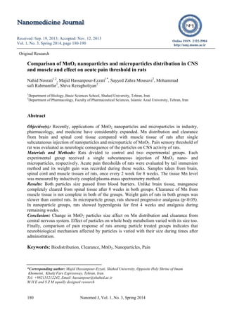
  • 3. Nosrati N, et al 182 Nanomed J, Vol. 1, No. 3, Spring 2014 during this period was compared between MnO2 microparticles and nanoparticles treated groups. Also, pain thresholds of rats in the two groups were determined by tail immersion methods during these weeks for comparison of acute neurological effects of these particles. Materials and Methods Animals Male albino Wistar rats (Pasteur’s institute, Tehran, Iran) weighing 150±5 g were housed in an air-conditioned colony room on a light/dark cycle (21-23°C and a humidity of 30-40%) and supplied with standard diet and tap water ad libitum. Procedures involving animals and their care were conducted in conformity with the NIH guidelines for the care and use of laboratory animals. Selecting the dose The dose of MnO2 nanoparticles and microparticles selected on the basis of You and coworkers report (21). Rats were divided into two groups and treated subcutaneously with solution containing nano- and microparticles of MnO2 (100 µg/kg) dissolved in saline, respectively. Control rats only received normal saline. MnO2 microparticles (Figure 1) used in this research were purchased from MERCK Company, Germany. Figure 1. A scanning electromicrograph of MnO2 micropaticles. The MnO2 nanoparticles preparation MnO2 nanoparticles were prepared via the hydrothermal procedure proposed by Zhang et al., with some modification (22). In practice, 20 ml of KMnO4 (0.2 mM/lit) were mixed with 16 ml MnO4 (0.125 mM/lit) for 5 minutes. The resulting mixture was taken directly into a steel autoclave with Teflon covering and kept for 16 hours at 160C and then was cooled at room temperature. The resulting brown product was collected, washed with distilled water and ethanol 3 times, and dried with hot air current 80C for 12 hours. The resulting particles were scrutinized by an electron microscope and it was ensured that they were 25 to 85 micrometers in size (Figure 2). Preparing MnO2 nanoparticles and microparticles injecting solution. First, the particles were dissolved in normal saline solution. The prepared solutions were sonicated for 30 minutes before injection. Figure 2. Scanning electromicrograph of MnO2 nanopaticles. Tissue Samples Preparation for ICP/MS measurement Male Wistar rats (140 ±10 g) were used in this experiment. Rats were divided into three groups: control, microparticle, and nanoparticle groups. The experimental groups injected subcutaneously with solution containing MnO2 (100 µg/kg) microparticles or nanoparticles, respectively. Control rats received only normal saline. Five rats were chose from each group every two weeks and were deeply anesthetized with diethyl-ether (Merck).
  • 4. Size dependent effect of MnO2 on acute pain threshold in rat Nanomed J, Vol. 1, No. 3, Spring 2014 183 Then, rats scarified and their brains transformed into ice-cold ACSF solution (4C). The spinal cords of rats were pushed from vertebral canal with perfusion of high-pressure of cold ACSF solution into the vertebral canal. Muscle tissue sample was taken from hind paw of rats (squadriceps femoris). All samples were frozen with liquid nitrogen after weighting and kept in a - 20C until ICP-MS measurement. The tissue Mn was extracted by the following procedure: 1/2 gram of each tissue sample was selected and one ml of HNO3 %63 (Merck, Germany) was added to it (23). Then, the samples were kept for 24 hours at 4C for complete digestion of the tissues. The digested samples were centrifuged at 4500 rpm for 5 minutes. The upper clear solution was diluted with extra-pure water and the MnO2 level was measured by ICP-MS (24). The ICP-MS measurement of tissue Mn level A standard Agilent 7500 ICP-MS, fitted with a crossflow nebulizer and a quartz spray chamber was used throughout (25). Operational parameters are given in Table 1. A solution containing 100 PPM of Mn (Merck) was used as the standard solution. Table 1. Operational parameters of ICP/MS for measurement of Mn level in liver tissue extract. Parameter Value Units Argon Gas Flow Rates Plasma 15.0 L/min Auxiliary 1.0 L/min Carrier 1.2 L/min RF Power 1350 W Sampling Depth 6.7 mm Nebulizer Cross-flow Spray Chamber Double-pass, quartz Spray Chamber Temp. 2 °C Sample Uptake 0.4 ml/min Tail immersion test The rat pain threshold was assessed using tail immersion test. After the adaptation, rat tail was immersed in warm water (49°C) and the tail flick response latency (withdrawal response of tail) was observed as the end-point response. Each experiment was repeated 4 times for each animal with an interval of 2 min and its average was reported. Cut-off time of 25 sec was considered (26, 27). Results Tissue Mn level The basal level of Mn in brain, spinal cord, and muscle tissues of control rats is 1 (µg/g tissue wet weight). The maximum content of Mn in brain tissue of microparticles group is higher than nanoparticles group. In MnO2 microparticles group, Mn level in the brain tissues reached to its maximum level 4 weeks after injection (Figure 3). Then, the Mn level in this tissue returned to 77.5% its maximal value after 8 weeks. In MnO2 nanoparticles group, Mn level in brain tissue was reached to its maximum level 2 weeks after injection and its level reduced to 42% its maximum valve after 8 weeks.Variation of Mn level in spinal cord tissues during 8 weeks after the injection of particles is shown in Figure 4. Mn concentration in spinal cord tissues following the injection of MnO2 microparticles attained its maximum level after 4 weeks, but this time for the nanoparticles receiving group was 2 weeks. The variation of Mn level in muscle tissues during 8 weeks after the injection of MnO2 microparticles and nanoparticles is shown in figure 5. In the MnO2 microparticle groups, Mn level in muscle tissue increase very slowly and reach to a plateau after 2th week. But in MnO2 nanoparticle group, the tissue level of Mn increased very fast and reached to high level in second weeks after this particle injection.
  • 5. Nosrati N, et al 184 Nanomed J, Vol. 1, No. 3, Spring 2014 But the tissue Mn level quickly decreased to a basal level at 4th week and remained stable till the end of experiment. Figure 3. Change in Mn level in brain tissues of rat during 8 weeks after single treatment with MnO2 (100µg/kg) microparticles and nanoparticles. Groups compared by ANOVA and tukey post-test, *p<0.05 (n=5). Figure 4. Comparison of Mn level in spinal cord tissues of rat (n=5) in MnO2 microparticles or nanoparticles (100 µg/kg) treated groups during 8 weeks. ANOVA and tukey post-test showed a significant (p<0.05) difference in Mn tissues level in between groups. Weight gain changes The rate of weight gain in rats in nanoparticle group in comparison with control rats significantly (p<0.05) reduced during 2th to 4th week after injection (Figure 6). Weight gain of rats receiving the same dose of microparticles only at the 4th weeks was significantly (p<0.05) lower than control rats (Figure 6). Tail immersion measurement of acute pain The results of tail withdrawal latency of rats in groups during 8 weeks after injections are shown in Figure 7. Figure 5. Change in Mn level in muscle tissues of rats (n=5) in MnO2 microparticles or nanoparticles (100µg/kg) groups during 8 weeks after injection. The ANOVA and Tukey post-test show a significant difference (p<0.05) between two groups. Figure 6. Rat weight gain changes during 8 weeks after single subcutaneous injections of MnO2 microparticles or nanoparticles (100 µg/kg) in comparison with control rats (n=5). *control vs. nanoparticle groups, # control vs. microparticle groups (p<0.05). The mean tail withdrawal latency of rats before the particles injections was 10 seconds. The cut-off time was chosen 25-second. The microparticles injection caused a significant (p<0.05) progressive increase in tail flick latency of rats during 8 weeks. This progressive increase in tail filck latency of rats continued to end of experiment. The rats treated with MnO2 nanoparticles showed a significant (p<0.05) hyperalgesia in the first four weeks after injection and analgesia (p<0.05) during the remaining weeks (Figure 7). Nosrati N, et al 184 Nanomed J, Vol. 1, No. 3, Spring 2014 But the tissue Mn level quickly decreased to a basal level at 4th week and remained stable till the end of experiment. Figure 3. Change in Mn level in brain tissues of rat during 8 weeks after single treatment with MnO2 (100µg/kg) microparticles and nanoparticles. Groups compared by ANOVA and tukey post-test, *p<0.05 (n=5). Figure 4. Comparison of Mn level in spinal cord tissues of rat (n=5) in MnO2 microparticles or nanoparticles (100 µg/kg) treated groups during 8 weeks. ANOVA and tukey post-test showed a significant (p<0.05) difference in Mn tissues level in between groups. Weight gain changes The rate of weight gain in rats in nanoparticle group in comparison with control rats significantly (p<0.05) reduced during 2th to 4th week after injection (Figure 6). Weight gain of rats receiving the same dose of microparticles only at the 4th weeks was significantly (p<0.05) lower than control rats (Figure 6). Tail immersion measurement of acute pain The results of tail withdrawal latency of rats in groups during 8 weeks after injections are shown in Figure 7. Figure 5. Change in Mn level in muscle tissues of rats (n=5) in MnO2 microparticles or nanoparticles (100µg/kg) groups during 8 weeks after injection. The ANOVA and Tukey post-test show a significant difference (p<0.05) between two groups. Figure 6. Rat weight gain changes during 8 weeks after single subcutaneous injections of MnO2 microparticles or nanoparticles (100 µg/kg) in comparison with control rats (n=5). *control vs. nanoparticle groups, # control vs. microparticle groups (p<0.05). The mean tail withdrawal latency of rats before the particles injections was 10 seconds. The cut-off time was chosen 25-second. The microparticles injection caused a significant (p<0.05) progressive increase in tail flick latency of rats during 8 weeks. This progressive increase in tail filck latency of rats continued to end of experiment. The rats treated with MnO2 nanoparticles showed a significant (p<0.05) hyperalgesia in the first four weeks after injection and analgesia (p<0.05) during the remaining weeks (Figure 7). Nosrati N, et al 184 Nanomed J, Vol. 1, No. 3, Spring 2014 But the tissue Mn level quickly decreased to a basal level at 4th week and remained stable till the end of experiment. Figure 3. Change in Mn level in brain tissues of rat during 8 weeks after single treatment with MnO2 (100µg/kg) microparticles and nanoparticles. Groups compared by ANOVA and tukey post-test, *p<0.05 (n=5). Figure 4. Comparison of Mn level in spinal cord tissues of rat (n=5) in MnO2 microparticles or nanoparticles (100 µg/kg) treated groups during 8 weeks. ANOVA and tukey post-test showed a significant (p<0.05) difference in Mn tissues level in between groups. Weight gain changes The rate of weight gain in rats in nanoparticle group in comparison with control rats significantly (p<0.05) reduced during 2th to 4th week after injection (Figure 6). Weight gain of rats receiving the same dose of microparticles only at the 4th weeks was significantly (p<0.05) lower than control rats (Figure 6). Tail immersion measurement of acute pain The results of tail withdrawal latency of rats in groups during 8 weeks after injections are shown in Figure 7. Figure 5. Change in Mn level in muscle tissues of rats (n=5) in MnO2 microparticles or nanoparticles (100µg/kg) groups during 8 weeks after injection. The ANOVA and Tukey post-test show a significant difference (p<0.05) between two groups. Figure 6. Rat weight gain changes during 8 weeks after single subcutaneous injections of MnO2 microparticles or nanoparticles (100 µg/kg) in comparison with control rats (n=5). *control vs. nanoparticle groups, # control vs. microparticle groups (p<0.05). The mean tail withdrawal latency of rats before the particles injections was 10 seconds. The cut-off time was chosen 25-second. The microparticles injection caused a significant (p<0.05) progressive increase in tail flick latency of rats during 8 weeks. This progressive increase in tail filck latency of rats continued to end of experiment. The rats treated with MnO2 nanoparticles showed a significant (p<0.05) hyperalgesia in the first four weeks after injection and analgesia (p<0.05) during the remaining weeks (Figure 7).
  • 6. Size dependent effect of MnO2 on acute pain threshold in rat Nanomed J, Vol. 1, No. 3, Spring 2014 185 Figure 7. Tail flick latency of rats (n=5) following single subcutaneous injections of MnO2 microparticles or nanoparticles (100 µg/kg) in comparison with the control group. *Control vs. experimental groups (p<0.05). Discussion Our results showed that basal amount of Mn in normal rat brain is very low. Previous research also has shown presence of very small amount of Mn in brain tissue of human and other animals (28, 29). The Mn level in brain tissue of control rats in this study was proportional with its level in previous studies (30). Measurement of Mn level in brain and spinal cord of rats following administrations of MnO2 micro and nanoparticles indicate that both types of particles can pass through blood barrier and entered to spinal cord. Previous research has shown Mn can enter and distributed in brain through olfactory bulb (31). It is shown that water soluble and insoluble forms of manganese particles can easily pass through the BBB (32). In this study, Mn clearance rate from brain and spinal cord is not the same for MnO2 nano- and microparticles receiving groups. The spinal cord tissues were completely cleared from Mn after 8 weeks in both treated groups but brain tissues were not completely cleared from Mn during this time. Previously, it was demonstrated that bioclearance of nanoparticles varies among different tissues; even in tissues have not blood barriers (33). The Mn clearance rate from brain tissues in microparticle group is faster than nanoparticles group, but Mn clearance rate from spinal cord tissues was the same for nanoparticles and microparticles treated groups. Sharma and colleagues evaluated permeability range of blood and spinal cord barrier in rats after hyperthermia stimulation with the use of nanoparticles. Their findings suggest that blood-spinal cord barrier permeability variation is more than blood-brain barrier (34). This report confirms our observation about difference of nanoparticles clearance rates from brain and spinal cord. Previously, it is reported that Mn distributed to all areas of rat brain after intracerebroventricular administration of MnO2 nanoparticles within three weeks and then concentration of Mn in brain tissue decreased gradually (35, 36). The time course of this report is equal to time that obtained in our study for reaching of Mn level in brain tissue to maximum value after MnO2 microparticle administration. Size-dependent differences in tissue bioclearance of particles were shown previously, for example, comparison of fluorescent polystyrene bioclearance microparticles (2 micrometers) with its nanoparticles (200 nanometers) after subconjunctival injections in rats showed that most of nanoparticles disappeared from the periocular tissue during 15 days and only %8 of their initial amount remained in tissues up to 7 days after the injection, but micro particles stayed in the injected tissue for more than two months (37). In nanoparticle group, the Mn slowly accumulated in muscle tissues and reached to plateau at fourth week. In micro particles group, the concentration of Mn in muscle tissue show an initial rise in the second week and then returned to a lower level and stayed at this level till the end of study.A similar situation was demonstrated for clearance of gold nanoparticles from liver and spleen tissues of rats (38). Thus, the accumulation and clearance pattern of Mn in each tissue special pattern and also changed with particles size. It should be noted that weight gain changes was measured as indicator of general Size dependent effect of MnO2 on acute pain threshold in rat Nanomed J, Vol. 1, No. 3, Spring 2014 185 Figure 7. Tail flick latency of rats (n=5) following single subcutaneous injections of MnO2 microparticles or nanoparticles (100 µg/kg) in comparison with the control group. *Control vs. experimental groups (p<0.05). Discussion Our results showed that basal amount of Mn in normal rat brain is very low. Previous research also has shown presence of very small amount of Mn in brain tissue of human and other animals (28, 29). The Mn level in brain tissue of control rats in this study was proportional with its level in previous studies (30). Measurement of Mn level in brain and spinal cord of rats following administrations of MnO2 micro and nanoparticles indicate that both types of particles can pass through blood barrier and entered to spinal cord. Previous research has shown Mn can enter and distributed in brain through olfactory bulb (31). It is shown that water soluble and insoluble forms of manganese particles can easily pass through the BBB (32). In this study, Mn clearance rate from brain and spinal cord is not the same for MnO2 nano- and microparticles receiving groups. The spinal cord tissues were completely cleared from Mn after 8 weeks in both treated groups but brain tissues were not completely cleared from Mn during this time. Previously, it was demonstrated that bioclearance of nanoparticles varies among different tissues; even in tissues have not blood barriers (33). The Mn clearance rate from brain tissues in microparticle group is faster than nanoparticles group, but Mn clearance rate from spinal cord tissues was the same for nanoparticles and microparticles treated groups. Sharma and colleagues evaluated permeability range of blood and spinal cord barrier in rats after hyperthermia stimulation with the use of nanoparticles. Their findings suggest that blood-spinal cord barrier permeability variation is more than blood-brain barrier (34). This report confirms our observation about difference of nanoparticles clearance rates from brain and spinal cord. Previously, it is reported that Mn distributed to all areas of rat brain after intracerebroventricular administration of MnO2 nanoparticles within three weeks and then concentration of Mn in brain tissue decreased gradually (35, 36). The time course of this report is equal to time that obtained in our study for reaching of Mn level in brain tissue to maximum value after MnO2 microparticle administration. Size-dependent differences in tissue bioclearance of particles were shown previously, for example, comparison of fluorescent polystyrene bioclearance microparticles (2 micrometers) with its nanoparticles (200 nanometers) after subconjunctival injections in rats showed that most of nanoparticles disappeared from the periocular tissue during 15 days and only %8 of their initial amount remained in tissues up to 7 days after the injection, but micro particles stayed in the injected tissue for more than two months (37). In nanoparticle group, the Mn slowly accumulated in muscle tissues and reached to plateau at fourth week. In micro particles group, the concentration of Mn in muscle tissue show an initial rise in the second week and then returned to a lower level and stayed at this level till the end of study.A similar situation was demonstrated for clearance of gold nanoparticles from liver and spleen tissues of rats (38). Thus, the accumulation and clearance pattern of Mn in each tissue special pattern and also changed with particles size. It should be noted that weight gain changes was measured as indicator of general Size dependent effect of MnO2 on acute pain threshold in rat Nanomed J, Vol. 1, No. 3, Spring 2014 185 Figure 7. Tail flick latency of rats (n=5) following single subcutaneous injections of MnO2 microparticles or nanoparticles (100 µg/kg) in comparison with the control group. *Control vs. experimental groups (p<0.05). Discussion Our results showed that basal amount of Mn in normal rat brain is very low. Previous research also has shown presence of very small amount of Mn in brain tissue of human and other animals (28, 29). The Mn level in brain tissue of control rats in this study was proportional with its level in previous studies (30). Measurement of Mn level in brain and spinal cord of rats following administrations of MnO2 micro and nanoparticles indicate that both types of particles can pass through blood barrier and entered to spinal cord. Previous research has shown Mn can enter and distributed in brain through olfactory bulb (31). It is shown that water soluble and insoluble forms of manganese particles can easily pass through the BBB (32). In this study, Mn clearance rate from brain and spinal cord is not the same for MnO2 nano- and microparticles receiving groups. The spinal cord tissues were completely cleared from Mn after 8 weeks in both treated groups but brain tissues were not completely cleared from Mn during this time. Previously, it was demonstrated that bioclearance of nanoparticles varies among different tissues; even in tissues have not blood barriers (33). The Mn clearance rate from brain tissues in microparticle group is faster than nanoparticles group, but Mn clearance rate from spinal cord tissues was the same for nanoparticles and microparticles treated groups. Sharma and colleagues evaluated permeability range of blood and spinal cord barrier in rats after hyperthermia stimulation with the use of nanoparticles. Their findings suggest that blood-spinal cord barrier permeability variation is more than blood-brain barrier (34). This report confirms our observation about difference of nanoparticles clearance rates from brain and spinal cord. Previously, it is reported that Mn distributed to all areas of rat brain after intracerebroventricular administration of MnO2 nanoparticles within three weeks and then concentration of Mn in brain tissue decreased gradually (35, 36). The time course of this report is equal to time that obtained in our study for reaching of Mn level in brain tissue to maximum value after MnO2 microparticle administration. Size-dependent differences in tissue bioclearance of particles were shown previously, for example, comparison of fluorescent polystyrene bioclearance microparticles (2 micrometers) with its nanoparticles (200 nanometers) after subconjunctival injections in rats showed that most of nanoparticles disappeared from the periocular tissue during 15 days and only %8 of their initial amount remained in tissues up to 7 days after the injection, but micro particles stayed in the injected tissue for more than two months (37). In nanoparticle group, the Mn slowly accumulated in muscle tissues and reached to plateau at fourth week. In micro particles group, the concentration of Mn in muscle tissue show an initial rise in the second week and then returned to a lower level and stayed at this level till the end of study.A similar situation was demonstrated for clearance of gold nanoparticles from liver and spleen tissues of rats (38). Thus, the accumulation and clearance pattern of Mn in each tissue special pattern and also changed with particles size. It should be noted that weight gain changes was measured as indicator of general
  • 7. Nosrati N, et al 186 Nanomed J, Vol. 1, No. 3, Spring 2014 health status of animals. The reduction in weight gain of microparticle receiving rats continued for two weeks after the injection, while this effect in nanoparticle-receiving group was longer and continued for four weeks after the injection. Our results show a relation between tissues level of Mn and weight changes of rats. Little information was available about the acute effect of metal oxide nanoparticles on weight changes of animals and most of the researchers have studied the effects of chronic administration of metal oxide particles on the weight changes of animals. Besides, the effect of nanoparticles on weight gain changes of animals varies from one metal oxide nanoparticle to another. For example, intraperitoneal injections of TiO2 nanoparticles for 3-month to rats have not any significant effects on its weight gain (39). On the contrary, inhalation of Fe2O3 nanoparticles significantly affects on rat weight gain (40). Scientists related these differences in nanoparticles effect on body weight to their oxidative power (41). In support of this statement, it is shown that manganese oxides can cause impaired mitochondrial redox activity (18). MnO2 oxidative power is higher than other form of Mn oxides, such as Mn3O4 (42). Therefore, our results indicate MnO2 particles in both size can influence on the whole body metabolism in rats, moreover the effect of the nanoparticle is slightly higher than the microparticles. Tail flick latency of rats was evaluated in order to compare effects of the particles on nervous system activity (43). Scientists previously are used tail immersion latency test for evaluation of analgesia duration after subcutaneously injected nanoparticles (44). Tail immersion test considered as a model of acute thermal pain (45). Acute pain results from disease, inflammation, or injury to tissue. Inflammatory processes contribute to nerve cell toxicity (46). Our results indicate that both size of MnO2 particles influenced on pain thresholds of rats. The administration of microparticles led to a gradual, irreversible increase in the pain thresholds of rats during eight weeks. However, nanoparticles produced biphasic response in rat, initial hyperalgesia for four weeks and late analgesic during last four weeks. On the other hand it has been reported that inhalation of MnO2 particles caused changes in the neurochemistry of brain dopaminergic system (47) and severely affect on expression of genes like MAOA, Park2, and SNCA (48). Dopamine also plays a clear role in pain control at the spinal cord level (49, 50) and dysfunctions in this system in spinal cord level can produce deficits in pain sensation (51). Patients with Parkinson's disease showed a progressive hyperalgesia similar to rats treated the MnO2 micro particles in our study (52, 53). As a conclusion we can say that MnO2 particles effects on acute pain threshold probably mediated via dopaminergic systems and progressive nature of their effect is similar to Parkinson's disease. As explanation for biphasic effect of MnO2 nanoparticles on rat pain responses and according to Wang’s finding, a direct relation exists between the level of Mn nanoparticles accumulation in central nervous system and its neurological symptoms (54). Tissue Mn concentration increased considerably after nanoparticle injection during the first four weeks and then decreased and remained constant during the remaining ones. Thus, it seems that the neural mechanisms that modulated by MnO2 nanoparticles in the first four weeks after injection are different from late-phase mechanisms. It is shown that manganese ions can interrupt the transmission of stimulatory signals in the nervous system through changes in the activity of calcium channels at pre-synaptic terminals (26). Also, neuronal evoked potentials of rats showed a significant reduction after long-
  • 8. Size dependent effect of MnO2 on acute pain threshold in rat Nanomed J, Vol. 1, No. 3, Spring 2014 187 term inhalation of MnO2 nanoparticles (55). The reduction in activity of descending pain modulation tract can induce hyperalgesia (56). Thus, it seems that during first four weeks after injection, nanoparticles activate central descending mechanisms that induced hyperalgesia in rats. Also these mechanisms can mask the MnO2 nanoparticle analgesic effects in this phase. But, at the end of first phase and termination of these fast effects, the long term analgesic effect of nanoparticles via effect on dopaminergic system is appeared. In support of our hypothesis that central dopaminergic system that mediate analgesic effect of MnO2 nanoparticles, it is reported that depletion of dopaminergic neurons in periaqueductal grey areas of rat brain can induce analgesia (20). Conclusions Administrations of microparticles or nanoparticles of MnO2 caused Mn accumulation in brain, spinal cord and muscle tissues of rats after single injection for long time. Both sizes of MnO2 have adverse effects on general metabolism of rats and reduce normal rate of rats weight gain. The MnO2 nanoparticle or microparticle has different effect on pain sensation. Probably, change in Mn level in CNS with effect on dopaminergic system mediates its effects on pain modulating systems of rats. Acknowledgment This work was supported by Islamic Azad University, Tehran, Iran and contribution of Shahed University of Iran. We also thanks from Dr Hassani in ICP/MS lab of Tarbiat Modares University for his helps. References 1. Allaker RP, Ren G. Potential impact of nanotechnology on the control of infectious diseases. Trans R Soc Trop Med Hyg. 2008; 102(1): 1-2. 2. Prasad B, Mondal KK. Environmental impact of manganese due to its leaching from coal fly ash. J Environ Sci Eng. 2009; 51(1): 27-32. 3. Najafpour MM, Rahimi F, Aro EM, Lee CH, Allakhverdiev SI. Nano-sized manganese oxides as biomimetic catalysts for water oxidation in artificial photosynthesis: a review. J R Soc Interface. 2012; 9(75): 2383-2395. 4. Davis CD, Wolf TL, Greger JL. Varying levels of manganese and iron affect absorption and gut endogenous losses of manganese by rats. J Nutr. 1992; 122(6): 1300-1308. 5. Na HB, Lee JH, An K, Park YI, Park M, Lee IS, et al. Development of a T-1 contrast agent for magnetic resonance imaging using MnO nanoparticles. Angew Chem Int Ed Engl. 2007; 46(28): 5397- 5401. 6. Roels H, Meiers G, Delos M, Ortega I, Lauwerys R, Buchet JP, et al. Lison, Influence of the route of administration and the chemical form (MnCl2, MnO2) on the absorption and cerebral distribution of manganese in rats. Arch Toxicol. 1997; 71(4): 223-230. 7. Oszlánczi G, Vezér T, Sárközi L, Horváth E, Kónya Z, Papp A. Functional neurotoxicity of Mn-containing nanoparticles in rats. Ecotoxicol Environ Saf. 2010; 73(8): 2004-2009. 8. Wang SH, Shih YL, Kuo TC, Ko WC, Shih CM. Cadmium toxicity toward autophagy through ROS-activatied GSK- 3beta in mesangial cells. Toxicol Sci. 2009; 108(1): 124-131. 9. Nel A, Xia T, Mädler L, Li N. Toxic potential of materials at the nanolevel. Science. 2006; 311(5761): 622-627. 10. Bowler RM, Roels HA, Nakagawa S, Drezgic M, Diamond E, Park R, et al. Dose-effect relationships between manganese exposure and neurological, neuropsychological and pulmonary function in confined space bridge welders. Occup Environ Med. 2007; 64(3): 167- 177. 11. Lison D, Lardot C, Huaux F, Zanetti G, Fubini B. Influence of particle surface area on the toxicity of insoluble manganese dioxide dusts. Arch Toxicol. 1997; 71(12): 725-729. 12. Schmidt C, Lautenschlaeger C, Collnot EM, Schumann M, Bojarski C, Schulzke JD, et al. Nano- and microscaled particles for drug targeting to inflamed intestinal
  • 9. Nosrati N, et al 188 Nanomed J, Vol. 1, No. 3, Spring 2014 mucosa-A first in vivo study in human patients. J Control Release. 2013; 165(2): 139-145. 13. J Kim JS, Yoon TJ, Yu KN, Kim BG, Park SJ, Kim HW, et al. Toxicity and tissue distribution of magnetic nanoparticles in mice. Toxicol Sci. 2006; 89(1): 338-347. 14. Nishiyama K, Suzuki Y, Fujii N, Yano H, Ohnishi K, Miyai T. Biochemical changes and manganese distribution in monkeys exposed to manganese dioxide dust. Tokushima J Exp Med. 1977; 24(3-4): 137-145. 15. Li Y, Tian X, Lu Z, Yang C, Yang G, Zhou X, et al. Mechanism for α-MnO2 nanowire-induced cytotoxicity in Hela Cells. J Nanosci Nanotechnol. 2010; 10(1): 397-404. 16. Muthu MS, Singh S. Targeted nanomedicines: effective treatment modalities for cancer, AIDS and brain disorders. Nanomedicine (Lond). 2009; 4(1): 105-118. 17. Sarkozy L, Horvath E, Szabo A, Horvath E, Sapi A, Konya Z, et al. Neurotoxic effects of metal oxide nanoparticles on the somatosensory system of rats following subacute intratracheal application. CEJOEM. 2008; 14(3): 277-290. 18. Yang Z, Liu ZW, Allaker RP, Reip P, Oxford J, Ahmad Z, et al. A review of nanoparticle functionality and toxicity on the central nervous system. J R Soc Interface. 2010; 7 Suppl 4: S411-422. 19. Wang J, Chen C, Liu Y, Jiao F, Li W, Lao F, et al. Potential neurological lesion after nasal instillation of TiO2 nanoparticles in the anatase and rutile crystal phases. Toxicol Lett. 2008; 183(1-3): 72-80. 20. Flores JA, El Banoua F, Galán-Rodríguez B, Fernandez-Espejo E. Opiate anti- nociception is attenuated following lesion of large dopamine neurons of the periaqueductal grey: critical role for D1 (not D2) dopamine receptors. Pain. 2004; 110(1-2): 205-214. 21. You H, Zhao J, Li L, Yuan J. Effects of MnO2 Nanoparticles on Liver and Kidney Cells of Rats. Proceedings of the 5th International Conference on Bioinformatics and Biomedical Engineering (iCBBE) 2011; May 10-12; Wuhan, China, p. 1-4. 22. Zhang Y, Yang Y, Zhang Y, Zhang T, Ye M. Heterogeneous oxidation of naproxen in the presence of α-MnO2 nanostructures with different morphologies. Appl. Catal. B. 2012; 127: 182-189. 23. Vanhoe H. A review of the capabilities of ICP-MS for trace element analysis in body fluids and tissues. J Trace Elem Electrolytes Health Dis. 1993; 7(3): 131- 139. 24. Niazi SB, Littlejohn D, Halls DJ. Rapid partial digestion of biological tissues with nitric acid for the determination of trace elements by atomic spectrometry. Analyst. 1993; 118(7): 821-825. 25. Pearson GF, Greenway GM. Recent developments in manganese speciation. Trends Anal Chem. 2005; 24(9): 803-809. 26. Baluchnejadmojarad T, Roghani M. Chronic oral epigallocatechin-gallate alleviates streptozotocin-induced diabetic neuropathic hyperalgesia in rat: involvement of oxidative stress. Iran. J Pharm Res. 2012; 11 (4): 1243-1253. 27. Yang In Kim, Heung Sik Na, Jung Soo Han, and Seung Kil Hong. Critical role of the capsaicin-sensitive nerve fibers in the development of the causalgic symptoms produced by transecting some but not all of the nerves innervating the rat tail. J Neurosci. 1995; 75(6): 4133-4139. 28. Takeda A. Manganese action in brain function: Review. Brain Res Brain Res Rev. 2003; 41(1): 79-87. 29. Santamaria AB. Manganese exposure, essentiality & toxicity. Indian J Med Res. 2008; 128(4): 484-500. 30. Reaney SH, Bench G, Smith DR. Brain accumulation and toxicity of Mn(ii) and Mn(iii) exposures. Toxicol Sci. 2006 Sep; 93(1):114-124. 31. Elder A, Gelein R, Silva V, Feikert T, Opanashuk L, Carter J, et al. Translocation of inhaled ultrafine manganese oxide particles to the central nervous system. Environ Health Perspect. 2006; 114(8): 1172-1178. 32. Brenneman KA, Wong BA, Buccellato MA, Costa ER, Gross EA, Dorman DC. Direct olfactory transport of inhaled manganese ((54)MnCl(2)) to the rat brain: toxicokinetic investigations in a unilateral nasal occlusion model. Toxicol Appl Pharmacol. 2000; 169(3): 238-248. 33. Gu L, Fang RH, Sailor MJ, Park JH. In Vivo clearance and toxicity of monodisperse iron oxide nanocrystals. ACS Nano. 2012; 6(6): 4947-4954. 34. Sharma HS, Sharma A. Breakdown of the blood-brain barrier in stress alters cognitive dysfunction and induced brain pathology, new perspective for neuroprotective strategies. In: Michel Ritsner, editor., Brain Protection in Schizophrenia, Mood and Cognitive Disorders, Volume 1. Netherlands:
  • 10. Size dependent effect of MnO2 on acute pain threshold in rat Nanomed J, Vol. 1, No. 3, Spring 2014 189 Springer; 2010. p. 243-304. 35. Gallez B, Demeure R, Baudelet C, Abdelouahab N, Beghein N, Jordan B, et al. Non invasive quantification of manganese deposits in the rat brain by local measurement of NMR proton T1 relaxation times. Neurotoxicology. 2001; 22(3): 387-392. 36. Takeda A, Sawashita J, Okada S. Localization in rat brain of the trace metals, zinc and manganese, after intracerebroventricular injection. Brain Res. 1994; 658(1-2): 252-254. 37. Amrite AC, Kompella UB. Size- dependent disposition of nanoparticles and microparticles following subconjunctival administration. J Pharm Pharmacol. 2005; 57(12): 1555-1563. 38. Balasubramanian SK, Jittiwat J, Manikandan J, Ong CN, Yu LE, Ong WY. Biodistribution of gold nanoparticles and gene expression changes in the liver and spleen after intravenous administration in rats. Biomaterials. 2010; 31(8): 2034-2042. 39. Warheit DB, Hoke RA, Finlay C, Donner EM, Reed KL, Sayes CM. Development of a base set of toxicity tests using ultrafine TiO2 particles as a component of nanoparticle risk management. Toxicol Lett. 2007; 171(3): 99-110. 40. Szalay B, Tátrai E, Nyírő G, Vezér T, Dura G. Potential toxic effects of iron oxide nanoparticles in in vivo and in vitro experiments. J Appl Toxicol. 2012; 32(6): 446-453. 41. Brieger K, Schiavone S, Miller FJ Jr, Krause KH. Reactive oxygen species: from health to disease. Swiss Med Wkly. 2012; 142: w13659. Available from URL: doi:10.4414/smw.2012.13659 42. Stefanescu DM, Khoshnan A, Patterson PH, Hering JG. Neurotoxicity of manganese oxide nanomaterials. J Nanopart Res. 2009; 11(8): 1957-1969. 43. van Rijn RM, Brissett DI, Whistler JL. Emergence of functional spinal delta opioid receptors after chronic ethanol exposure. Biol Psychiatry. 2012; 71(3): 232-238. 44. Yin QQ, Wu L, Gou ML, Qian ZY, Zhang WS, Liu J. Long-lasting infiltration anaesthesia by lidocaine- loaded biodegradable nanoparticles in hydrogel in rats. Acta Anaesthesiol Scand. 2009; 53(9): 1207-1213. 45. Ahmadi A, Khalili M, Mihandoust F, Barghi L. Synthesis and determination of acute and chronic pain activities of 1- [1-(3-methylphenyl) (tetralyl)]piperidine as a new derivative of phencyclidine via tail immersion and formalin tests. Arzneimittelforschung. 2010; 60(1): 30- 5. 46. Fernandes A, Brites D. Contribution of inflammatory processes to nerve cell toxicity by bilirubin and efficacy of potential therapeutic agents. Curr Pharm Des. 2009; 15(25): 2915-2926. 47. Bird ED, Anton AH, Bullock B. The effect of manganese inhalation on basal ganglia dopamine concentration in rhesus monkey, Neurotoxicology. 1984; 5(1): 59-65. 48. Wang J, Rahman MF, Duhart HM, Newport GD, Patterson TA, Murdock RC, et al. Expression changes of dopaminergic system-related genes in PC12 cells induced by manganese, silver, or copper nanoparticles. Neurotoxic ology. 2009; 30(6): 926-933. 49. Liu QS, Qiao JT, Dafny N. D2 dopamine receptor involvement in spinal dopamine-produced antinociception. Life Sci. 1992; 51(19): 1485-1492. 50. Barasi S, Ben-Sreti MM, Clatworthy AL, Duggal KN, Gonzalez JP, Robertson J, et al. Dopamine receptor- mediated spinal antinociception in the normal and haloperidol pretreated rat: effects of sulpiride and SCH 23390. Br J Pharmacol. 1987; 90(1): 15-22. 51. Gao X, Xin BM, Zhu CB, Wu GC, Xu SF. Effect of intrathecal injection of dopamine receptor agonists/antagonists on pain and acupuncture analgesia in rats. Sheng Li Xue Bao. 1998; 50(1): 43-48. 52. Greco R, Tassorelli C, Armentero MT, Sandrini G, Nappi G, et al. Role of central dopaminergic circuitry in pain processing and nitroglycerin-induced hyperalgesia. Brain Res. 2008; 1238: 215-223. 53. Borsook D, Upadhyay J, Chudler EH, Becerra L. A key role of the basal ganglia in pain and analgesia - insights gained through human functional imaging. Mol Pain. 2010; 6: 27. 54. Wang J, Liu Y, Jiao F, Lao F, Li W, Gu Y, et al. Time-dependent translocation and potential impairment on central nervous system by intranasally instilled TiO2 nanoparticles. Toxicology. 2008; 254(1-2): 82-90. 55. Sárközi L, Horváth E, Kónya Z, Kiricsi I, Szalay B, Vezér T, et al. Subacute intratracheal exposure of rats to
  • 11. Nosrati N, et al 190 Nanomed J, Vol. 1, No. 3, Spring 2014 manganese nanoparticles: behavioral, electrophysiological, and general toxicological effects. Inhal Toxicol. 2009; 21 Suppl 1: 83-91. 56. Hudspith MJ, Harrisson S, Smith G, Bountra C, Elliot PJ, Birch PJ, Hunt SP, Munglani R. Effect of post-injury NMDA antagonist treatment on long- term Fos expression and hyperalgesia in a model of chronic neuropathic pain. Brain Res. 1999; 822(1-2): 220-227.